Vitrectomy may prevent the occurrence of diabetic macular oedema
Abstract.
Purpose: This study aimed to demonstrate that vitrectomy may prevent the occurrence of diabetic macular oedema (DMO).
Methods: Three patients with diabetes type 1 underwent vitrectomy in one eye to treat complications of proliferative diabetic retinopathy.
Results: During follow-up, all patients suffered unilateral macular oedema in the non-vitrectomized eye as a result of general metabolic changes. In two of these patients, the DMO resolved with management of the underlying medical condition.
Conclusions: These case reports suggest the vitreous may play a role in the occurrence of DMO associated with general risk factors. Further studies are needed to increase understanding of the mechanisms involved in the development and progression of DMO.
Introduction
Macular oedema is the main cause of visual loss in diabetes patients. The increase in retinal capillary permeability that leads to diabetic macular oedema (DMO) is mainly caused by a breakdown of the blood–retinal barrier (Lewis 2001). Several studies have demonstrated vascular endothelial growth factor (VEGF) involvement in the mechanisms that modulate vascular permeability and angiogenesis (Aiello et al. 1994; Boyd et al. 2002). Upregulation of VEGF has been reported in the vitreous of the diabetic eye, along with a reciprocal decrease in pigment epithelium-derived factor (PEDF) (Patel et al. 2006). Recently, Hayreh (2008) suggested that retinal hypoxia may play a role in DMO. Oxygenation has an effect on arteriolar constriction and production of VEGF.
Furthermore, systemic factors such as unbalanced glycaemia, arterial hypertension and pregnancy may increase macular oedema.
We report three cases of young, diabetes type 1 patients who had been vitrectomized in one eye previously and who developed macular oedema in the fellow eye as a result of systemic factors.
Case Reports
Case 1
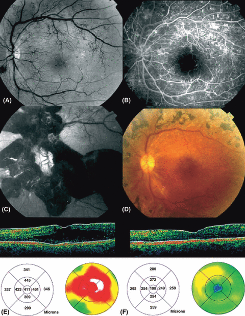
A 21-year-old woman with an 11-year history of insulin-treated diabetes type 1 was referred by her endocrinologist for ocular evaluation. On presentation, best corrected visual acuity (BCVA) was 20/20 in the right eye and 20/25 in the left. She was diagnosed as having proliferative diabetic retinopathy (Fig. 1A, B) and received panretinal photocoagulation (PRP) in both eyes. Six months later, the patient presented with acute visual loss in the left eye. Funduscopy showed an extensive subhyaloid haemorrhage (Fig. 1C).

Fundus photography and optical coherence tomography (OCT) in patient 1. (A) Red-free fundus photography and (B) fluorescein angiography show proliferative diabetic retinopathy in the left eye. (C) Subhyaloid haemorrhage is observed in the left eye. (D) Fundus photography of the left eye after vitrectomy. OCT scans and mapping images of the (E) right and (F) left eyes demonstrate unilateral diabetic macular oedema in the right eye.
The patient underwent vitrectomy with incision and peeling of the posterior hyaloid, and photocoagulation was completed (Fig. 1D). This treatment was effective and BCVA in the left eye improved to 20/25. At that time, BCVA in the right eye was 20/20.
Three years later, the subject presented with visual loss in her right eye (20/50). Optical coherence tomography (OCT) demonstrated cystoid macular oedema in the right eye (Fig. 1E). Central foveal thickness (CFT) was 411 μm. No vitreoretinal traction was demonstrable. In her left, previously vitrectomized eye, BCVA was 20/25 and CFT was 198 μm (Fig. 1F).
Systemic findings showed high blood pressure (systolic arterial pressure of 220 mmHg) and unbalanced diabetes (glycosylated haemoglobin [HbA1c] 12%). With medical treatment of systemic abnormalities, macular oedema progressively decreased and BCVA improved to 20/32.
Case 2
A 30-year-old woman with a 22-year history of insulin-treated diabetes type 1 presented with proliferative diabetic retinopathy with fibrovascular tractional retinal detachment in the right eye. She underwent combined surgery including vitrectomy, fibrovascular dissection and endolaser associated with phacoemulsification. Proliferative diabetic retinopathy in the left eye was treated by PRP. Best corrected VA was 20/125 in the right eye and 20/60 in the left.
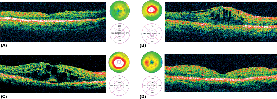
Five years after surgery, the patient presented with a 6-week unplanned pregnancy. Her BCVA was 20/32 in the right eye and 20/25 in the left. Fundus examination showed inactivated diabetic retinopathy without macular oedema. The patient’s HbA1c level was 8.0% and intensive insulin therapy was begun; a few weeks later HbA1c was 6.6%. At 22 weeks of pregnancy, the patient complained of visual loss in the left eye. Clinical and OCT examinations disclosed macular oedema in the left eye, but not in the right. In the left eye, BCVA was 20/40 and CFT was 530 μm without a tractional component (Fig. 2A, B). In the right eye, BCVA was 20/32 and CFT was 233 μm. After delivery, macular oedema resolved spontaneously and BCVA improved to 20/30 in the left eye.

Optical coherence tomography scans and mapping images in (A, B) patient 2 and (C, D) patient 3. Diabetic macular oedema is present in the left eye of patient 2 (B), and the right eye of patient 3 (C).
Case 3
A 30-year-old man with an 18-year history of diabetes type 1 treated by insulin received bilateral PRP and macular grid for proliferative diabetic retinopathy combined with macular oedema. Two years later, vitrectomy for vitreous haemorrhage was performed in the left eye. Two years after this, the patient complained of acute visual loss in the right eye (BCVA 20/100) caused by macular oedema without traction (CFT = 659 μm) (Fig. 2C, D). In the left eye, BCVA was 20/25 and CFT was 210 μm. Systemic findings showed unbalanced diabetes (daily glycaemia of 7–22 mmol/l).
Discussion
In the present study, DMO developed in three patients as a result of systemic factors, including high blood pressure, unbalanced diabetes and a worsening of diabetic retinopathy caused by unplanned pregnancy. No vitreomacular traction component was identified, and medical management alone improved the DMO in two patients. That DMO occurred in only one eye in each case, and not in the fellow eye, which had been vitrectomized previously, suggests that vitrectomy may protect against the occurrence of DMO.
The vitreous could be implicated in the development or exacerbation of DMO through several mechanisms. Many reports have suggested that vitrectomy may be beneficial in the treatment of macular oedema with vitreomacular traction (Kaiser et al. 2001). Nevertheless, some studies report beneficial effects of vitrectomy for diffuse DMO, even without any visible tractional component at the vitreoretinal interface (Otani & Kishi 2002; Recchia et al. 2005; Stolba et al. 2005; Yanyali et al. 2005).
Vitreous gel contains several growth factors, including VEGF, which increase retinal vascular permeability (Aiello et al. 1994). Its removal during vitrectomy could reduce their presence in the eye, decreasing viscosity and thus facilitating the circulation of intraocular molecules. Vitrectomy increases the diffusion of growth factors and their clearance (Stefánsson & Loftsson 2006). Furthermore, vitrectomy with surgically induced posterior vitreous detachment may not only release any unobserved vitreous traction on the macula but may also eliminate the condensed fluid containing growth factors.
Oxygenation has a direct influence on DMO (Nguyen et al. 2004). Increased oxygenation induces arteriolar constriction, which decreases hydrostatic pressure in the capillaries and venules and the outward flux of fluid. Increased oxygenation also decreases the production of VEGF (Stefánsson 2006). Kadonosono et al. (2000) showed that vitreous surgery may improve perifoveal microcirculation in the eyes of diabetes patients with cystoid macular oedema and resolve macular oedema. Vitrectomy appears to increase the oxygen levels in the eye and reduces hypoxia in ischaemic areas of the retina (Stefánsson & Landers 2006). However, this time-limited increase in vitreous cavity oxygen pressure has been correlated to continuous perfusion of balanced salt solution during vitrectomy in rabbits (Barbazetto et al. 2004) and miniature pigs (Petropoulos IK, et al. IOVS 2006; 47: ARVO E-Abstract 4665).
These case reports suggest that the vitreous plays a role in the evolution of DMO and that vitrectomy may prevent the occurrence of DMO. Further studies are needed to understand the mechanisms involved in the development and progression of DMO.


