Relationship between pancreatic and/or extrapancreatic lesions and serum IgG and IgG4 levels in IgG4-related diseases
Abstract
OBJECTIVE: We aimed to investigate the relationship between the number of involved organs or regions and serum immunoglobulin G (IgG) and immunoglobulin G4 (IgG4) levels.
METHODS: The number of pancreatic and/or extrapancreatic lesions and serum IgG and IgG4 levels were examined by groups in 46 patients with IgG4-related diseases at diagnosis prior to the initiation of steroid treatment: group A (one region involved, n = 7), group B (two regions involved, n = 11), group C (three regions involved, n = 12), group D (four regions involved, n = 9) and group E (five to seven regions involved, n = 7).
RESULTS: Both serum IgG and IgG4 levels increased with the number of inflamed regions. Mean serum IgG levels were 15.11, 18.65, 20.92, 23.29 and 30.98 g/L while the mean IgG4 levels were 3.99, 4.70, 4.70, 9.86 and 16.49 g/L in group A, B, C, D and E, respectively. Regression analysis also suggested that IgG4 was positively correlated with the number of regions involved. Additionally, serum IgG4 was higher in patients with multiple lesions when accompanied by sclerosing sialadenitis.
CONCLUSION: Patients having IgG4-related disease with high serum IgG and IgG4 levels should be systematically examined for involved lesions.
INTRODUCTION
Autoimmune pancreatitis (AIP) is a disease concept which was proposed in Japan, and the autoimmune mechanism is believed to be involved in its onset.1,2 Kamisawa et al.3 suggested that AIP is a systemic sclerosing disease, as it is sometimes accompanied by extrapancreatic lesions (EPLs) including sclerosing cholangitis, sclerosing sialadenitis, retroperitoneal fibrosis, abdominal/hilar lymphadenopathy, chronic thyroiditis and interstitial nephritis.4 In recent years the concept of immunoglobulin G4 (IgG4)-related disease has been put forward, proposing that they are disorders of unknown causes characterized by simultaneous or heterochronous swelling and nodular or thickened lesions in multiple organs resulting from diffuse lymphoplasmacytic infiltration and fibrosis.3 And Hamano et al.5 reported increased serum IgG4 levels in Japanese patients with AIP. Therefore, AIP may be an IgG4-related pancreatic disease.6
Some researchers have suggested that serum IgG4 level is an indicator for both the diagnosis7,8 and disease activity9 of IgG4-related disease; however, more detailed analysis on the clinical significance of serum IgG4 levels is required.
Immunological examinations of AIP patients have shown a high incidence of hypergammaglobulinemia and increased serum immunoglobulin G (IgG) as well as IgG4 levels.10 The sensitivity and specificity of serum IgG were 70% and 75%, respectively, which were lower than those of IgG4. In IgG4-related disease, the clinical significance of serum IgG for both diagnosis and disease activity is not well understood either.
In the present study we focused on the number of organs and regions involved in IgG4-related diseases and investigated the relationship between the number of IgG4-related pancreatic lesion (PL)/EPLs and serum IgG and IgG4 levels.
PATIENTS AND METHODS
The study protocol was approved by the Ethics Committee of Kyushu University Hospital (Fukuoka, Japan), within which the work was undertaken and conformed to the World Medical Association's Declaration of Helsinki.
Patients
A total of 53 patients (48 AIP and 5 IgG4-related diseases without PL) diagnosed in Department of Medicine and Bioregulatory Sciences, Graduate School of Medical Sciences, Kyushu University from June 2003 to September 2010 were enrolled in the study. Patients with a serum IgG4 level < 1.35 g/L and idiopathic duct-centric pancreatitis (IDCP) were excluded.
Diagnosis
Diagnosis of AIP was made based on the Japanese diagnostic criteria for AIP (proposed in 200210, revised in 200611), the Asian criteria12 or HISORt criteria13. Diagnosis of IgG4-related diseases without PL was made based on elevated serum IgG4 with a biopsy of swollen salivary glands or imaging findings.
The number of lesions and serum IgG and IgG4 levels of each patient were determined at diagnosis and prior to the initiation of steroid treatment. PL and EPLs were comprehensively examined by abdominal ultrasound, computed tomography (CT) of head, neck, chest and abdomen, abdominal magnetic resonance imaging (MRI), endoscopic retrograde cholangiopancreatography (ERCP), endoscopic ultrasound (EUS), positron emission tomography (PET)-CT and/or gallium scintigraphy. Malignant diseases were excluded by cytological examination of pancreatic juice obtained during ERCP or EUS-guided fine-needle aspiration (FNA).
Altogether 7 patients were excluded from the study: one young woman with ulcerative colitis (UC) was histologically diagnosed with IDCP (or type 2 AIP14), and the other 6 were diagnosed as seronegative AIP15 (serum IgG4 level < 1.35 g/L).
Serum levels of IgG and IgG4 were examined prior to the initiation of steroid treatment, and the relationship between serum IgG and IgG4 levels and the number of regions involved (PL and/or EPLs) was examined. Patients were grouped based on the number of regions involved as follows: group A, only one region involved (n = 7, AIP only); group B, two regions involved (n = 11; e.g., AIP and sclerosing cholangitis or AIP and hilar lymphadenopathy); group C, three regions involved (n = 12; e.g., AIP, sclerosing cholangitis and retroperitoneal fibrosis); group D, four regions involved (n = 9; e.g., sclerosing sialadenitis, pseudotumor of lacrimal gland, hilar lymphadenopathy and interstitial pneumonia); group E, five to seven regions involved (n = 7; e.g., AIP, sclerosing cholangitis, sclerosing sialadenitis, pseudotumor of lacrimal gland and hilar lymphadenopathy).
Statistical analysis
Data were analyzed using SPSS 17.0 (SPSS Inc., Chicago, IL, USA). Serum IgG and IgG4 levels were expressed as mean ± standard deviation (SD). Differences between groups were analyzed using Scheffe's test. Regression analysis was performed to investigate the relationship between serum IgG4 and the number of regions involved. A Mann–Whitney U-test was performed to compare the differences between groups with or without sclerosing sialadenitis. P < 0.05 was considered as statistically significant.
RESULTS
Clinical characteristics at diagnosis
Table 1 shows the clinical characteristics of the patients at diagnosis. Most patients were elderly (mean age 67 years) with a male predominance. In all, 41 patients were diagnosed as AIP with mean serum IgG and IgG4 levels of 21.25 g/L and 6.68 g/L, respectively, in which 34 (82.9%) had EPLs. The other five had IgG4-related diseases without PL, among which biopsies of the swollen salivary gland were performed in 4 with sclerosing sialadenitis, while the other one was diagnosed with retroperitoneal fibrosis by abdominal CT.
| Number of patients | 46 |
| Gender (n) | |
| Male | 33 |
| Female | 13 |
| Age [years, mean (range)] | 67 (35–84) |
| Autoimmune pancreatitis (n) | 41 |
| PL only | 7 |
| EPLs coexisting | 34 |
| Serum IgG level (g/L, mean ± SD) | 21.25 ± 9.85 |
| Serum IgG4 level (g/L, mean ± SD) | 6.68 ± 6.27 |
| IgG4-related diseases without PL (n) | 5 |
- PL, pancreatic lesion; EPLs, extrapancreatic lesions; IgG, immunoglobulin G; IgG4, immunoglobulin G4; SD, standard deviation.
The involved lesions were examined. AIP (89.1%) was the most common IgG4-related disease, while sclerosing cholangitis, sclerosing sialadenitis, lacrimal gland pseudotumor and retroperitoneal fibrosis were also observed in a number of patients (Table 2). The lymph nodes were divided into cervical, hilar/mediastinal and abdominal ones and were analyzed regardless of the number of relevant ones, and hilar/mediastinal lymphadenopathy was observed in 47.8% of the patients. In a number of cases, inflammation had spread to multiple regions. In 28 patients at least 3 regions were involved, for example, pancreatitis, sclerosing cholangitis and sclerosing sialadenitis.
| Involved lesions | n (%) |
|---|---|
| Pancreas: autoimmune pancreatitis | 41 (89.1) |
| Biliary duct: sclerosing cholangitis | 22 (47.8) |
| Salivary gland: sclerosing sialadenitis | 17 (37.0) |
| Lacrimal gland: pseudotumor | 9 (19.6) |
| Retroperitoneal fibrosis | 8 (17.4) |
| Hilar/mediastinal lymph node | 22 (47.8) |
| Cervical lymph node | 5 (10.9) |
| Abdominal lymph node | 4 (8.7) |
| Thyroid gland: chronic thyroiditis | 1 (2.2) |
| Lung: interstitial pneumonia | 3 (6.5) |
| Kidney: interstitial nephritis | 5 (10.9) |
| Stomach | 1 (2.2) |
| Spleen | 1 (2.2) |
| Soft tissue | 1 (2.2) |
Relationship between the number of PL/EPLs and serum IgG and IgG4 levels
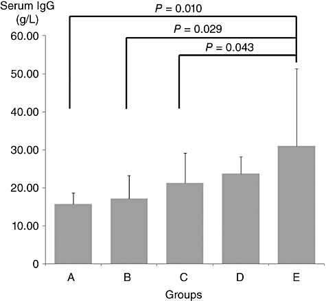
Serum IgG levels in groups A, B, C, D and E were 15.11 ± 3.22, 18.65 ± 7.49, 20.92 ± 7.64, 23.29 ± 4.35 and 30.98 ± 19.25 g/L, respectively. There was a trend towards higher IgG levels when more regions were affected. Significant differences were observed between group A and E (P = 0.010), group B and E (P = 0.029) and group C and E (P = 0.043), but not between group D and E (Fig. 1).

Serum immunoglobulin G (IgG) levels in groups A, B, C, D and E (mean ± SD). Differences are shown between group E and group A, B and C. SD, standard deviation.
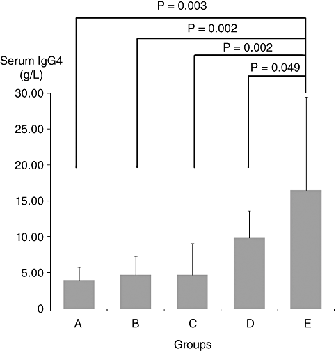
A similar analysis was performed on serum IgG4 levels (Fig. 2). Serum IgG4 levels in groups A, B, C, D and E were 3.99 ± 1.75, 4.70 ± 2.62, 4.70 ± 4.32, 9.86 ± 3.78 and 16.49 ± 12.95 g/L, respectively. Significant differences were observed between group A and E (P = 0.003), group B and E (P = 0.002), group C and E (P = 0.002) and group D and E (P = 0.049) (Fig. 2), indicating that the serum IgG4 levels of the patients with five or more involved regions was significantly higher than those of patients in the other groups.

Serum immunoglobulin G4 (IgG4) levels in groups A, B, C, D and E (mean ± SD). Significant differences are shown between group E and all the other four groups. SD, standard deviation.
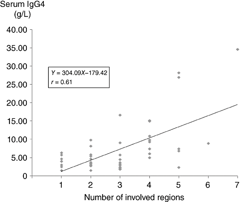
A regression analysis suggested a correlation (r = 0.61) between serum IgG4 levels and the number of regions involved (P = 0.001, Fig. 3). A similar regression analysis was performed for serum IgG levels and the number of PL/EPLs (r = 0.58, data not shown).

Regression analysis shows that there is a correlation between serum immunoglobulin G4 (IgG4) levels and the number of pancreatic lesion/extrapancreatic lesions.
We also examined the number of patients with sclerosing sialadenitis in each group (Table 3), and found that patients with sclerosing sialadenitis all had other lesions involved. As shown in Table 4, patients with sclerosing sialadenitis had significantly higher serum IgG4 levels than those without. There was no significant difference in the IgG4 levels of patients with or without any other lesion involved.
| Group | Patients with sclerosing sialadenitis, n (%) |
|---|---|
| A (n = 7) | 0 (0) |
| B (n = 11) | 0 (0) |
| C (n = 12) | 3 (25.0) |
| D (n = 9) | 7 (77.8) |
| E (n = 7) | 7 (100.0) |
| Sclerosing sialadenitis | Serum IgG4 level (g/L, mean ± SD) |
|---|---|
| Present (n = 17) | 12.51 ± 9.50* |
| Absent (n = 29) | 4.48 ± 3.05 |
- * P < 0.01, Mann–Whitney U-test. SD, standard deviation.
DISCUSSION
Researchers have been discussing the possible diagnostic significance of the increased serum IgG4 levels observed in patients with IgG4-related diseases and whether the increase could be used as an indicator of disease activity or a predictor of recurrence.8
The study by Hamano et al.5 was the first to demonstrate the significance of serum IgG4 for the diagnosis of AIP. However, three issues must be considered regarding the diagnostic significance of serum IgG4 for AIP: (i) elevated serum IgG4 levels are not necessarily specific for AIP; they can also be observed in other diseases (such as atopic dermatitis and bronchial asthma);16 (ii) elevated serum IgG4 levels are also observed, although rarely, in pancreatic cancer,17 so differentiation of pancreatic cancer from AIP is required.18 In addition, cases of pancreatic cancer concurrent with AIP have recently been reported;19 (iii) although AIP is considered to be an IgG4-related disease, seronegative cases without IgG4 elevation20 and IDCP14 have been included. Seronegative patients who were clinically characterized by pancreatic enlargement, narrow pancreatic duct and multiple concurrent EPLs responded to steroids and were not very different from the seropositive patients. Further explanations for the seronegative cases of AIP are expected.
Tabata et al.9 examined the serial changes in elevated serum IgG4 levels of patients with IgG4-related diseases, including AIP and Mikulicz's disease, concluding that the measurement of serial serum IgG4 levels is useful to determine the disease activity of IgG4-related diseases, and persistent elevation or re-elevation of serum IgG4 levels after steroid therapy might predict a relapse. But the following questions should be considered while judging whether serum IgG4 levels at diagnosis reflects the disease activity: (i) does serum IgG4 level reflect the extent of lesion enlargement? (ii) does serum IgG4 level reflect the number of involved organs or regions? (iii) is there a gap between the decrease in serum IgG4 levels and the resolution in inflammation shown by diagnostic imaging, as reported for the spontaneous remission of AIP?15
In this study, we found that serum IgG levels tended to be elevated with the increasing number of PL/EPLs, and there were significant differences in serum IgG levels between group E and group A or C. As for serum IgG4, significant differences were observed when group E was compared with all the other four groups. Furthermore, a regression analysis suggested that a correlation existed between serum IgG4 levels and the number of PL/EPLs.
Serum IgG4 levels were significantly increased in patients with sclerosing sialadenitis, which was concurrently accompanied by PL or other EPLs. This finding might be attributed to the characteristics of sclerosing sialadenitis. Naitoh et al.21 reported that the presence of sclerosing sialadenitis at clinical onset as EPL was a significant predictive factor for the relapse of AIP. Another possible explanation was that patients with sclerosing sialadenitis tended to have other EPLs, resulting in the involvement of multiple regions. In this study, we had neither patients with sclerosing sialadenitis alone, nor those with PL and only sclerosing sialadenitis as EPL; therefore, further investigation is required.
There were some limitations of this study. First, each group had only a small number of cases, which was not sufficient for a full analysis. Second, the imaging modalities for the detection of inflamed regions have different sensitivities, and PET-CT, which could have been useful for the detection of lymph node and lesions, was not used on all the patients.22 Third, many regions diagnosed as inflamed in this study were not confirmed by histopathological examination. Fourth, because high serum IgG and IgG4 levels may indicate the disease activity of IgG4-related diseases at diagnosis, comparisons of IgG and IgG4 before and after steroid therapy is required. The improvement of PL/EPLs in all patients in this study was confirmed by imaging to some degree after steroid therapy and serum IgG levels decreased (data not shown). However, in most patients, changes in serum IgG4 levels were not measured. Finally, with an even smaller number of patients with IgG4-related disease without PL, we could not determine the characteristics specific to AIP.
However, the results in this study suggest that IgG4-related disease patients with high levels of serum IgG and IgG4 often have multiple lesions including sclerosing sialadenitis; therefore, the entire body should be examined to investigate the pathophysiology of this disease. To clarify the clinical significance of IgG and IgG4 serum levels, a more detailed analysis is required in the future.
ACKNOWLEDGMENT
This work was partially supported by a grant from the Research Program of Intractable Disease to Kazuichi OKAZAKI and a grant from the Research Committee of Intractable Pancreatic Diseases to Tooru SHIMOSEGAWA, both provided by the Ministry of Health, Labor and Welfare of Japan.


