Laparoscopically assisted resections of small bowel stromal tumors are safe and effective
Conflict of interest: none.
Abstract
OBJECTIVE: To compare the efficacy of laparoscopically assisted and open resections in treatment of small bowel stromal tumors (SBST).
METHODS: A retrospective study of 85 patients who underwent curative resections for SBST (38 by laparoscopically assisted procedures and 47 by open procedures) was performed.
RESULTS: There were no differences between open and laparoscopically assisted approaches in terms of patients' age, gender, presenting symptoms, histological risk or extent of resection (P > 0.05). The median tumor size for laparoscopically assisted resections was 4.0 cm (range 1.2–7.0 cm), which was the same as that for the open resections (range 2.0–10.0 cm). There were fewer complications in the laparoscopic group than those in the open resection group (7.9% vs 17.0%), but no significant difference was observed (P > 0.05). The 2-year survival of the two patient groups was almost the same (86.8% vs 89.4%). Laparoscopically assisted procedures required on average 22.5 min less of operating time (87.5 min vs 110.0 min, P = 0.006), 1.0 day less of bowel recovery time (3.0 days vs 4.0 days, P = 0.001) and 5.0 days less in hospital stay (8.0 days vs 13.0 days, P < 0.001).
CONCLUSION: Laparoscopically assisted resection of SBST is a safe alternative to open resection.
INTRODUCTION
Gastrointestinal stromal tumors (GISTs) arise from the interstitial cells of Cajal.1–3 The overall incidence of GIST is 10–20 cases per million person per year, in which 10–30% are malignant.4 Most GIST arise from the stomach, while about 20–30% arise from the small bowel.2 The prognosis of small bowel stromal tumors (SBST) depends mainly upon tumor size and completeness of resection,5 and a histological risk classification has been devised.3,5–9 The goals of surgical resection are the extirpation of visible and microscopic lesions and the avoidance of tumor rupture with subsequent intra-abdominal contamination by neoplastic cells.10 Standard recommendations include restriction of laparoscopic resections to tumors smaller than 2 cm,11 but some investigators suggest that the techniques are useful for larger neoplasms.12–21 We hypothesized that a comparison of laparoscopically assisted and open procedures might lend weight to the notion that laparoscopically assisted resections are as safe and effective as open resections of SBST.
PATIENTS AND METHODS
Patients
From December 2002 to December 2007, 90 consecutive patients underwent resections for SBST at Department of General Surgery, Ruijin Hospital, Shanghai Jiaotong University School of Medicine. Five patients who had metastases at the time of diagnosis were excluded. All 85 operations were done by one surgical team. In general, the procedures were not performed laparoscopically if the lesion was found to be larger than 5 cm in size by preoperative non-traumatic diagnostic modalities. Most of those patients with a tumor size larger than 5 cm who underwent a laparoscopically assisted procedure were underestimated preoperatively. The patients were advised to undergo open surgery if a laparoscope was contraindicated in their case. A lymphadenectomy is not imperative because GIST rarely metastasize to the lymph nodes.22–25 Diagnosis were confirmed by two pathologists who placed the tumors in risk categories using Fletcher's system.3 If the margin of the tumor specimen was microscopically positive, showing tumor cells in the frozen section, then extensive resection was indicated. For all patients, antibiotics were administrated the day prior to the operation. Written informed consents were obtained from each patient before the operation and the institutional review board approval was obtained before the initiation of this review. The study was approved by the Ethics Committee of Ruijin Hospital, Shanghai Jiaotong University School of Medicine.
Laparoscopically assisted technique
The laparoscopic explorations were accomplished after a pneumoperitoneum was established under general anesthesia. A 30-degree laparoscope was inserted via a 10-mm optical trocar placed above the umbilicus. Non-traumatic graspers were inserted into a 5-mm port in the right subcostal region, at a point midway between the umbilicus and the xyphoid process. After inspecting the liver, gallbladder, stomach, spleen, mesentery, colon and pelvis, the ligament of Treitz was retrieved to allow a detailed evaluation of duodenum, jejunum and ileum. With the results of the inspection at hand, the extent of resection required was determined. A small intestinal resections and subsequent anastomosis were accomplished extra-corporeally after the bowel segment involved was pulled out through a small abdominal wall incision.
Open resection technique
Open resections were accomplished via an upper middle incision under general anesthesia. The organs were examined in the same order as above to determine the extent of resection require. Thereafter the small intestinal resection and subsequent reconstruction were performed.
Statistical analysis
SPSS 13.0 (SPSS Inc., Chicago, IL, USA) was used for statistical analysis. Quantitative variables, such as patients' age, tumor size, distance from upper margin and lower margin, total hospital charges, medical and surgical charges were tested for normality assumption using Kolmogorov–Smirnov tests. Normal variables were listed as mean ± standard deviation (SD) unless otherwise specified. Non-normal quantitative variables were shown as median (minimum, maximum). Statistical comparisons were made using a two-tailed Student's t-test or Fisher's exact test for normal variables and by Wilcoxon rank–sum test for ordinal variables, whereas gender, presenting symptoms, risk groups comparisons were analyzed using the χ2 test. Actuarial survival was determined by a Kaplan–Meier analysis using log–rank. The multivariate analysis was performed using the Cox proportional hazards model, and only variables deemed to be statistically significant were included in the final Cox model. P < 0.05 was considered as statistically significant.
RESULTS
The patients' characteristics are shown in Table 1. The median age of the open resection group was the same with that of the laparoscopically assisted group. There was no difference in gender or symptomatology between these two groups (P = 0.922 and P = 0.296, respectively).
| Laparoscopically assisted group (N = 38) | Open resection group (N = 47) | P value | |
|---|---|---|---|
| Age (years, median [range]) | 56 (21–79) | 56 (33–81) | 0.925 |
| Gender | |||
| Male (n, %) | 19 (50.0) | 23 (48.9) | 0.922 |
| Female (n, %) | 19 (50.0) | 24 (51.1) | |
| Symptoms | |||
| Melena (n, %) | 32 (84.3) | 31 (65.9) | 0.296 |
| Hematochezia (n, %) | 1 (2.6) | 0 (0.0) | |
| Abdominal mass (n, %) | 1 (2.6) | 1 (2.1) | |
| Anemia related (n, %) | 1 (2.6) | 1 (2.1) | |
| Dyspepsia (n, %) | 0 (0.0) | 2 (4.3) | |
| Upper abdominal discomfort (n, %) | 0 (0.0) | 2 (4.3) | |
| Lower abdominal pain (n, %) | 3 (7.9) | 8 (17.0) | |
| Incidental discovery (n, %) | 0 (0.0) | 2 (4.3) |
In this study, all margins were histologically free of tumor cells. No significant difference was obtained in term of the histology and gross appearance of the tumor, as shown in Table 2. Differences between Fletcher histological risk and the median tumor size were not significant (P = 0.235 and P = 0.235, respectively). There were no differences in distances from the upper (W = 1379.50, Z = −0.76, P = 0.445) and lower margin (W = 1398.50, Z = −0.58, P = 0.563).
| Laparoscopically assisted group (N = 38) | Open resection group (N = 47) | P value | |
|---|---|---|---|
| Fletcher histological high-risk groups | |||
| Very low, n (%) | 1 (2.7) | 0 (0) | 0.235 |
| Low, n (%) | 22 (57.9) | 22 (46.8) | |
| Intermediate, n (%) | 11 (28.9) | 13 (27.7) | |
| High, n (%) | 4 (10.5) | 12 (25.5) | |
| Tumor size (cm, median [range]) | 4.0 (1.2–7.0) | 4.0 (2.0–10.0) | 0.235 |
| Distance from upper margin (cm, median [range]) | 3.5 (1.0–13.0) | 4.0 (1.0–11.0) | 0.445 |
| Distance from lower margin (cm, median [range]) | 3.0 (1.0–14.0) | 3.8 (1.0–24.0) | 0.563 |
The peri-operative parameters and costs are shown in Table 3. No differences in the extent of resection were observed (P = 0.63). The median operative time for laparoscopically assisted procedures was 22.5 min less than that for open procedures (W = 1320.00, Z = −2.66, P = 0.006). The median abdominal incision for the open procedures was 11.0 cm longer than that for laparoscopically assisted procedures (W = 829.00, Z = −7.19, P < 0.001). The median bowel recovery time for the laparoscopically assisted procedures was 1.0 day less than that for open procedures (W = 1251.00, Z = −3.43, P = 0.001). The median postoperative hospital stay for laparoscopically assisted procedures was 5.0 days shorter than that for open procedures (W = 1073.50, Z = −5.00, P < 0.001). Although the median surgical charge for laparoscopically assisted procedures cost RMB 784 more than that for open procedures (W = 1701.00, Z = −2.83, P = 0.005), the median medical charge for open procedures was RMB 3584 more than that for laparoscopically assisted procedures (W = 1217.00, Z = −3.69, P < 0.001), yielding a RMB 3808 difference in median total hospital charges which was in favor of laparoscopy (W = 1423.00, Z = −1.87, P = 0.062). Complication rates in the two groups were similar (P = 0.213). Only one patient required a conversion from a laparoscopically assisted procedure to an open one. There were no operative deaths.
| Laparoscopically assisted group (N = 38) | Open resection group (N = 47) | P value | |
|---|---|---|---|
| Morbidity | |||
| Complications, n (%) | 3 (7.9) | 8 (17.0) | 0.213 |
| No complications, n (%) | 35 (92.1) | 39 (83.0) | |
| Operative time (min, median [range]) | 87.5 (40.0–255.0) | 110.0 (55.0–300.0) | 0.006 |
| Abdominal incision (cm, median [range]) | 4.0 (2.0–15.0) | 15.0 (3.0–200.0) | 0.000 |
| Bowel recovery time (days, median [range]) | 3.0 (1.0–13.0) | 4.0 (2.0–15.0) | 0.001 |
| Postoperative hospital stay (days, median [range]) | 8.0 (6.0–21.0) | 13.0 (7.0–130.0) | 0.000 |
| Total hospital charges (RMB, median [range]) | 17 346 (9 548–75 495) | 21 154 (8 778–120 253) | 0.062 |
| Medical charges (RMB, median [range]) | 3 325 (1 547–52 962) | 6 909 (1 561–57 995) | 0.000 |
| Surgical charges (RMB, median [range]) | 6 349 (3 122–14 525) | 5 565 (2 702–19 509) | 0.005 |
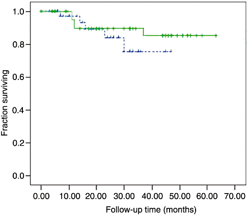
The follow-up time was at a median of 26 months (range 0–63 months). Among all 85 patients, 10 had recurrences, of which 9 had hepatic metastases and one had diffuse peritoneal seeding. During the whole follow-up period the survival rate of laparoscopically assisted group and the open resection group was 86.8% and 89.4%, respectively, with no significant statistical difference between the two operative modalities (χ2 = 0.76, d.f. = 1, P = 0.383, Fig. 1).

Disease-free survival (laparoscopically assisted vs open resection). During the whole follow-up period, the survival rate of the laparoscopically assisted group and open resection group was 86.8% and 89.4%, respectively, with no significant difference between the two operative modalities (χ2 = 0.76, d.f. = 1, P = 0.383). Approach: ( ) laparoscopy; (
 ) open; (
 ) laparoscopy-censored; (
 ) open-censored.
DISCUSSION
In this study, two different approaches to the resection of SBST were compared. No difference was found between open and laparoscopically assisted procedures in terms of the patients' age, gender, presenting symptoms, tumor size, histological risk and complication rate. The results of the survival analysis favored laparoscopical procedures. Although the surgical costs were greater for laparoscopically assisted procedures than those for open ones, there was no difference in the total cost that favored laparoscopic approach.
Surgery remains the therapeutic mainstay for SBST.10,11,23 In recent years function-preserving and minimally invasive surgeries have also been performed as treatment strategies for submucosal tumors, including GISTs which are clinically diagnosed as being low-risk tumors.14 This indicates that a laparoscopic approach should be considered in all patients with gastric GIST who do not have a contraindication to this approach.15,17,18,20 We suggest this view might also be applied to the SBST. The National Comprehensive Cancer Network Clinical Practice Guidelines suggested that laparoscopy should be restricted to patients with tumors smaller than 2 cm,11 but there is reason to believe that this recommendation lacks an evidentiary base.13–15,18,20,21 Several reports of safe resections of larger tumors have been published.13–15,18,20,21 Our study lends support to this notion, given that tumors removed with laparoscopical assistance averaged 4 cm and ranged from 1.2 cm to 7 cm.
Although laparoscopic procedures are usually less traumatic than open procedures, this generalization does not always confer minimal morbidity.16 Because our surgical team had already completed a review of laparoscopic surgery before the beginning of this study, there was no significant difference in the postoperative morbidities between the laparoscopically assisted group and the open resection group. Laparoscopic surgery requires more surgical experience than open surgery. Laparoscopic procedure must sometimes be converted to an open one, and skills in the latter are the prerequisite to success of the former. A laparoscopy requires sophisticated manipulative techniques because non-touch criteria and the prevention of tumor rupture are vital.
The laparoscopy was especially advantageous with respect to peri-operative parameters. Only one patient in our study required conversion to an open procedure due to a suspicious liver mass, which was proved to be a hepatocellular adenoma by histological examination. Laparoscopically assisted resections showed decreased operative time, bowel recovery time and postoperative hospital stay compared with open resections. These advantages reduced costs and neutralized the difference in operative charges. Extracorporeal anastomoses eliminated anastomotic stapling charges.


