The orientation of cutaneous ridges influences the appearance of longitudinal stripes on the skin surface of bottlenose dolphins (Tursiops truncatus)
The integument of the bottlenose dolphin (Tursiops truncatus, Montagu 1821) has been described as being furrowed by small ridges, with a general annular disposition relative to the longitudinal axis of the body (Shoemaker and Ridgway 1991, Ridgway and Carder 1993).
Shoemaker and Ridgway (1991) gave a detailed description of the cutaneous ridges in terms of size and orientation. The ridges have a spatial period between 0.56 and 0.71 mm and a height between 14 and 41 μm. They are vertically oriented around the head and more obliquely oriented in the caudal portion of the body. The transition from vertical to oblique orientation of the ridges apparently occurs gradually along the body. Ridgway and Carder (1993) presented a “rough sketch” of the orientation of the cutaneous ridges in T. truncatus, which is consistent with the results from Shoemaker and Ridgway (1991). Shoemaker and Ridgway (1991) also noted the appearance of longitudinal stripes along the dolphin's body, and hypothesized that they “appear to be due to variations in ridge shape or height along the circumferential direction; these variations are in register from ridge to ridge and give the appearance of longitudinal features.” We investigated this hypothesis by photographically documenting both the (1) orientation of the cutaneous ridges and (2) appearance of longitudinal stripes in captive bottlenose dolphins. The results of this study suggest that the longitudinal stripes are an optical effect, which is likely due to specular reflection patterns caused by differences in the angle of inclination of the cutaneous ridges.
Photographic data were collected at the Genoa Aquarium between 2002 and 2004. Beta, a wild caught female, approximately 20 yr old, was the focus of the study. Opportunistic observations of longitudinal stripe patterns were also gathered on three others individuals: Blu (Beta's daughter), a captive born female (Delfinario di Rimini, June 1997); Bonnie, a wild caught female, approximately 27 yr old; Achille (Bonnie's son), a captive born male (Acquario di Genova, August 2003). To describe the appearance of the longitudinal stripes, we collected photographs and video sequences from all three dolphins through the large transparent wall of the dolphin pool. A digital video camera (Sony DCR-PC100E, Sony Communication, Tokyo, Japan) was used to collect both photographs (using the “photo mode” on a memory card) and video sequences (on digital mini-DV tape).
To describe the orientation of the cutaneous ridges, we divided Beta's body surface (between the eye and the anus) into 16 sections and collected photomacrographs for each section (Fig. 1). All the sections were identified by anatomical landmarks that could be easily located by the photographer during the data collection. Because the dolphin skin structure is delicate and can be easily deformed by mechanical stress and drying, all photographs were taken underwater. The digital camera, housed within a watertight case, had a transparent hollow cylinder positioned around the objective. The cylinder, which was in contact with the dolphin body, acted to maintain a constant distance (2.5 cm) between the lens and the skin surface. The dolphin was trained to position underwater along the poolside and the camera was kept at a constant orientation, perpendicular to the longitudinal axis of the dolphin. We collected a minimum of 16 photographs for each section, for a total of 327 photographs. Four more photographs were taken in the ventral area (labeled “belly” in Fig. 1). For each photograph we measured the angle between the cutaneous ridges and the circumferential body axis (the inclination angle) (Fig. 2) using an image analysis software program (Adobe Photoshop, Adobe Systems Inc., San Jose, CA). This program reports an angle measurement precision of 0.1°. The inclination angle is reported as the mean ± SE calculated from the 16 photographs of each section.

The dolphin body surface was divided into 16 sections. All the sections are referred to anatomical elements of the dolphin body that can be easily located by the photographer during the data collection.
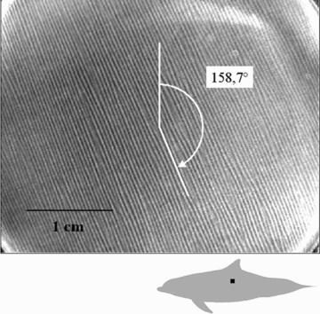
A photomacrograph of the dolphin skin, showing the cutaneous ridges. The black square on the dolphin shows the sampling area.
To visually present the orientations of the cutaneous ridges across the body, we developed a 3D model of Beta, using computer aided design (CAD) software (Thinkdesign, Think3 Inc., Cincinnati, OH). The model was created in a 1:1 scale using photos and video frames of Beta. Selected video frames were used to draw the three basic profiles of the dolphin: longitudinal, transverse, and frontal. Starting from these three profiles, we drew a series of 28 transverse sections of the dolphin body, which were then covered with a virtual skin. The ridges were drawn on the model as uni-dimensional lines (i.e., not properly as ridges in relief), using the mean inclination angle for each body region (Table 1) resulting from the photographic analysis, with an arbitrary spatial period of 1 cm (Fig. 3). The connection between contiguous regions belonging to the same vertical section (for example Fig. 1, sections 5A, 5B, 5C) was obtained with a cubic spline curve (Bartels et al. 1987). The spaces between contiguous vertical sections (Fig. 1, whole body sections 2, 3, 4, 5) were filled in with intermediate conjunction lines (i.e., a line which had a curvature angle which was intermediate between the two equidistant closest lines).
| Inclination angle (on average) of the ridges in each section | ||||
|---|---|---|---|---|
| 2An: 24Angle: 180.8°SE: ±0.84 | 3An: 19Angle: 175.2°SE: ±0.96 | 4An: 29Angle: 173.7°SE: ±0.5 | 5An: 29Angle: 170.0°SE: ±0.73 | 6An: 22Angle: 188.8°SE: ±0.67 |
| 2Bn: 24Angle: 161.2°SE: ±1.08 | 3Bn: 21Angle: 160.7°SE: ±0.95 | 4Bn: 21Angle: 166.0°SE: ±0.52 | 5Bn: 26Angle: 157.0°SE: ±0.34 | 6Bn: 17Angle: 175.7°SE: ±0.69 |
| 2Cn: 25Angle: 189.8°SE: ±1.53 | 3Cn: 16Angle: 163.8°SE: ±1.3 | 4Cn: 17Angle: 170.6°SE: ±0.91 | 5Cn: 20Angle: 171.6°SE: ±0.73 | 6Cn: 17Angle: 164.5°SE: ±0.64 |
| BELLYn: 4Angle: 178.2°SE: ±0.21 | ||||
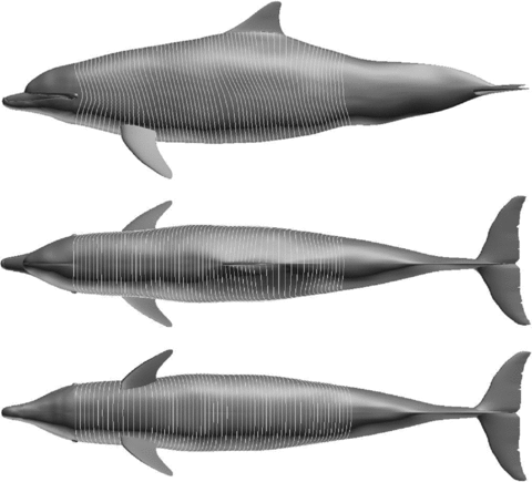
Orientation map of the ridges on the 3D model.
The lateral body surfaces of all the adult dolphins in this study were marked by longitudinal stripes, although the stripes were not visible on the newborn (Achille) until he was about 3 mo old (Fig. 4A). Depending on the position of the dolphin in relation to the camera, though, the stripes changed in appearance. The pattern of light and dark gray stripes appeared to be inverted, depending upon whether the dolphin was moving towards or away from the camera (Fig. 4B). When the dolphin was perpendicular to the camera, the stripes were barely visible.
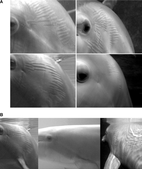
(A) The body walls of all the adult dolphins in this study were marked by longitudinal stripes. In Achille the stripes appeared when it was about 3 mo old (top left: Beta; top right: Blu; bottom left: Bonnie; bottom right: Achille). (B). The pattern of light and dark gray stripes appeared to be inverted, depending upon whether the dolphin (Bonnie) was moving towards or away from the photographer.
This effect was particularly evident in the region spanning from the neck to the level of the pectoral flipper (Fig. 1, sections 2C and 3C). Here, there was a distinct border where the light and dark longitudinal stripes appeared to be interdigitated (see Fig. 4). These finger-like projections were each about 0.6 cm wide. In this region, ridge systems with different inclination angles meet (Table 1). In contrast, in the upper sections (Fig. 1, sections 2B, 3B) the two cutaneous ridge systems had similar inclination angles. Close up video of sections 2C and 3C (the skin was first washed with freshwater and dried) also showed that between a dark stripe and a light one, the inclination angle of individual cutaneous ridges changes (Fig. 5). This zigzag pattern was likely accentuated on the dried skin. Nonetheless, dark stripes appear to be characterized by cutaneous ridges that are oriented caudo-dorsally, while ridges in the light stripes are oriented caudo-ventrally. The cutaneous ridges seem to behave like the rubber ridges of the hot-water bottles (Fig. 6) producing a specular reflection (i.e., a reflection where most of the rays are reflected in the same direction) according to the Snell's Law of reflection (Whewell 1837); those reflecting the light to the observer's eye look bright, while the ridges reflecting the light to the opposite direction look dark. The zigzag produces a dark–light alternating pattern, which produce the optical effect of dark and light alternating longitudinal stripes. Integrating data from photographic and video analyses, we created a morphological hypothesis of the relationship between the longitudinal stripes and the orientation of the cutaneous ridges (Fig. 7).
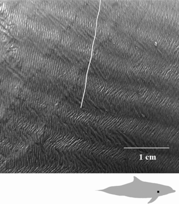
The zigzagging of the cutaneous ridges across the border. The black square on the dolphin shows the sampling area.
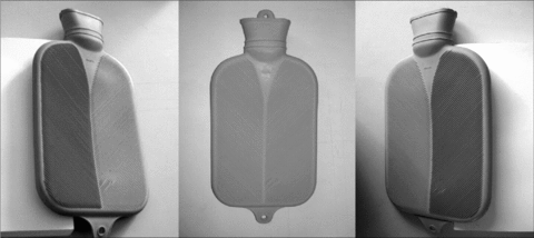
The “boule effect,” resembling the change in the dark–light pattern of the dolphin body.
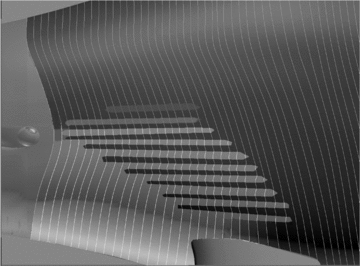
A detail of the 3D model showing the border area. The ridges were drawn with an arbitrary period of 1 cm. The dark–light pattern was reconstructed to better show the mechanism generating the dark–light pattern itself and the longitudinal stripes origin.
In conclusion, the results of this study supports the hypothesis of Shoemaker and Ridgway (1991) that the longitudinal stripes could be due to variations in cutaneous ridge shape or height along the dolphin's circumference. This pattern is slightly different in each dolphin (see Fig. 4A), likely due to individual differences in the patterns of their cutaneous ridges. Because our results are based upon a single, adult female bottlenose dolphin, our representation of the cutaneous ridge orientations must be considered as a general, simplified model. The similarities in the overall patterns of longitudinal stripes across individuals, and the photographic analyses presented here though, suggest that cutaneous ridge orientation likely plays a key role in producing the visual effect of longitudinal stripes across the body walls of bottlenose dolphins.
Acknowledgments
Thanks to the Marine Mammal Staff of Acquario di Genova for training Beta, Mario Dalpiaz for his contribution to the English version, Maurizio Wurtz and Giovanni Caltavuturo for their suggestions.


