Brain-derived neurotrophic factor-like neuropeptide is secreted as a neurohormone from specific brain neurons into the corpus allatum in the silkworm Bombyx mori
Abstract
The aim of this study was to investigate the secretion of brain-derived neurotrophic factor (BDNF)-like neuropeptide in the silkworm, Bombyx mori , by using immunocytochemical techniques on the brain and retrocerebral complex of fifth instar larvae. In the brain, four pairs of median neurosecretory cell (MNC) bodies and six pairs of lateral neurosecretory cell (LNC) bodies had distinct immunoreactivities to this peptide, suggesting that this peptide is produced from two types of brain neuron. These reactivities were much stronger in the MNC than in the LNC. Labeled MNC projected their axons into the contralateral corpora allata, to which axons of labeled MNC were eventually innervated, through decussation in the median region, contralateral nerve corporis cardiaci I and nerve corpora allata I. Labeled LNC extended their axons into the ipsilateral corpora allata to be innervated through the ipsilateral nerve corporis cardiaci II and nerve corpora allata I. These results suggest that BDNF is secreted as a neurohormone from MNC and LNC of the brain into the corpora allata.
Introduction
Neurotrophins are a family of structurally and functionally related peptides that play pivotal roles in the survival, growth, differentiation, energy metabolism, and maintenance of vertebrate neurons in the peripheral and central nervous systems (Snider 1994; Spenger et al. 1995; Lewin & Barde 1996; Schwartz et al. 1997; Ringstedt et al. 1998; Berardi et al. 2000; Tucker et al. 2001; Burkhalter et al. 2003; Gorski et al. 2003). There are four members of this family: nerve growth factor (NGF), brain-derived neurotrophic factor (BDNF), neurotrophin (NT)-3 and NT-4/5. These peptides have six conserved cysteine residues and have a 55% sequence identity at the amino acid level (Ilag et al. 1995).
Following exposure to BDNF in vitro , there is a marked enhancement in neurite outgrowth and a significant increase in the number of neurites in the neural precursor cells of mouse embryonic and adult striatum (Ahmed et al. 1995). BDNF, together with NT-4/5, enhances neurite extension and survival of differentiated cerebellar granule cells (Gao et al. 1995). It also influences the morphological differentiation of dopaminergic neurons in cultured rat substantia nigra (Studer et al. 1995). In addition, BDNF supports the survival of axotomized developing spinal motor neurons both in vitro and in vivo (Friedman et al. 1995), and prevents excessive loss of sensory neurons in mouse peripheral sensory ganglia (Ernfors et al. 1995).
Analysis using polyclonal guinea pig antibodies against recombinant BDNF in has shown widespread neuronal localization of BDNF in many areas of rat brain, including all areas of the hippocampus (Dugich-Djordjevic et al. 1995). BDNF is synthesized by a population of small- to medium-sized neurons in all sensory ganglia except in the mesencephalic nucleus of the trigeminal nerve (Wetmore & Olson 1995). In these ganglia, a widespread population of neurons and scattered satellite cells produce BDNF.
Even though extensive studies on the various aspects of vertebrate BDNF have been carried out, there is currently little information regarding the secretion, molecular and biochemical characteristics, and functional aspects of BDNF in invertebrates, including insects. Therefore, it is necessary to determine which cells normally produce the BDNF-like peptide in insects. In the present paper we describe the localization of the BDNF-like neuropeptide-producing neurons in the brain and retrocerebral complex of the silkworm, Bombyx mori .
Materials and methods
Animals
Cold-treated eggs of the silkworm, Bombyx mori (Lepidoptera, Insecta), which were obtained from the National Institute of Agricultural Science and Technology (Suwon, Korea), were hatched after approximately 10 days of incubation at 27–28°C with a relative humidity of 60–70%. Hatched larvae were reared on fresh mulberry leaves under a long-day photoperiod regimen (17 h light : 7 h dark [LD 17:7]) at 27°C and approximately 60% humidity, and most larvae routinely developed into first, second, third and fourth and fifth (final) instar larvae.
Whole-mount immunocytochemistry
Tissue preparation and whole-mount immunohistochemistry were performed according to Park et al. (2002). The fifth instar larvae were collected from stock colonies in a rearing chamber, and used for immunocytochemical studies of the brains and retrocerebral complexes. After larvae were kept at 4°C for 1 h, brains and retrocerebral complexes were dissected in a 0.1 mol/L sodium phosphate buffer (pH 7.4) using a stereoscope, and fixed in 4% paraformaldehyde dissolved in a 0.1 mol/L phosphate buffer for 6 h at 4°C. Fixed tissues were immersed overnight in a 0.01 mol/L phosphate buffer saline solution with 1% Triton X-100 at 4°C. Peroxidase activity was blocked in 10% methanol in 3% H2O2 for 25 min.
Tissues were washed in a 0.1 mol/L Tris-HCl buffer (pH 7.6–8.6) containing 1% Triton X-100 and 4% NaCl, and incubated with primary antiserum anti-human BDNF (monoclonal antibody; Sigma, St Louis, MO, USA), diluted to 1:1000 with a dilution buffer (0.01 mol/L phosphate-buffered saline with 1% Triton X-100 and 10% normal goat serum) for 4–5 days with gentle shaking. It is known that this primary antibody has a <2% cross-reactivity with rhâ-NGF, rhNT-3 and rhNT-4 based on enzyme-linked immunosorbent assay (ELISA) and immunoblotting (manufacturer's data sheet).
Following washes in 0.01 mol/L phosphate-buffered saline with 1% Triton X-100, tissues were incubated in peroxidase-conjugated mouse IgG (Dako, Glostrup, Denmark) diluted to 1:200, for 2 days at 4°C. After a pre-incubation stage in 0.03% diaminobenzidine (DAB) in a 0.05 mol/L Tris-HCl buffer for 1 h at 4°C, tissues were treated with 0.03% DAB in a 0.05 mol/L Tris-HCl buffer containing 0.01% H2O2 for 5–10 min. After being rinsed in a 0.05 mol/L Tris-HCl buffer, tissues were mounted in glycerin, examined and then photographed with a digital camera attached to an interference microscope (Zeiss, Dublin, Ireland).
Results
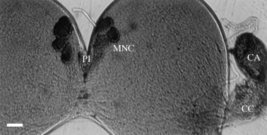
Two types of specific brain neuron were labeled by human BDNF peptide. The first type was the median neurosecretory cells (MNC). The brains of the fifth instar larvae each had four pairs of MNC in the pars intercerebralis, of which cell bodies were all intensively immunoreactive to the human BDNF. Therefore, the larval brain had eight MNC in total, four in each hemisphere. These cell bodies were very large in size (approximately 25 µm in diameter) and were localized as a cluster in the pars intercerebralis (Fig. 1).

Four pairs of median neurosecretory cells (MNC) in the brain and retrocerebral complex of fifth instar larva of Bombyx mori showing immunolabeling with anti-human brain-derived neurotrophic factor (BDNF) antiserum. Four pairs of large MNC cell bodies that are located as a cluster in the pars intercerebralis (PI) are intensively immunostained. Thus, two clusters of MNC are found in each brain. In the right side of the brain, the retrocerebral complex that consists of the corpus cardiacum (CC) and corpus allatum (CA) is structurally connected with the brain (Br). The corpus allatum is intensively immunostained with the BDNF-like peptide (arrow) but the corpus cardiacum is not labeled, suggesting transportation of the BDNF-like peptide to the corpus allatum. Scale bar, 30 µm.
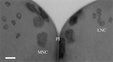
The second type of brain neuron that was immunoreactive to the human BDNF was the lateral neurosecretory cells (MNC). As shown in Figure 2, cell bodies of six pairs of LNC, six in each hemisphere, were found to be weakly labeled by the peptide in the pars lateralis. Cell bodies of these LNC were grouped as a cluster in the pars lateralis and had a small diameter, approximately 15 µm (Fig. 2).

Six pairs of lateral neurosecretory cells (LNC) in the pars lateralis of the Bombyx mori fifth instar larval brain, with four pairs of median neurosecretory cells (MNC) in the pars intercerebralis (PI). Twelve cell bodies of LNC in the two hemispheres are weakly immunostained and small (approximately 15 µm in diameter), but cell bodies of MNC, of which some are strongly labeled, are all large (approximately 25 µm). Scale bar, 30 µm.
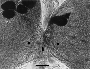
Labeled MNC projected their axons into the medial region between the two cerebral hemispheres and their axons decussated there. After this point, the MNC axons extended to the contralateral nerve corporis cardiaci (NCC) I with an axonal bundle through the contralateral hemisphere. Through nerve corpora allata (NCA) I, these axons were eventually innervated to the contralateral corpora allata that accumulated the transported BDNF-like peptide before its release (Fig. 3). Labeled LNC, of which cell bodies were located in the pars lateralis, extended to the ipsilateral NCC II without decussation in the medial region. NCC II joined NCC I before arrival in the corporis cardiaci, and the joined nerve was then followed by NCA I. These axons finally terminated in the ipsilateral corpora allata (data not shown).

Axonal pathways of median neurosecretory cells (MNC) and lateral neurosecretory cells (LNC) in the medial region of the Bombyx mori fifth instar larval brain. Axons of MNC, of which cell bodies are located in the pars intercerebralis (PI), are decussated (small, thick arrow) in the medial region, and then extend to the contralateral hemispheres, whereas axons of LNC, of which cell bodies are located in the pars lateralis, project into the medial region and then extend to the ipsilateral hemisphere without decussation (arrowheads). Therefore, MNC project their axons into the contralateral corpora allata after decussation in the medial region, and axons of LNC extend to the ipsilateral corpora allata without decussation in the medial region. Scale bar, 30 µm.
As shown in Figure 4, axon terminals of MNC and LNC were intensively labeled by the human BDNF peptide in the corpora allata, suggesting that MNC and LNC projected into the corpora allata to be eventually innervated. However, none of the axons innervated the corporis cardiaci.

Labeling of brain-derived neurotrophic factor (BDNF)-like peptide transported from labeled median neurosecretory cells (MNC) and lateral neurosecretory cells (LNC) to the corpora allata of the retrocerebral complex in Bombyx mori . A labeled axonal bundle (arrowhead) of MNC and LNC can be seen in the brain (Br), which extends from the cell bodies of MNC and LNC and seems to be seen in the NCC I+II, and finally innervates the corpus allatum (CA) with no termination in the corpus cardiacum (CC) or through the NCA I. The corpus allatum accumulates transported neuropeptide (arrow) before it is released as a neurohormone. Scale bar, 30 µm.
Discussion
In an immunocytochemical study using a guinea pig BDNF polyclonal antibody, a variety of neurons in many areas of rat brain were found to produce BDNF (Dugich-Djordjevic et al. 1995). All areas of the hippocampus show widespread neuronal localization of BDNF (Solum & Handa 2002). Immunocytochemistry has also been used to detect both BDNF in the cell bodies and dendrites of the Purkinje cells of normal mouse cerebellum, and low levels of BDNF immunoreactivity in the granule cells (Schwartz et al. 1997). These findings suggest that BDNF is translated into its protein in the developing cerebellar cells and that it is appropriately expressed to regulate various aspects of granule and Purkinje cell development. In addition to the brain neurons in vertebrates, some neurons in the sensory ganglia have also been reported to synthesize BDNF (Wetmore & Olson 1995; Onda et al. 2003).
Despite abundant studies in vertebrates, how brain neurons in invertebrates, including insect species, produce BDNF remained unknown until recently. Therefore, the present study is probably the first to describe the secretion of BDNF from the brain neurons to the corpora allata in insects.
Silkworm brains had two types of neuron that were immunoreactive to the BDNF-like peptide: four pairs of MNC, of which cell bodies were localized in the pars intercerebralis, and six pairs of LNC, of which cell bodies were found in the pars lateralis.
The BDNF-like neuropeptide, which was synthesized by four pairs of the MNC cell bodies, was transported from the brain into the contralateral corpora allata through decussated axons in the median region, contralateral NCC I, and contralateral NCA I. It was also synthesized by six pairs of the LNC cell bodies, which transported the BDNF-like peptide from the brain to the ipsilateral corpora allata via the ipsilateral NCC II and NCA I. It has also been shown that this BDNF-like neuropeptide, which was secreted from the corpora allata to the hemolymph, and has a molecular weight of approximately 38 kDa, might play an important role as a growth factor that promotes the development and survival of particular brain neurons in B . mori (Park et al. 2004).


