Enhanced proliferation and efficient transmission of Candidatus Liberibacter asiaticus by adult Diaphorina citri after acquisition feeding in the nymphal stage
Abstract
We carried out a quantitative detection of Candidatus Liberibacter asiaticus, the bacterium associated with the disease of huanglongbing, in the vector psyllid Diaphorina citri by using a TaqMan real-time PCR assay. The concentration of the bacterium was monitored at 5-day intervals for a period of 20 days after psyllids were exposed as fifth instar nymphs or adults to a Ca. L. asiaticus-infected plant for an acquisition access period of 24 h. When adults fed on Ca. L. asiaticus-infected plant, the concentration of the bacterium did not increase significantly and the pathogen was not transmitted to any citrus seedlings. In contrast, when psyllids fed on infected plant as nymphs, the concentration of the pathogen significantly increased by 25-, 360- and 130-fold from the initial acquisition day to 10, 15 and 20 days, respectively. Additionally, the pathogen was successfully transmitted to 67% of citrus seedlings by emerging adults. Our data suggested that multiplication of the bacterium into the psyllids is essential for an efficient transmission and show that it is difficult for adults to transmit the pathogen unless they acquire it as nymphs.
Introduction
Huanglongbing (HLB) or citrus greening is one of the most serious diseases of citrus plants in many countries of Asia, Africa and North and South America (da Graça, 1991; Halbert & Manjunath, 2004). The phloem-limited, non-culturable bacteria associated with this disease have been provisionally categorised according to the International Code of Nomenclature of Bacteria and named as follows: Candidatus Liberibacter asiaticus (isolates mainly from Asia); Candidatus Liberibacter africanus (isolates from Africa) and Candidatus Liberibacter americanus (isolates from Brazil) (Garnier et al., 2000; Texeira & Ayres, 2005). In Japan, this disease occurs in southwestern islands with subtropical climate and causes great economical damage to citrus cultivation in this region. The disease has apparently spread northward since its first appearance in one of the southernmost islands of Japan in 1988 (Miyakawa & Tsuno, 1989; Okuda et al., 2005). Infected citrus trees show various types of symptoms, such as yellow shoots, blotchy mottle leaves and small and lopsided fruits; however, these symptoms are non-specific and similar to the symptoms associated with other diseases or trace element deficiency (Halbert & Manjunath, 2004; Bové, 2006). Therefore, disease diagnosis is currently confirmed by sensitive molecular methods including conventional PCR (Planet et al., 1995; Jagoueix et al., 1996; Subandiyah et al., 2000), real-time quantitative PCR (Q-PCR) (Li et al., 2006, 2007) and loop-mediated isothermal amplification (Okuda et al., 2005).
The Asian citrus psyllid Diaphorina citri Kuwayama (Hemiptera: Psylloidea: Psyllidae) is the principal insect vector of the disease in Asian countries, Brazil and USA, whereas another psylloid species, Trioza erytreae (Del Guercio) (Triozidae), is the main vector in the Middle East, Mauritius, Réunion and African countries (Aubert, 1987; Halbert & Manjunath, 2004). D. citri adults and nymphs can acquire Ca. L. asiaticus by feeding on infected citrus trees (Hung et al., 2004). Studies have been conducted on the transmission of Ca. L. asiaticus using psyllids that acquired the pathogen at different stages and performing transmission assays using healthy citrus plants as indicators (Capoor et al., 1974; Xu et al., 1988). According to such investigations, fourth to fifth instar nymphs and adults can acquire Ca. L. asiaticus and emerged adults that fed on infected plants as nymphs can transmit the pathogen in a shorter latent period than psyllids that fed on infected plants only as adults. Furthermore, infected psyllids can transmit the pathogen to citrus plants during their entire life. A transmission electron microscopy study (Xu et al., 1988) showed that the pathogen invades cells of the midgut and salivary glands, suggesting that it could multiply within the vector. However, the multiplication of the pathogen in the psyllid body has not yet been confirmed. A basic understanding of the dynamics, especially multiplication, of the pathogen in psyllids will provide a clearer insight into the interaction between psyllids and the pathogen.
The objective of this study was to investigate the multiplication of Ca. L. asiaticus in individual D. citri. We first developed a Q-PCR method for a relative quantification of Ca. L. asiaticus within D. citri. We then compared the multiplication of the pathogen in psyllids that acquired the bacterium by feeding as adults or fifth instar nymphs, and we tested for their ability to transmit the pathogen to healthy citrus seedlings.
Materials and methods
Insect and plant materials
A colony of D. citri, originally collected on the Amami-Ôshima Island, Kagoshima, Japan, was established and maintained on potted trees of Murraya paniculata (L.) Jack (Rutaceae). Rough lemon trees (Citrus jambhiri Lush, approximately 40 cm in height), grafted with Ca. L. asiaticus-infected scions originating from the Ishigaki Island, Okinawa, Japan, were used as source for pathogen acquisition by insects. Plants were maintained in a temperature-controlled greenhouse at 30°C during daytime and at 25°C during night-time. Seedlings of Citrus reticulata Blanco, on which the longevity of D. citri adult is relatively long (H. Inoue, unpublished data), approximately 10 cm in height were prepared and maintained in the same greenhouse and used as post-acquisition plants. In addition, healthy seedlings of Citrus junos Sieb. ex Tanaka, which seem to show relatively distinct symptoms (T. Iwanami, personal observation), approximately 10 cm in height were prepared for transmission test.
Acquisition by adults
All experiments were conducted within growth chambers maintained at 25°C with a 16L:8D photoperiod. Approximately thirty 10-day-old post-emerging adults were placed on a leaf of an infected citrus tree within a plastic tube (9 cm in length and 3 cm in diameter). After an acquisition access period (AAP) of 24 h, five adults were collected for DNA extraction and the others were transferred to healthy citrus seedlings by using an aspirator in groups of five per plant. Five adults were collected at 5, 10, 15 and 20 days after acquisition for DNA extraction. The experiment was replicated five times.
Acquisition by fifth instars
Fifty to 100 fifth instar nymphs were transferred to an infected citrus tree with a fine brush. After an AAP of 24 h, five nymphs were collected for DNA extraction and the others were transferred to healthy citrus seedlings. The cohort was checked daily for adult emergence, and newly emerged adults were collected and transferred to a new healthy citrus seedling. Adults that emerged within 5 days after AAP were used in the experiment. Five adults were collected from the seedling at intervals of 0, 5, 10, 15 and 20 days for DNA extraction. The experiment was replicated three times.
DNA extraction and conventional PCR
DNA was purified from the entire body of single psyllids using the DNeasy Blood and Tissue Kit (Qiagen, Tokyo, Japan). Individual insects were placed in microcentrifuge tubes containing the buffer provided by Qiagen and homogenated by grinding with a plastic pestle (Bel-Art Products, Pequannock, NJ, USA). DNA isolation was carried out according to the manufacturer’s instructions, and 100 μL of DNA suspension was finally recovered from a spin column and used for conventional PCR and Q-PCR assays.
Conventional PCR was conducted with all DNA samples using a OI1/OI2c primer pair that targets a sequence in the 16S rDNA gene from Ca. L. asiaticus and is commonly used for PCR detection of the pathogen in infected plants (Jagoueix et al., 1994, 1996). DNA extract from an infected fifth instar nymph that was given an AAP of 24 h and then tested positive for Ca. L. asiaticus was used as positive control and that from a healthy fifth instar nymph was used as negative control. The reactions were performed using a PCR Thermal Cycler MP TP3000 (TaKaRa, Shiga, Japan) in 20 μL reaction volumes containing 4 μL of DNA template, 0.2 μm of each primer, 200 μm of dNTP mixture, 1× PCR buffer and 0.5 U of Ex Taq HS DNA polymerase (TaKaRa). The thermal cycling condition included an initial denaturation stage at 94°C for 3 min followed by 35 cycles of denaturation at 94°C for 30 s, annealing at 59°C for 30 s and an extension at 72°C for 80 s and a final extension at 72°C for 7 min. Amplified DNA fragments (5 μL) were separated by electrophoresis on a 1% agarose gel (1× Tris-acetate/EDTA), stained with ethidium bromide and visualised under a ultraviolet transilluminator.
Q-PCR
The individual psyllid DNA samples that tested positive for Ca. L. asiaticus in conventional PCR assay were subjected to Q-PCR analysis. The oligonucleotide primers and TaqMan probes targeting a portion of the tufB gene from Ca. L. asiaticus (Okuda et al., 2005) and the wg gene from D. citri (Thao et al., 2000) were designed using Primer Express software (Applied Biosystems, Foster City, CA, USA). Sequences of primers and probes are listed in Table 1. The TaqMan probes were labelled at the 5′-end with the fluorescent reporter dye 6-carboxyfluorescein and labelled at the 3′-end with the quencher dye 6-carboxy-tetramethyl-rhodamine. To prepare plasmid-based dilution series for the tufB gene of Ca. L. asiaticus and the wg gene of D. citri as reference standards for Q-PCR, the PCR products of the respective DNA fragments were cloned into pCR 2.1 vectors using a TA Cloning Kit (Invitrogen, Carlsbad, CA, USA). Recombinant plasmid DNAs were purified by a QIAprep Spin Miniprep Kit (Qiagen), and the sequences of the insert DNAs were confirmed using a DNA Sequencer SQ5500 (Hitachi, Tokyo, Japan). The concentration of the plasmids was estimated using a spectrophotometer DU 640 (Beckman Coulter, Fullerton, CA, USA), and the number of target DNA copies in the plasmid solution was calculated on the basis of plasmid and respective insert sizes.
| Primers/probes | Sequence (5′–3′) | Position |
|---|---|---|
| Candidatus Liberibacter asiaticus (gene tufBa) | ||
| Cloning and Q-PCR primers | ||
| tb-122F | CGA CTG CGC ATG TTA GCT ATG | 122–142 |
| tb-225R | CTG CGT CGC ACC AGT AAT CAT | 205–225 |
| TaqMan probe | ||
| tb-167T | FAM-ACA TTG ACT GTC CTG GGC ATG CTG ATT ATG-TAMRA | 167–196 |
| Diaphorina citri (gene winglessb) | ||
| Cloning primers | ||
| wg-21F | GTT TGA CGG CGC GTC CAG AGT | 21–41 |
| wg-337R | GGA ATG TGC AGG CAC ACC GTT C | 316–337 |
| Q-PCR primers | ||
| wg-164F | TGG AAA CCT CGC CAG GAT T | 164–182 |
| wg-240R | GTC ATT GCA TTG TCG TCC ATG | 220–240 |
| TaqMan probe | ||
| wg-184T | FAM-TGT GAG AAG AAT CCC GCA CTG GGA ATA-TAMRA | 184–210 |
- FAM, 6-carboxyfluorescein, Q-PCR, real-time quantitative PCR; TAMRA, 6-carboxy-tetramethyl-rhodamine.
- a GenBank accession number AY342001.
- b GenBank accession number AF231365.
Q-PCR assays were conducted with the TaqMan PCR Reagent Kit (Applied Biosystems) and each of the primers and probe set for Ca. L. asiaticus or D. citri (Table 1) by using an ABI Prism 7700 Sequence Detection System (Applied Biosystems) in 25 μL reaction volumes containing 5 μL of DNA template. The thermal cycling conditions were as follows: 50°C for 2 min, 95°C for 10 min, 40 cycles of 95°C for 15 s and 60°C for 1 min. All reactions were performed in duplicate. The data were analysed using Sequence Detector software version 1.6.3 (Applied Biosystems). Tenfold dilutions of the plasmid DNA inserted with a part of the tufB gene of Ca. L. asiaticus and the wg gene of D. citri were used as standard samples for the quantitative analysis of Ca. L. asiaticus and D. citri, respectively. For each DNA sample, the concentration of Ca. L. asiaticus in D. citri was evaluated by dividing the mean copy number of the tufB gene by that of the wg gene.
The sensitivity of Q-PCR targeting the tufB gene of Ca. L. asiaticus was evaluated by comparison with the result of agarose gel electrophoresis of conventional PCR amplification products targeting 16S rDNA of Ca. L. asiaticus with OI1/OI2c primers. A 10-fold dilution series (100–10−5) of a total DNA sample extracted from an adult psyllid that tested positive for Ca. L. asiaticus was used as template DNA. The psyllid adult was given an AAP of 24 h in the fifth instar stadium and then reared on a healthy C. reticulata seedling for 10 days after emergence in an insect-proof growth chamber.
Transmission test
Transmission tests were carried out independently of the experiments on the temporal change of the pathogen concentration in psyllids. Adult 12-day-old psyllids, after an AAP of 24 h on infected citrus trees as either adult or fifth instar in insect-proof growth chambers at 25°C with 16L:8D, were used for transmission tests. Groups of three adults were transferred to healthy C. junos seedlings for an inoculation access period (IAP) of 30 days. At the end of the 30-day IAP, psyllids were collected and individually tested by conventional PCR assays using OI1/OI2c primer pair. The concentration of the pathogen in each psyllid was estimated by Q-PCR assays as described above. Test plants were maintained for 3 months in a temperature-controlled greenhouse. To determine whether plants were infected, total DNA was extracted from a midrib part of a leaf using a DNeasy Plant Mini Kit (Qiagen) according to the manufacturer’s instruction. Conventional PCR assay was conducted using OI1/OI2c primer pair. The test plants were also screened for the pathogen using healthy D. citri nymphs, which can freely feed on phloem sap of the plants, as ‘psyllid diagnosis’, in case a leaf midrib from an infected plant possesses such a low titre of the pathogen that it falls below the detection limit of the conventional PCR. In this method, a group of three healthy nymphs (fifth instar) of D. citri was allowed to feed freely on each test plant for 24 h, and total DNA was then extracted from the three nymphs using the DNeasy Blood and Tissue Kit (Qiagen) and examined by the PCR assay with OI1/OI2c primers. The experiment was replicated five and six times for acquisition by adults and fifth instars, respectively.
Statistical analysis
Statistical analysis was performed using JMP statistical software (SAS Institute, Cary, NC, USA). The data on the temporal changes of the concentration of the pathogen in psyllids were not normally distributed (Shapiro–Wilk test, P < 0.05); therefore, differences in the mean concentration among sampling times were evaluated with Kruskal–Wallis test, and Mann–Whitney U-test was used for the pairwise comparisons between the end of AAP and each sampling time after AAP.
Results
Sensitivity of Q-PCR assay
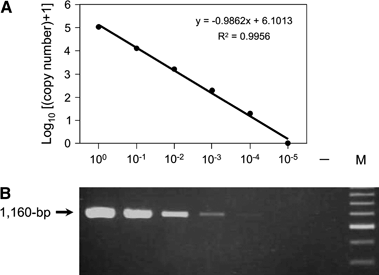
The Q-PCR assay targeting the tufB gene of Ca. L. asiaticus showed a linear relationship (R2 = 0.996) between the 10-fold dilution series of the total DNA sample and the logarithm of the estimated copy number of the target gene (Fig. 1A). In addition, the copy number of the tufB gene was also reduced in proportion to the intensity of the products of 16S rDNA amplified by conventional PCR with OI1/OI2c primers (Fig. 1A and Fig. 1B). In conventional PCR, the sample with the lowest concentration that we could evaluate as positive for Ca. L. asiaticus was obtained after 1:1000 dilution of the original DNA preparation. This corresponds to approximately 190 copies of the tufB gene in one reaction mixture in our Q-PCR reaction system. Although our conventional PCR assay showed only a faint band at 10−4 dilution, the Q-PCR assay was able to evaluate tufB copy number of the dilution sample as 18 copies per 5 μL of the template DNA. At the lowest concentration, that is 10−5, Ca. L. asiaticus DNA was not detectable with either assay.

Detection of the target DNA in 10-fold dilution series (100–10−5) of a total DNA sample extracted from a Diaphorina citri individual positive for Candidatus Liberibacter asiaticus. (A) Quantities (copy numbers) of the tufB gene of Ca. L. asiaticus analysed by real-time quantitative PCR using ABI Prism 7700 sequence detector with the primer pair tb-122F/tb-225R and TaqMan probe tb-167T (Table 1). Means of two replicates are shown. The target DNA was not detected in 10−5 dilution. (B) Agarose gel electrophoresis of conventional PCR amplification products (16S rDNA) of Ca. L. asiaticus with OI1/OI2c primers. –, No template control; M, 200-bp ladder marker (Toyobo, Osaka, Japan).
Temporal changes in the concentration of Ca. L. asiaticus in psyllids after acquisition feeding by adults
The percentage of Ca. L. asiaticus-positive psyllids detected by conventional PCR assays at 0, 5, 10, 15 and 20 days after the AAP of 24 h was 88%, 68%, 48%, 56% and 50%, respectively.
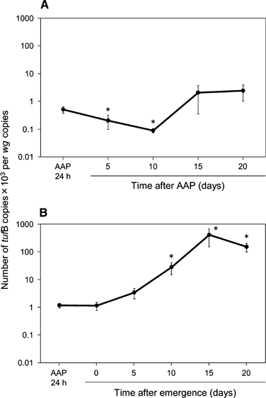
As revealed by Q-PCR assay, the mean concentration of the pathogen in the positive psyllids did not increase over time (Fig. 2A). There were significant differences in the mean concentration of the pathogen among sample dates (P = 0.011, Kruskal–Wallis test). The mean concentration of the pathogen at 5 and 10 days after AAP was significantly lower than that at the end of AAP (P = 0.0037 and 0.0018, respectively, Mann–Whitney U-test). The two highest concentration samples reached 2.4 × 10⁻² and 1.6 × 10⁻² (tufB copies/wg copies) at 15 and 20 days after AAP, respectively; however, neither of these mean concentrations differed significantly from the concentration at the end of AAP (P = 0.16 and 0.89, respectively, Mann–Whitney U-test).

Concentration of Candidatus Liberibacter asiaticus in Diaphorina citri evaluated by real-time quantitative PCR (Q-PCR). (A) Acquisition in the adult stage. (B) Acquisition in the nymphal stage (fifth instar). Psyllids were maintained on healthy citrus seedlings after an acquisition access period (AAP) of 24 h. Means and standard errors are presented. Asterisks indicate a significant difference from AAP of 24 h (Mann–Whitney U-test, P < 0.01). tufB, the tufB gene of Ca. L. asiaticus and wg, the wingless gene of D. citri. Samples that tested positive for Ca. L. asiaticus by conventional PCR were subsequently analysed by Q-PCR.
Temporal changes in the concentration of Ca. L. asiaticus in psyllids after acquisition feeding by fifth instars
The percentage of Ca. L. asiaticus-positive psyllids based on conventional PCR assays at the end of the AAP of 24 h and at 0, 5, 10, 15 and 20 days after emergence was 100%, 80%, 80%, 67%, 87% and 85%, respectively.
As revealed by Q-PCR assay, the mean concentration of the pathogen in the positive psyllids increased over time (Fig. 2B). There were significant differences in the mean concentration of the pathogen (P < 0.0001, Kruskal–Wallis test). Although the mean concentration at 0 and 5 days after emergence was not significantly different from the concentration at the end of AAP (P = 0.68 and 0.71, respectively, Mann–Whitney U-tests), concentration at 10, 15 and 20 days after emergence was significantly higher than the concentration at the end of AAP (P = 0.0043, 0.0015 and 0.0002, respectively, Mann–Whitney U-tests). In one psyllid, the concentration reached 3.3 × 100 (tufB copies/wg copies) at 15 days after AAP.
Transmission test
In the transmission tests by psyllids that fed as fifth instar nymphs on Ca. L. asiaticus-infected plant, the percentage of Ca. L. asiaticus positive at the end of IAP was approximately 78% (seven of nine individuals positive in PCR assays). Three months after inoculation, four of six inoculated test plants tested positive for Ca. L. asiaticus by both PCR assays using leaves and psyllid diagnoses (Table 2). Seedlings that were positive in PCR assays showed foliar ‘blotchy mottle’-like symptoms that were closely associated with Ca. L. asiaticus infection.
| Plant no. | Nymphal acquisition (fifth instar) | Plant no. | Adult acquisition | ||||||||
|---|---|---|---|---|---|---|---|---|---|---|---|
| Psyllids for inoculation | PCR detectiona | Psyllids for inoculation | PCR detectiona | ||||||||
| IAP 15 daysb | PCR-positive/total testedc | Q-PCRd | Leaf midrib | Psyllid diagnosis | IAP 15 daysb | PCR-positive/total testedc | Q-PCRd | Leaf midrib | Psyllid diagnosis | ||
| 1 | 2 | 1/1 | 2 | − | − | 1 | 1 | nt | nt | − | − |
| 2 | 1 | 1/1 | 6 | + | + | 2 | 2 | 0/2 | 0 | − | − |
| 3 | 3 | nt | nt | + | + | 3 | 1 | nt | nt | − | − |
| 4 | 3 | 2/2 | 3806 | + | + | 4 | 3 | 1/3 | 238 | − | − |
| 5 | 3 | 1/3 | 393 | − | − | 5 | 3 | 0/3 | 0 | − | − |
| 6 | 3 | 2/2 | 851 | + | + | ||||||
- IAP, inoculation access period; Q-PCR, real-time quantitative PCR.
- a PCR with OI1/OI2c primers examined at 3 months after IAP; +, positive; −, negative. Leaf midrib: inoculated plants were examined on the basis of a PCR assay using leaf midrib. Psyllid diagnosis: three individuals of healthy fifth instars of D. citri were fed on each test plant for 24 h and then examined together by PCR for the presence of Ca. L. asiaticus.
- b Number of psyllids alive on the 15th day of IAP of 30 days.
- c Inoculative psyllids collected at the end of the IAP of 30 days on each test plant were individually tested by conventional PCR with OI1/OI2c primers and Q-PCR assays; nt, not tested, all insects died.
- d Mean concentration of Ca. L. asiaticus in positive D. citri analysed by Q-PCR [(copy number of the tufB gene of Ca. L. asiaticus) × 103 per copy number of the wingless gene of D. citri].
In the transmission tests by psyllids that acquired the pathogen as adults, the percentage of Ca. L. asiaticus-positive psyllids at the end of an IAP of 30 days was approximately 13% (one of eight individuals positive in PCR assays). The concentration of the pathogen in the only positive psyllid was as high as some psyllids that acquired the pathogen during the nymphal stage. However, all test plants were negative for the pathogen assessed by both PCR assays (Table 2) and showed no symptoms.
Discussion
Our Q-PCR assay targeting the tufB gene of Ca. L. asiaticus DNA was found to be more sensitive in detecting the pathogen than conventional PCR assays targeting 16S rDNA using OI1/OI2c primers. In the case of a DNA sample in which the tufB copy number was estimated to be less than 10, cycle threshold (Ct) values were often unstable (data not shown) in the Q-PCR assay, and a clear band was not detected in conventional PCR assay of such a sample. Therefore, we supposed that the quantitative result of more than 10 copies should be considered to be positive for the pathogen.
Adult psyllids showed different vector competence characteristics depending on when during their developmental stage they fed on infected plants for pathogen acquisition. When psyllids fed on infected plants as adults, the percentage of Ca. L. asiaticus-positive psyllids declined continuously after an AAP of 24 h until it reached approximately 50% after 20 days, and the concentration of the pathogen in the positive psyllids did not significantly increase over the same period. Furthermore, the bacterium was not transmitted to any test plants, and the percentage of Ca. L. asiaticus-positive psyllids at the end of an IAP of 30 days (which corresponded to 42 days after an AAP of 24 h) was only 13%. Hung et al. (2004) reported that the percentage of Ca. L. asiaticus-positive adults ranged from 55% to 70% at 42 days after an AAP of 14 days as adult. These different observations regarding percentage of adults that positively acquired the pathogen are thought to be a consequence of differences in the length of AAPs, suggesting that longer periods of an AAP may result in higher percentage of Ca. L. asiaticus-positive psyllids. The great decline in the percentage of Ca. L. asiaticus-positive psyllids after the transmission test may have been caused by death of positive psyllids and/or a temporal infection shift from being positive to negative because of excretion of Ca. L. asiaticus population from the alimentary canal. Our results were not consistent with those of an earlier psyllid transmission study in which persistent infectivity of D. citri occurred after an AAP of 24 h in the adult stage (Capoor et al., 1974). We suspect that the different outcomes were related to differences in techniques for detecting the pathogen in the previous report (Capoor et al., 1974), which was performed by transmission assays using indicator citrus plants. Another possible reason for the different results may be differences in the origin and strain of the pathogen and/or psyllid population used in the respective studies; this issue should be further investigated. Our data suggested that in the case of an acquisition by adults, ingested bacterial cells were unable to propagate remarkably and therefore they were not persistently retained in the psyllid body; thus, such psyllids would not transmit the pathogen. By taking this mode of transmission into consideration, we speculate that adult psyllids temporarily visiting and feeding on infected citrus trees would become PCR positive for Ca. L. asiaticus but not high-risk infective vectors.
In the case of nymphal acquisition, the percentage of Ca. L. asiaticus-positive psyllids was maintained at a high level over time, and the concentration of the pathogen in the positive psyllids conspicuously increased during the experimental period of 20 days. These results are the first molecular evidence of the propagation of the pathogen in psyllids. Successful transmission to 67% of test plants by inoculative adult psyllids that were given acquisition feeding in the nymphal developmental period suggested that Ca. L. asiaticus was present in the salivary glands of these psyllids. Capoor et al. (1974) reported that the pathogen ingested by nymphs will be persistently retained in the psyllid body after emergence, and it is unnecessary for such psyllid adults to have additional acquisition feeding on infected citrus trees to maintain infectivity. Our results are in accordance with this report (Capoor et al. 1974). We conclude that nymphal acquisition results in higher risk infective vectors than adult acquisition. Therefore, we believe the control of the nymphs on infected citrus trees is important for efficient reduction of the potential risk of disease spread rather than that of adults.
Some plant pathogenic viruses are transmitted similarly to Ca. L. asiaticus. Several hemipteran insects, for example aphids, leafhoppers and planthoppers, can acquire circulative and propagative viruses more efficiently as nymphal stages than adults (Sylvester, 1980; Ammar, 1994). In the case of Frankliniella occidentalis (Pergande), Frankliniella fusca (Hinds) and Thrips setosus (Moulton) (Thysanoptera: Thripidae), all three insect species can transmit tospoviruses, only if they acquire the pathogen during the larval stage (German et al., 1992; Ullman et al., 1992; Van de Wetering et al., 1996; Ohnishi et al., 2001; Assis Filho et al., 2002). In the aforementioned vector–virus combination, a transmission barrier that blocks the escape of the virus from the midgut tissue has been characterised. Similarly, we assume that Ca. L. asiaticus ingested by adults cannot cross their alimentary canals, for example the midgut barrier; therefore, the bacterium does not reach and propagate within the salivary glands. To test this hypothesis, we are now investigating the distribution of the pathogen in the body of infective and non-infective psyllids.
Acknowledgements
We thank T. Takahara and F. Kawamura for providing various plant materials and A. Imai for useful suggestions on statistics. This work was supported by National Agriculture and Food Research Organization Research Project no. 166 ‘Establishment of Agricultural Production Technologies Responding to Global Warming’.


