EFFECT OF BREED, AGE, WEIGHT AND GENDER ON RADIOGRAPHIC RENAL SIZE IN THE DOG
Abstract presented at the 2010 International EVDI meeting, Giessen, Germany.
Abstract
In the adult dog, kidney length has been reported as 2.98 ± 0.44 times the length of L2 on ventrodorsal views and 2.79 ± 0.46 times the length of L2 on lateral radiographs. Our aim was to test the hypothesis that the suggested maximum normal left kidney size is too high, and to evaluate the effect of breed type, gender, weight and age of the dog on kidney size. Abdominal radiographs of 200 dogs with no evidence of concurrent disease that might have an effect on renal size were included in the study. The mean ratio of kidney length to the second lumbar vertebra length was similar to previous reports. For the right lateral view it measured 2.98 ± 0.60 and for the ventrodorsal view 3.02 ± 0.66. Significant differences of this ratio between skull type were present, especially between brachycephalic and dolichocephalic dogs. On the right lateral view brachycephalic dogs had the highest median LK/L2 ratio of 3.1 (3.20 ± 0.40), whereas for dolichocephalic dogs it was 2.8 (2.82 ± 0.50), and for mesaticephalic dogs it was 2.97 (3.01 ± 0.6). A ratio >3.5 was found only in mesaticephalic dogs on the ventrodorsal view. There was a significant difference in the LK/L2 ratio between small (≤10kg) and large breed dogs (>30kg) where small dogs had a significantly higher LK/L2 ratio. There was no statistically significant relation between this ratio and age or gender. The previously reported ratios for kidney size seem valid, but because skull type has an impact on the LK/L2 ratio, a single normal ratio should not be used for all dogs.
Introduction
maging techniques used for assessing renal size and shape in animals and humans include ultrasonography, radiography, contrast studies, computed tomography, magnetic resonance imaging, and renal scintigraphy.1-8 Traditionally, comparison of kidney length to the second lumbar vertebra (L2) on ventrodorsal radiographs has been accepted for mensuration of the canine kidney, where in the adult dog, a length between 2.5 and 3.5 times the length of L2 has been reported as normal.3 However, ventrodorsal projections often require sedation, whereas lateral projections are generally easier to obtain in the conscious patient.
Survey radiographs rarely provide a good image of the right kidney, though it is slightly more visible in the right lateral projection, so calculation of that renal length can be problematic.3, 4 The normal range of kidney length to L2 ratio on lateral projection has been reported to be 2.38–3.10 with a mean (±2 SD) of 2.79 (±0.46) in a study performed on 27 male dogs of unknown breed.3 In another study with a larger sample of dogs a mean (±2 SD) of 2.86 (±0.66) was given.4
Assessment of both kidneys can be improved by intravenous contrast medium injection. However, ratios calculated from nephrograms are larger than those obtained from survey radiographs or aortography.3, 9, 10
Alterations in size, shape, and contour of the kidneys may suggest underlying disease, several of which have been recognized as causing radiographically detectable renomegaly, atrophy or change of renal shape.11-14
To our knowledge there are no previous reports comparing renal size on radiographs of dogs with different skull types and of different reproductive status. Our aim was to test the hypothesis that the suggested maximum normal kidney size of 3.5 × L2 obtained from ventrodorsal radiographs is too high, and to evaluate on the lateral view whether skull type as a measure of conformation (brachycephalic, dolichocephalics, mesaticephalic), age, gender, neutering status, and weight of the dog have an impact on kidney size, using a large group of dogs with no evidence of renal disease.
Materials and Methods
Retrospectively, abdominal radiographs from 200 dogs were evaluated. Inclusion criteria were normal; renal blood profile, electrolytes and glucose levels, urinary tract ultrasound, lumbar vertebral column, absence of medication known to cause hypertension, and no evidence of concurrent disease that could have had an effect on renal size, shape, or contour. In addition, the left kidney had to be measurable on the right lateral or ventrodorsal view. One hundred eighty-eight dogs met the inclusion criteria.
To comply with legislation all patients were sedated or under general anesthesia, and the majority received intravenous fluids before or during imaging. To our knowledge, there are no reports suggesting that anesthesia or fluid therapy could impact renal size. None of the patients had evidence of pelvis or ureteric dilation on ultrasound suggesting an osmotic effect of fluids on the kidneys, nor shown signs of hypertension. There was no specific patient preparation such as starving or enema before radiography, as diet does not have impact on renal size.15 However, food was withhold for at least 12 h before imaging. All radiographs were obtained at a focus-film distance of 100 cm with the beam centered at the level of the L2 to minimize magnification and geometric effects on renal silhouettes. The projections included right lateral, left lateral and ventrodorsal views, although the number of projections depended on the reason for clinical investigation. The images were assessed using a DICOM workstation with electronic calipers (Visbion Ltd, Surrey, UK) or conventional films. Kidney measurements were only done when the margins were clearly visible.3, 4
On lateral recumbent radiographs, the length of L2 was measured at the level of the origin of the transverse processes parallel to the long axis of the vertebral body, and on the ventrodorsal view the midline length. The radiographic kidney length was defined as the maximum distance between the outline of the cranial and caudal poles of the kidney.
The ratios of kidney length to L2 length (K/L2) of each dog in all available views were calculated. Care was taken to discount the partial en face projection of the vertebral end plate when measuring the length of the vertebral body, if this occurred due to positioning.
The dogs were grouped according to skull type16—group 1: brachycephalic, group 2: mesaticephalic, group 3: dolichocephalic and gender category: group 1 = male, group 2 = neutered male, group 3 = female, group 4 = neutered female.
Dogs were also categorized according to weight: group 1 (0–10 kg), group 2 (>10–30 kg), group 3 (>30kg) (one dog weight unknown); and age: group 1 (12 weeks <≤ 1 year), group 2 (1 <≤ 7 years), group 3 (>7 years).
Some of the radiographs 29 of 200 right lateral views and 116 of 200 ventrodorsal views were not included because the kidney was not measurable due to poorly defined margins or organ superimposition or there was only one of the views available.
Standard descriptive measures were calculated for all measurements of the left and right kidneys in all available views and are presented as the mean ± 2SD on each view. Box and whisker plots of the distribution of the measurements are also presented. As nonnormal distribution for some of the measurements emerged, 2.5 and 97.5 percentiles of each measure are also provided. Given concerns about the nonnormality of the data, hypotheses concerning the association between breed/type, gender, weight and age were tested using the Kruskal–Wallis test. Statistical significance was accepted at P < 0.05. All statistical analyses were done using Minitab v161.
Results
One hundred eighty-eight dogs had a right lateral view and on 171 (91%) the left kidney was visible whereas the right kidney was measurable in only 65 images (34.6%). One hundred and five dogs had a ventrodorsal view and on 84 (80%) the left kidney was well defined, but the right kidney was only seen on only 21 (20%). Information from the left lateral view was not pursued as it was rarely acquired due to lack of clinical justification. Consequently, evaluation of the possible impact of breed type, gender and neutering status, weight and age of the dog on K/L2 ratio were done using right lateral view for the left kidney.
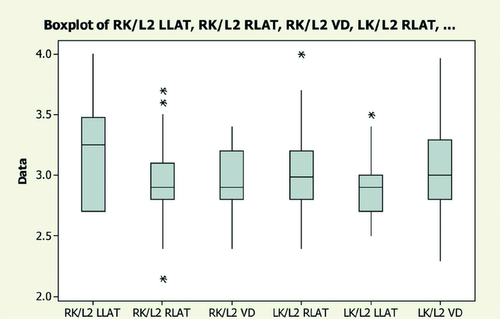
The mean ratio for LK/L2 on the right lateral view was 2.98 ± 0.60 and for the ventrodorsal view the mean ratio was 3.02 ± 0.66. The descriptive statistics for all views as well as the reference ranges for the measurements and boxplot of all the ratios on different views for left and right kidney are presented in Fig. 1 and Tables 1 and 2.
| Variable | N | N* | Mean | SE mean | SD | Minimum | Q1 | Median | Q3 | Maximum |
|---|---|---|---|---|---|---|---|---|---|---|
| RK/L2 LLAT | 6 | 144 | 3.2 | 0.197 | 0.482 | 2.7 | 2.7 | 3.25 | 3.475 | 4.0 |
| RK/L2 RLAT | 65 | 135 | 2.9291 | 0.0355 | 0.2859 | 2.15 | 2.8 | 2.90 | 3.10 | 3.7 |
| RK/L2 VD | 21 | 179 | 2.9471 | 0.0646 | 0.2960 | 2.4 | 2.8 | 2.9 | 3.20 | 3.4 |
| LK/L2 RLAT | 171 | 29 | 2.9848 | 0.0229 | 0.3021 | 2.4 | 2.8 | 2.985 | 3.20 | 4.0 |
| LK/L2 LLAT | 23 | 177 | 2.8983 | 0.0553 | 0.2654 | 2.5 | 2.7 | 2.9 | 3.00 | 3.5 |
| LK/L2 VD | 84 | 116 | 3.0208 | 0.0362 | 0.3316 | 2.3 | 2.8 | 3.0 | 3.29 | 3.96 |
- N*, number not measureable for whatever reason; RK, right kidney; LK, left kidney; L2, length of lumbar vertebra 2; LLAT, left lateral abdomen view; RLAT, right lateral abdomen view; VD, ventrodorsal abdomen view.
| RK/L2 RLAT | RK/L2 VD | LK/L2 RLAT | LK/L2 LLAT | LK/L2 VD | |
|---|---|---|---|---|---|
| Mean | 2.93 | 2.95 | 2.98 | 2.90 | 3.02 |
| 2 SD | 0.58 | 0.60 | 0.60 | 0.54 | 0.66 |
| Mean – (1.96 × SD) | 2.37 | 2.37 | 2.39 | 2.38 | 2.37 |
| Mean + (1.96 × SD) | 3.49 | 3.53 | 3.58 | 3.42 | 3.67 |
| 2.5 percentile | 2.40 | 2.40 | 2.48 | 2.50 | 2.51 |
| 97.5 percentile | 3.54 | 3.40 | 3.60 | 3.45 | 3.60 |
- Mean and SD for K/L2 ratios on different radiographic views. RK, right kidney; LK, left kidney; L2, length of lumbar vertebra 2; LLAT, left lateral abdomen view; RLAT, right lateral abdomen view; VD, ventrodorsal abdomen view.
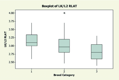
Analysis of potential influences was done using LK/L2 ratios on the right lateral views because on that view, the kidneys were most often measurable. The left kidney was measurable in 28 of 30 brachycephalic dogs, 110 of 126 mesaticephalic dogs, and 33 of 38 dolichocephalic dogs. The relationship between renal length and second lumbar vertebrae length as a function of different skull types, especially between brachy- and dolichocephalic dogs (P = 0.000) is shown in Fig. 2. Some mesaticephalic breeds such as West Highland White Terrier, English Bull Terrier, Scottish Staffordshire Bull Terrier had a bigger LK/L2 ratio, approximately 4.0, 3.7, and 3.6, respectively, but there were not enough individuals to allow statistical confirmation for predisposition of those breeds to have larger K/L2 ratios. The LK/L2 mean ratio in dolichocephalic dogs was 2.82 ± 0.50 with median of 2.8, in mesaticephalic dogs was 3.01 ± 0.6 with median of 2.97 and brachycephalic dogs had the highest median ratio of 3.1 with mean of 3.20 ± 0.40.
Although not statistically significant, on ventrodorsal views where measurable the LK/L2 ratio never reached 3.5 in dolichocephalic dogs (12 views) and brachycephalic dogs (11 views), but did in 7/58 mesaticephalic dogs.
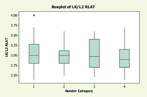
One hundred sixty-seven dogs (32/36 male, 65/74 neutered male, 15/21 neutered female, 55/69 female) were assessed for gender influence. There was no significant effect of gender (P = 0.58), although a marginally wider range was present in intact dogs compared to neutered dogs (Fig. 3).
Regarding the weight category 18 of 26 dogs were included within group 1 (0–10 kg), 89 of 105 in group 2 (>10–30 kg), and 62 of 69 dogs in group 3 (>30 kg). A significant difference of LK/L2 ratio between weight categories (P = 0.030) was found and it was most striking between the lightest and heaviest groups (Fig. 4).
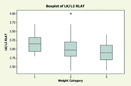
As far as age was concerned there were 13 of 16 dogs in the group 1, 81 of 88 dogs in group 2, and 74 of 84 dogs in group 3. There was not a significant relationship between age and the LK/L2 ratio (P = 0.07), although the ratio was greater in older dogs (Fig. 5).

Discussion
In the adult dog, a normal left kidney length has been reported as 2.98 ± 0.44 (range 2.54–3.45) times the length of L2 on ventrodorsal views and 2.79 ± 0.46 (range 2.38–3.10) for LK/L2 in lateral recumbency.3 However, in what has been considered a key study the authors acknowledged that the population was small and homogenous with respect to gender (only male), age, diet and body condition, which is not representative of the situation encountered in general veterinary practice. Consequently, the range quoted might be unrepresentative of the general healthy canine population.3 Other reports quote slightly different mean ratios ± 2 SD, for ventrodorsal (2.92 ± 0.62) and lateral projections (2.86 ± 0.66), and a range of 2.95–3.01 on survey ventrodorsal views.4, 5 Our results are similar to those reported previously. An element of breed variation was apparent where four of five West Highland White Terriers had a high LK/L2 ratio, which in three dogs was 4.0. These lie outside of the normal population. However, whether this is a real breed variation requires the study of a larger group of West Highland White Terriers.
Here from a large population, there were significant differences (P = 0.000) in the ratios for the three skull types, most notable between brachycephalic and dolichocephalic dogs. The brachycephalic group had the highest median ratio of 3.1 (mean 3.20 ± 0.40) whereas dolichocephalic dogs had the lowest median ratio of 2.8 (mean 2.82 ± 0.50). The LK/L2 ratio was widest in the mesaticephalic group, possibly due to this group being composed of a larger number of dogs (110), whereas the brachycephalic group had 28 dogs and the dolichocephalic group had 33. However, it must be accepted that referral hospital populations are biased towards pure breeds, whereas in general practice cross-breeds are seen commonly.
In people, the kidney is longer in the male, probably due to larger body size.17, 18 In mice exogenous injection of testosterone causes cellular hypertrophy and thus increasing renal size has been reported.19 In cats there is no significant difference between renal size in males vs. females but a higher ratio was found in intact vs. neutered cats.10 In male dogs the kidneys are slightly larger than in the female.4 Our study population included a mix of gender groups and a significant difference between males and females was not detected, although the range was slightly bigger in intact animals compared to neutered animals, but this was not statistically significant. A limitation was the small number of intact females, but that is a reflection of the dog population generally. The relationship between kidney and body weight in the dog seems to be linear and directly proportional although the influence of body weight on K/L2 ratio has not been investigated before, but by implication weight should be related to size for a soft tissue organ.3, 20 There was a significant difference between the LK/L2 ratio of small vs. large dogs. Small dogs up to 10 kg had a significantly higher LK/L2 ratio than the large dogs over 30 kg weight (P = 0.03). This may be due to many small dogs being chondrodystrophoid and having shorter vertebrae compared to large dogs. However, breed and weight are interrelated.
In people, the relationship between age and kidney length is linear and the length of the kidney increases proportionally with age until 16 years of age, where body height is accepted as the most reliable comparator for measuring renal length. After that age kidney size is fairly constant and has a mild predisposition to decrease with time.18 In dogs an age factor has been identified in the relationship between renal length and L2, where it is bigger in dogs under 1 year of age whereas after 1 year no significant changes were noted.4 For newborn and 1-week-old dogs, renal length was on average four to five times the length of the L2; this was explained by the difficulty in radiographic detection of the vertebral body endplates. In the same study, subjects between 2 and 8 weeks had a renal length approximately 3–3.5 times the length of the L2. This was significantly greater than for subjects over 12 weeks and adult dogs.21 After 12 weeks of age renal size was proportionate to body size and was not significantly different from adult dogs.21 We did not find a significant difference in the median LK/L2 ratio on right lateral radiographs between adult age groups. The ratios were greater in older dogs (>7 years) compared to dogs under 1 year of age in our study however it was not significant (P = 0.68). Most of the dogs in oldest group were mesaticephalic (58) including West Highland White Terrier (4), Scottish Bull Terrier (4) and English Staffordshire Bull Terrier (3) which had the highest LK/L2 ratios, up to 4.0, 3.6, and 3.7, respectively, whereas the numbers of brachycephalic dogs (14) and dolichocephalic dogs (13) included in oldest group category were similar. Animals over 1 year of age are skeletally mature but we cannot rule out the possibility that the kidneys continue to grow beyond this age. From our results we conclude that in dogs the K/L2 ratio stays fairly constant after the age of 1 year.
Our hypothesis that the previously suggested maximum normal K/L2 ratio of 3.5 obtained from radiographs is too high was not supported in this large heterogeneous population of dogs, where a maximum ratio can be as high as 3.96 on the ventrodorsal view with median value of 3.0. Nevertheless, we found that the skull type has a significant impact on the LK/L2 ratio with brachycephalic breeds having the highest median LK/L2 ratio whereas in dolichocephalic breeds it was significantly lower. For that reason assessment of the LK/L2 ratio should be performed separately for different skull type dogs. The ratio of 3.5 on the ventrodorsal view in our study was never reached for dolichocephalic and brachycephalic skull types but was found on several occasions in mesaticephalic type dogs. It would appear that the right lateral view is the most suitable for measurement of the left kidney as it is more likely to be visible. In this view the mean LK/L2 value for brachycephalic dogs was 3.20 ± 0.40 with median of 3.1, for dolichocephalic dogs it was 2.82 ± 0.50 with median of 2.8 and for mesaticephalic dogs it was 3.01 ± 0.6 with median of 2.96. In addition, significant differences between weight categories were found and it was most striking between the dogs up to 10 kg weight and those over 30 kg.
In conclusion, the previously reported ratios for kidney size seem valid, at least on the basis of this large study group. Because skull type has a significant impact on the LK/L2 ratio, we recommend that a single normal ratio not be used for all dogs.
ACKNOWLEDGEMENT
The authors acknowledge Sheilla Wills for medical advice.


