IMAGING DIAGNOSIS: PITUITARY APOPLEXY IN A CAT
Abstract
A 7-year-old male neutered domestic short-haired cat had depression for 5 months and acute blindness. A lesion at the level of the rostral and middle cranial fossae was suspected. A large pituitary mass compressing the optic chiasm was detected in magnetic resonance images and there was also evidence of recent intratumoral hemorrhage, leading to a diagnosis of pituitary apoplexy; these findings were confirmed at postmortem examination. Pituitary apoplexy is a clinical syndrome characterized by acute neurologic signs related to hemorrhagic infarction within a pituitary tumor. Pituitary apoplexy should be considered in patients with acute onset of blindness and altered mental status.
Signalment, History, and Clinical Findings
A 7-year-old neutered male domestic short-haired cat had depression for 5 months, recent episodes of restlessness and acute blindness. The cat was depressed and disorientated. Postural reactions were decreased on all limbs but spinal reflexes were normal. There was bilaterally absent menace response, direct and consensual pupillary light reflexes and bilaterally decreased vestibulo-oculocephalic reflexes. A lesion at the level of the rostral and middle cranial fossae was suspected.
Imaging
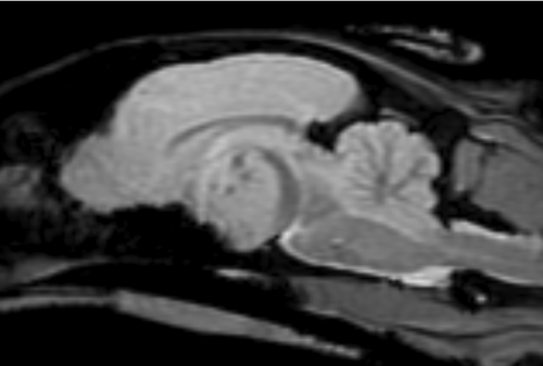
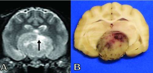
Magnetic resonance (MR) images were acquired at 1.5 T and there was a large, well-defined, globular suprasellar mass measuring 22 mm transversely, 15 mm rostrocaudally, and 16 mm dorsoventrally. The mass compressed the optic chiasm, and thalamus, and elevated the dorsal part of the third ventricle, causing caudal transtentorial herniation. The mass was T2-isointense to normal gray matter with T2-hyperintense foci within (Fig. 1). The mass was T1-hyperintense to normal gray matter and contained T1-hypointense foci that corresponded to the focal T2-hyperintense areas (Fig. 2). There was heterogeneous contrast enhancement. A crescentic rim around the caudal border of the mass was markedly T2-hypointense and mildly T1-hyperintense to normal gray matter. The gradient echo (GE) sequences showed signal void in the caudal crescentic rim and hypointense foci in the mass (Fig. 3). The imaging findings were consistent with a pituitary mass with recent hemorrhage. The main diagnoses were adenoma, adenocarcinoma and, less likely, meningioma.


Outcome
The owner declined treatment and elected to have the cat euthanized. At postmortem examination a large (15 mm diameter), well-circumscribed and nonencapsulated tumor replaced the normal pituitary gland and compressed the surrounding parenchyma. The tumor was formed by sheets of polyhedral cells, sometimes subdivided into packets or cords by fine fibrovascular septa. The cells contained moderate amounts of nongranular eosinphilic cytoplasm. Nuclei were finely stippled to vesiculated, with mild anisokaryosis and rare mitotic activity. Several focal areas of acute hemorrhage were present within the center of the tumor, sometimes associated with local coagulative necrosis at the margins of the hemorrhagic zone. Occasional psammoma bodies were present within the hemorrhagic zones. The histologic diagnosis was pituitary adenoma with acute hemorrhage (Fig. 4).

Discussion
Pituitary apoplexy is a syndrome characterized by acute neurologic signs related to sudden compression of parasellar structures, usually due to hemorrhagic infarction of a pituitary tumor.1 The main clinical features of pituitary apoplexy in humans are sudden headache, nausea, vision loss, and internal ophthalmoplegia.2 Pituitary apoplexy has been described in dogs and was characterized mainly by acute visual disturbances and altered consciousness.3, 4 The main findings in the present of disorientation, acute blindness, dilated nonresponsive pupils and decreased vestibulo-oculocephalic reflexes in both eyes indicated combined deficits of cranial nerves II, III, IV, and VI bilaterally and thalamus/midbrain dysfunction. In MR images there was lateral, rostral, dorsal and caudal extension of the tumor with the sudden vision loss being due to acute hemorrhagic expansion of the pituitary tumor. Decreased vestibulo-oculocephalic movement in a nonrestricted, centrally positioned ocular globe can be explained by dysfunction of cranial nerves III, IV, and VI that innervate the extraocular muscles, bilateral medial longitudinal fasciculus, or vestibular dysfunction. In this cat, the reduced vestibulo-oculocephalic reflex could be explained by compression of the rostral medulla oblongata, pons, and midbrain due to the caudal transtentorial herniation and therefore dysfunction of the medial longitudinal fasciculus.
Imaging features of canine pituitary apoplexy have been described using computed tomography, although it was difficult to differentiate subacute and chronic hemorrhage from necrosis with cystic changes.4
In MR images, feline pituitary adenomas are characterized mainly by a heterogeneous appearance on both T1W and T2W images, with uneven contrast enhancement. 5 The tumor in this cat had some unusual imaging features characterized by T2-hyperintense/T1-hypointense foci that could have represented small fluid pockets or acute hemorrhage. Histopathology identified these central areas within the tumor as recent hemorrhage. The crescentic rim around the caudal border of the mass, which was markedly T2-hypointense and T1-hyperintense to normal gray matter was thought to represent early subacute hemorrage. The susceptibility artifact seen in the GE images could have been due to hemosiderin, methemoglobin, deoxyhemoglobin, oxyhemoglobin, ferritin, calcium, other metals or air6 but in this cat was caused by recent hemorrhage (intracellular oxyhemoglobin and deoxyhemoglobin/methemoglobin).
The pathophysiology of pituitary apoplexy in people has not been fully elucidated. Pituitary adenomas may have an intrinsic vasculopathy and be more susceptible to bleeding than are other intracranial tumors.7 Alternatively, rapid expansion of the tumor could result in hemorrhagic necrosis.8 Whether these are relevant causes of pituitary hemorrhage in cats is not known.
In summary, pituitary apoplexy should be considered in dogs and cats that present with acute onset of blindness and altered mental status. MR imaging features of pituitary apoplexy are mainly characterized by T2-hyperintense, T1-hypointense and GE signal void areas within a pituitary mass which can be attributed to recent hemorrhage. The GE sequence is useful in detecting and delineating hemorrhage and it should be included in the MR protocol for patients with acute neurological deterioration.


