COMPUTED TOMOGRAPHIC FEATURES OF FELINE NASOPHARYNGEAL POLYPS
Abstract
The computed tomographic (CT) findings of histopathologically confirmed nasopharyngeal polyps are described in 13 cats. Most polyps were mildly hypoattenuating to adjacent muscles and isoattenuating to soft-tissue (n= 13), homogeneous (n = 12) and with ill-defined borders (n = 10) on precontrast images. After contrast medium administration, the polyps were homogeneous (n = 11), with well-defined borders (n = 13), oval (n = 13), and had rim enhancement (n = 13). Nasopharyngeal polyps were pedunculated in 11 cats with a stalk-like structure connecting the polyp through the auditory tube to an affected tympanic bulla. All cats had at least one tympanic bulla severely affected, with CT images identifying: (1) complete (n = 12) or partial (n = 1) obliteration of either the dorsal or ventral compartments with soft-tissue attenuating material; (2) pathologic expansion (n = 13) with wall thickening (n = 10) that was asymmetric in nine cats; and (3) identification of a polyp-associated stalk-like structure (n = 11). Nine cats had unilateral tympanic bulla disease ipsilateral to the polyp, and four cats had bilateral tympanic bulla disease, most severe ipsilateral to the polyp with milder contralateral pathologic changes. Two cats had minimal osteolysis of the tympanic bulla. Enlargement of the medial retropharyngeal lymph node was seen commonly (n = 8), and in all cats it was ipsilateral to the most affected tympanic bulla. One cat had bilateral lymphadenopathy. CT is an excellent imaging tool for the supportive diagnosis of nasopharyngeal polyps in cats. CT findings of a well-defined mass with strong rim enhancement, mass-associated stalk-like structure, and asymmetric tympanic bulla wall thickening with pathologic expansion of the tympanic bullae are highly indicative of an inflammatory polyp.
Introduction
Inflammatory polyps of the feline middle ear and nasopharynx are nonneoplastic masses that are presumed to originate from the epithelial lining of the tympanic bulla or auditory tube.1 The etiology is unknown but has been linked to chronic inflammation and upper respiratory tract infection.2 Other suggested causes include congenital abnormality of the fetal branchial or pharyngeal arches and otitis media.3, 4 Histologically, the polyps consist of a core of loosely arranged fibrovascular tissue covered by a stratified squamous to ciliated columnar epithelial layer containing scattered lymphocytes, plasma cells, and macrophages. The mucosa is commonly ulcerated.4, 5 Presumptive diagnosis of inflammatory polyps is based on history and physical examination, supported by imaging and endoscopic evaluation, and confirmed histopathologically.3
The identification of nasopharyngeal masses can be made by oropharyngeal examination, digital palpation of the soft palate, skull radiography, endoscopy, computed tomography (CT), magnetic resonance imaging, nasopharyngeal biopsy, or any combination of these.2, 4 Radiographs can be used to identify a soft-tissue mass in the nasopharynx and to evaluate the tympanic bullae for thickening of the wall and loss of the normally contained air. CT allows assessment of the external ear canal, tympanic membrane, osseous bulla and nasopharynx.6 In addition to localizing the affected osseous bulla and potentially earlier detection of a mass, CT has other valuable characteristics including the ability to reformat images in other planes, lack of superimposition and good soft tissue contrast resolution.7
Given the sensitivity for detecting and characterizing nasopharyngeal lesions, CT has potential as an ideal imaging modality for supporting the diagnosis of inflammatory nasal polyps in cats. However, the CT features of nasopharyngeal polyps have only been described once and in that report only two cats were described and postcontrast images were not available.7 Our goal was to further characterize the CT features of histologically confirmed nasopharyngeal polyps in cats.
Materials and Methods
After a review of medical records between 2006 and 2011, 13 cats with a histologically confirmed nasopharyngeal polyp that underwent CT examination of the head that included pre- and postcontrast CT images were identified. One cat had been included in a previous report.8 The median age was 2.7 years (range 0.5–17.5 years) and median weight was 4.2 kg (range 1.5–8.2 Kg). There were nine neutered males, three neutered females, and one intact female. Breeds included domestic short hair (n = 9), Maine Coon (n = 2), domestic long hair (n = 1), and Siamese (n = 1). The most common presenting complaints were: increased respiratory effort (n = 7), nasal discharge (n = 5), noise while sleeping (n = 3), and sneezing (n = 3). Eight cats had clinical signs for more than 1 month, one cat had signs for 24 h and in four cats the duration of clinical signs was unknown. The most common physical findings were: stertor (n = 11), dyspnea (n = 7), external ear canal mass (n = 5), and head-tilt (n = 4). Other findings included ocular and nasal discharge (n = 3), Horner's syndrome (n = 2), circling (n = 1), and ataxia (n = 1). Ten cats had surgery and in all of these a ventral bulla osteotomy was performed to sever the polyp base the nasopharyngeal mass was removed by laryngeal traction. In the remaining three cats only laryngeal traction avulsion was performed.
Eight cats had CT examinations performed using a 16-slice helical scanner1 and five were evaluated with a single slice helical scanner2. The scan parameters for the 16-slice machine were: small display field of view of 25 cm, 2.0 mm slice thickness with 1 mm slice interval and one of the two techniques: 80 kV with 16 mAs, or 100 kVp with 7 mAs, and for the single slice were: small display field of view, 3.0 mm slice thickness with 2 mm interval, 120 kV, and 80 mAs. Postcontrast images were obtained immediately after injection of iohexol3 at 2 ml/kg. Reformatted images in sagittal and dorsal planes were available for nine cats. For the remaining four cats, reformatting was performed using the PACS workstation4.
All images were reviewed by a board certified radiologist (IC) and a radiology resident (CRO) in a PACS workstation in bone (window width [WW] = 2000, window level [WL] = 800) and soft tissue (WW = 360, WL = 60) windows. These values were adjusted as needed to better evaluate the images and to evaluate the tympanic wall thickening the images were sharpened. Information was recorded of the nasopharyngeal polyps, the ear system, the nasal cavity and the regional lymph nodes. For all structures evaluated, the term mild was applied when the abnormality affected less than 25% of the involved structure(s). Abnormalities affecting 25–50% of the involved structure(s) was defined as moderate, and when the majority of involved structure(s) was pathologically altered it was classified as severe. Tympahic bulla expansion was diagnosed when its overall diameter was larger than the contralateral bulla.
The distance of the mass from the hard palate was calculated in a rostrocaudal direction, starting at the beginning of the mass. Attenuation values were calculated in the center and in the outer margins (rim enhancement) of the polyps, and the numbers were compared. For the center of the polyp, three regions of interest ROIs) of 10 mm2 were placed in the most central part of the polyp in three consecutive slices pre- and postcontrast medium administration. The rim enhancement was calculated by placing three ROIs of 0.6 mm2 on the outer margins of the polyps on postcontrast images in three consecutive slices. The mean Hounsfield unit (HU) was calculated and used for statistical analysis. The left and right medial retropharyngeal lymph nodes were measured in all cats. The maximum width and height were measured on transverse images and the length on sagittal reformatted images. Left and right mandibular lymph nodes were evaluated subjectively and were not measured.
Statistical analysis was performed using commercial software5. Normality of the data was assessed using a D'Agostino–Pearson test. Nonnormally distributed data were logarithmic transformed for future calculations and the mean, standard deviation (SD), 95% confidence interval (CI) and range (minimum–maximum) reported. A repeated measures ANOVA test was used to compare the precontrast, postcontrast, and rim enhancement HU of the nasopharyngeal polyps. A paired sample t-test was used to compare the size of the medial retropharyngeal lymph node ipsilateral to the side of origination of the polyp to the contralateral lymph node in each cat. A one-sample t-test was used to compare the mean measurements of ipsilateral to contralateral lymph nodes. A P-value < 0.05 was considered statistically significant.
Results
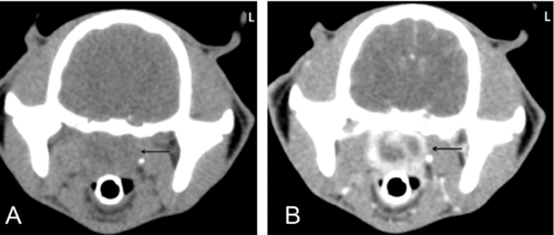
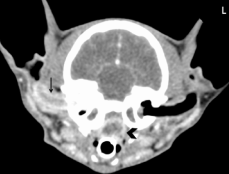
A soft-tissue attenuating nasopharyngeal mass located caudal to the hard palate, representing the polyp, was present in all cats (Table 1). Precontrast, the polyps were minimally hypoattenuating to adjacent musculature and isoattenuating to soft-tissue (range: 26.49–39.95 HU) (n = 13), homogeneous (n = 12) with ill-defined borders (n = 10), an ill-defined shape (n = 13), and could not be delineated from adjacent structures (n = 13) (Fig. 1A). After contrast medium administration, the polyps were hypoattenuating to soft-tissue (n = 13), homogeneous (n = 11), with well-defined borders (n = 13), oval (n = 13), and with rim enhancement (n = 13) (Fig. 1B). The rim enhancement provided clear definition of the margins the polyp, which appeared to be pedunculated with a stalk-like structure connecting the polyp through the auditory tube to the tympanic bulla in 11 cats (Figs. 2–C). In these 11 cats the auditory tube was subjectively widened, while the contralateral auditory tube could not be identified. The polyps were well defined in all imaging planes (Figs. 3 and B) and the stalk-like structure connecting the polyp with the tympanic bulla was easier to identify on transverse and sagittal images. In some cats, the rim enhancement surrounding the polyp could be traced clearly into the auditory tube. The central region of each polyp had homogenous enhancement in 11 cats and heterogeneous enhancement in 2 cats. The objective parameters of the polyps are summarized in Table 2. In nine cats the polyp caused complete obliteration of the nasopharynx at their widest transverse dimension. In two cats there was approximately 75% obliteration, in one cat 50% and in one cat 30%. The mean postcontrast attenuation of the peripheral portion of the polyps was significantly higher than postcontrast attenuation in the more central region. (Table 3).
| Precontrast (N) | Postcontrast (N) | |
|---|---|---|
| Borders | ||
| Ill-defined | 10 | 0 |
| Well-defined | 3 | 13 |
| Attenuation (center of polyp) | ||
| Homogeneous | 12 | 11 |
| Heterogeneous | 1 | 2 |
| Shape | ||
| Ill-defined | 13 | 0 |
| Oval | 13 | |
| Pedunculated (stalk seen) | 11 | |
| Extension | ||
| Into auditory tube | Not visible | 11 |
| Into middle ear | 11 | |
| Into external ear canal | 4 | |
| Contrast Pattern | ||
| Rim enhancing Mild | – | 2 |
| Moderate | 1 | |
| Strong | 10 | |
| Tympanic bulla | ||
| Soft-tissue attenuating material | 13 | 13 |
| Ipsilateral | 9 | 9 |
| Bilateral | 4 | 4 |
| Thick-walleda | 10 | 10 |
| Expandeda | 13 | 13 |
| Lysisa | 2 | 2 |
| Medial retropharyngeal lymph node | ||
| Enlarged | 8 | 8 |
| Ipsilateral | 7 | 7 |
| Bilateral | 1 | 1 |
| Heterogeneous enhancement. | – | 13 |
- a Always ipsilateral. N = number of cats.
| Mean | SD | 95% CI of Mean | Range | |
|---|---|---|---|---|
| Size (mm) | ||||
| Width | 14.68 | 5.33 | 11.45 to 17.9 | 6.28–25.15 |
| Height | 11.59 | 4.34 | 8.96 to 14.2 | 2.92–18.04 |
| Length | 20.57 | 10.03 | 14.51 to 26.6 | 6.49–42.11 |
| Distance from | 11.79 | 6.25 | 7.81 to 15.76 | 4.95–26.17 |
| hard palatea (mm) | ||||
- a Measured rostrocaudally. Excluding one cat in which the mass was in contact with the hard palate. SD, standard deviation; CI, confidence interval.
| Precontrast | Postcontrast | |||||
|---|---|---|---|---|---|---|
| Attenuation (HU) | Mean | 95% CI of mean | Range | Mean | 95% CI of mean | Range |
| Center | 32.53a | 26.49 to 39.95 | 18.32 – 48.08 | 43.49b | 33.62 to 56.25 | 25.06 – 102.93 |
| Rim | 148.37c | 126.12 to 174.55 | 99.33 – 228.89 | |||
- Among the same line different letters indicate statistically significant difference: a and b: P = 0.0015; a and c: P < 0.0001; b and c: P < 0.0001. CI, confidence interval.


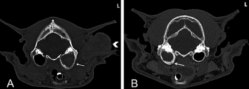
All cats had at least one tympanic bulla severely affected, with CT images identifying: (1) complete (n = 12) or partial (n = 1) obstruction of either the dorsal or ventral compartments of the tympanic bulla with soft-tissue attenuating material; (2) pathologic expansion (n = 13) with wall thickening (n = 10) (Figs. 4 and B); and (3) identification of a polyp-associated stalk-like structure (n = 11). Nine cats had unilateral severe tympanic bulla disease on the same side of the polyp. Four cats had bilateral bullae disease, severe on the same side of the polyp and mild on the contralateral side with partial obstruction with soft-tissue attenuating material. In all cats with wall thickening, the thickening appeared as a smoothly marginated increase in the width of the wall, which was asymmetric in all but one cat, and present only on the ventromedial aspect of the bulla in eight cats, and on the ventromedial and ventrolateral aspects in one cat. No periosteal reaction was seen in any tympanic bulla. Two cats had mild osteolysis with smooth margins of the ventral wall of the affected tympanic bulla. All affected tympanic bulla were characterized homogeneous mild enhancement of the soft-tissue attenuating material after contrast medium administration. The cochlea was visible in all cats and considered to be normal.

A mild amount of soft-tissue attenuating material was present in the horizontal ear canal in two cats and thought to represent exudate, and both cats had tympanic bulla disease on the same side. Five other cats had an external ear canal polyp that was continuous with the material in the tympanic bulla and had similar CT characteristics of the nasopharyngeal polyp (Figs. 4 and 5). In four of these cats, a stalk-like structure was seen communicating the nasopharyngeal polyp with the tympanic bulla.

Seven cats had nasal cavity disease characterized by soft-tissue or fluid attenuating material in the nasal cavity that was mild (n = 4), moderate (n = 2) or severe (n = 1), and bilateral in five cats. The distribution was: rostral and middle (n = 3), generalized (n = 3), middle (n = 1) and caudal (n = 1). Ethmoidal turbinate destruction was present in four cats and there was fluid in the frontal sinus in one cat. In one cat images of the complete nasal cavity were not available for review.
The mandibular and medial retropharyngeal lymph nodes were elliptical and exhibited heterogeneous contrast enhancement with multiple enhancing linear structures in the center and periphery. Enlargement of the medial retropharyngeal lymph node was present in eight cats. In seven cats the enlargement was unilateral and on the same side as the affected tympanic bulla. One cat had bilateral enlargement. The mean size of the widest dimension of normal lymph nodes was 23.6 mm (SD = 5.3, 95% CI = 20.83–26.27, range = 16.24–38.99), and of the abnormal lymph nodes was 29.4 mm (SD = 8.5, 95% CI = 21.54–37.20, range = 19.1–44.5). There was a statistically significant difference between abnormal and normal medial retropharyngeal lymph nodes within cats (P = 0.003) and among cats (P < 0.001). No difference was found between left and right medial retropharyngeal lymph nodes in cats with no lymphadenopathy (P = 0.437). Two mandibular lymph nodes were consistently identified on the left and right side and were symmetric in all cats.
Discussion
All nasopharyngeal polyps had a characteristic postcontrast appearance of a well-circumscribed pedunculated mass with strong rim enhancement. In most cats, the polyp could be seen extending through the auditory tube into one tympanic bulla, which was completely obliterated by soft-tissue attenuating material, thick walled and with pathologic expansion. The auditory tube was identified based on its expected location on the ventral aspect of the skull, caudal border of the soft-palate and immediately rostral to the tympanic bulla, connecting the tympanic bulla to the nasopharynx.9 It could be seen only because of the presence of the polyp that could be traced from the nasopharynx into the expected location of the auditory tube, and then into the tympanic bulla. The normal canine auditory tube has been identified using virtual CT otoscopy but in the same dogs it was not visible using high resolution CT.10 In another study, the auditory tube was identified with CT after administration of contrast medium directly into the tympanic cavity.9
Rim enhancement was present in all polyps. Histopathologically, inflammatory polyps consist of a core of loosely arranged fibrovascular tissue covered by a stratified squamous to ciliated columnar epithelial layer that is commonly ulcerated. Variable lymphoplasmacytic, lymphoid aggregates/follicles, and pleocellular inflammation are often present throughout the stromal tissues.5 This inflammatory nature is likely the cause of the rim enhancement. Human nasal polyps, which have the same histologic characteristics as feline inflammatory polyps, have increased vascularization induced by inflammatory and epithelial cells.11 Mucosal enhancement of human nasal polyps is an occasional MR imaging finding in recently developed polyps.12 We found no reports of enhancement of human choanal polyps in CT images.
Five cats had concurrent nasopharyngeal and external ear canal polyps and in four of these there was a connection between the nasopharyngeal mass, the tympanic bulla and the external ear canal mass. Coexisting nasopharyngeal, tympanic bulla and ear canal lesions have been reported as uncommon.1 CT was beneficial in identifying the multicompartmental nature of the disease in these cats.
Differential diagnosis for a feline nasopharyngeal soft-tissue mass seen in CT images includes lymphoma, squamous cell carcinoma, melanoma, lymphocytic-plasmacytic inflammation, adenocarcinoma, rhabdomyosarcoma and cryptococcosis.14 Based on our findings, the CT features that may be most predictive of a polyp as the cause of a nasopharyngeal mass are the presence of a stalk-like structure, visualization of the auditory tube, strong rim enhancement and pathologic expansion with thickening of the affected tympanic bulla. Features previously described in cats with malignant nasopharyngeal masses, but seen in no cat in our group include extension of the mass into another region of the skull including the brain, and severe effacement of the tympanic bulla and adjacent structures.
All cats in this study had soft-tissue attenuating material and pathologic expansion of the affected tympanic bulla. In 37 cats with a nasopharyngeal polyp, soft-tissue opacification and periosteal proliferation was present in the tympanic bulla of 17 of 19 cats that underwent radiographic examination.18 Tympanic bulla abnormalities are likely related to the location of the primary disease in the caudal aspect of the nasopharynx, near the opening of the auditory tube. Obstruction of the caudal portion of the nasopharynx therefore could contribute to physical or functional alteration in the auditory tube resulting in effusive tympanic bulla disease.19 All polyps in this study were located caudal to the hard palate. Feline nasopharyngeal polyps may be a consequence of otitis media, or predispose to otitis media because of obstruction of the auditory tube with accumulation of fluid and debris in the bulla. Other mechanisms by which the polyp can predispose to otitis media are increased negative pressure within the auditory tube causing transudation from blood vessels within the tympanic cavity and distortion of the pharyngeal opening of the auditory tube, resulting in ascending infection.6
The affected tympanic bulla was considered to be abnormally thick in 10 cats, a possible sign of bulla osteitis. The wall of a fluid-filled tympanic bulla may appear artificially thicker than that of an air-filled tympanic bulla, especially when images are acquired as thick slices (>5 mm), reconstructed with a soft tissue algorithm, or displayed with a narrow window (<250 HU).20 This artifact is unlikely in this study as all images were acquired at a maximum of 3 mm slice thickness, and reviewed using a wide window (2000 HU) and sharp algorithm. The tympanic bulla wall thickening was asymmetric in nine cats and seen only on the ventromedial or ventral aspect of the tympanic bulla in eight cats whereas artifactual thickening would affect the entire tympanic bulla.20 It is unknown why the tympanic bulla wall thickening was predominantly ventromedial. One explanation is that the entry of the auditory tube is ventral and the presence of the polyp in the auditory tube and extending into the tympanic bulla could resulted in increased inflammation and osteitis in that region.
Increased width of the medial retropharyngeal lymph node on the side of the polyp was common. It is not known if the asymmetry was due to reactive changes associated with the polyp, changes within the tympanic bulla or a combination of these factors.
In most previous studies, the majority of cats with nasopharyngeal polyps were less than 3 years of age whereas cats in this study had a wider range, with four being 6–7 years old, and one being 17.5 years old.4, 6, 7 This is similar to one study that also reported an older mean age at onset of clinical signs.5 It is possible that some of these cats developed the polyp at an early age but clinical signs did not develop until later. Thus, a nasopharyngeal polyp should not be excluded as a differential in older cats with upper airway obstruction and it also highlights the importance of knowing the CT findings to be expected with a polyp versus a malignant mass.


