MRI FEATURES OF GASTROCNEMIUS MUSCULOTENDINOPATHY IN HERDING DOGS
Poster presentation at the 15th Congress of the IVRA, July 26–31, 2009, Buzios, Brazil.
Abstract
Pelvic limb lameness that was localized clinically to the lateral gastrocnemius head was observed in dogs without history of trauma. The aim of this retrospective study was to describe magnetic resonance imaging (MRI) findings of this condition. Nine dogs were identified, eight Border Collies and one Australian Shepherd. They all had chronic pelvic limb lameness; no signs of joint effusion or instability were present. In MR images there was high signal intensity in the lateral head of the gastrocnemius muscle around the sesamoid bone in T2-weighted, T2*-weighted, and STIR images and an iso- to mildly hyperintense signal in T1-weighted images with marked contrast enhancement. The abnormal signal intensity most likely represents a myotendinous strain. The breed affiliation to Border Collies is striking, and a relation to biomechanical forces or motion pattern may be possible. Except for the dog with the most extensive lesion all dogs had an excellent outcome.
Introduction
The gastrocnemius muscle is divided into a lateral and a medial head, which are covered by strong tendinous leaves and infiltrated by tendinous strands. Both heads arise out of a large tendon from the lateral and medial supracondylar tuberosity of the femur, and contain a large sesamoid bone that articulates with the corresponding condyle. The medial and lateral heads fuse distally, with the tendon attaching at the tuber calcanei. The lateral head of the superficially located gastrocnemius muscle almost completely encloses the superficial digital flexor tendon, which originates from the femur and attaches at the medial phalanx of the digits.1 The main action of the gastrocnemius muscle is the extension of the tarsus, but it also flexes the knee in the nonweight bearing state2 resulting in action throughout the stance phase and during walk, trot, or gallop.1
Injuries of the gastrocnemius muscle origin in dogs include total or partial avulsion of the lateral or medial head with or without known prior trauma.3–9 Affected dogs have high grade lameness, sometimes with a plantigrade stance. Distal displacement of the corresponding sesamoid bone of the gastrocnemius head is usually apparent in radiographs of affected animals.3–8 We observed injury of the gastrocnemius muscle in a group of herding dogs with pelvic limb lameness without history of trauma. The aim of this retrospective study was to describe the magnetic resonance imaging (MRI) findings associated with this condition.
Materials and Methods
Medical records of two referral centers (Vetsuisse Faculty Berne and Tierärztliches Überweisungszentrum, Switzerland) were reviewed for dogs that underwent MRI of the stifle joint between January 2001 and June 2009. Nine dogs with abnormal MRI findings in the gastrocnemius muscle were identified. There were eight Border Collies and one Australian Shepherd. Six were used or trained for herding of sheep or for search and rescue purposes and two were doing agility. Three were male (one neutered) and six female (four neutered), with a mean age of 5.3 years (range 0.3–8.3 years) and a mean weight of 15.8 kg (range 9.5–22.5 kg). Two were skeletally immature (4 and 6 months old).
All dogs had chronic (1– 4 months) right (five dogs) or left (four dogs) low to moderate grade pelvic limb lameness, which was most obvious during the first steps of walking. In dog #6 intermittent high grade lameness was reported. Trauma associated with the onset of lameness was not reported. In six dogs the lameness was localized to the region of the lateral gastrocnemius head. There were no signs of stifle joint effusion or instability.
Stifle radiographs were available in five dogs and all were characterized by mineralizations at the sesamoid bone and in the surrounding tissue (Fig. 1).
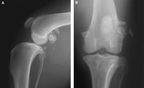
Dog #6: Mediolateral (A) and caudocranial (B) radiographs of the left stifle joint. There is an abnormal lateral sesamoid bone with mineralization in the surrounding tissues. The sesamoid bones are in a normal position.
All dogs underwent MRI examination of one (seven dogs) or both (two dogs) stifle joints. MRI was performed under general anesthesia induction with propofol after sedation and anesthesia maintenance with inhalation of isoflurane. Dogs were in dorsal recumbency with the pelvic limbs extended. In eight dogs MR images were obtained using a low-field system* in one dog with a high-field system† (Table 1).
| Dog | STIR | FSE T2 | GE T2* | SE T1 | GE T1 |
|---|---|---|---|---|---|
| 1 | Dor 4400/25 | Tra 4600/90 | Sag 1600/30Dor 1600/30 | Dor 510/24 | Sag 30/12 |
| 2 | — | — | Sag 1600/30Tra 1600/30 | Tra 472/27 | Sag 30/12 |
| 3 | Dor 4000/30 | — | Sag 1600/30 | — | Dor 30/12 |
| 4 | Tra 4100/30 | Sag 5652/125Tra 6080/125 | — | Sag 97/20Tra 703/20 | — |
| 5 | Sag 4690/30Dor 5360/30 | — | Sag 350/17Dor 400/17 | Sag 50/20Dor 50/20 | — |
| 6 | Sag 4690/30Dor 4106/30 | Tra 4000/120 | Sag 400/17Dor 400/17 | Sag 50/20Dor 50/20 | — |
| 7 | Sag 4020/30Dor 6700/30 | — | Sag 400/17Dor 400/17 | Sag 50/20Dor 40/16 | — |
| 8 | Dor 4690/30 | Tra 4428/120 | Sag 400/17Dor 400/17 | Sag 97/20Dor 49/20 | — |
| 9 | — | Sag 2195/60Dor 4996/110 | — | Sag 600/10Dor 600/10 | — |
- Dogs 1–3, Hitachi Airis 2.2, 0.3 T; dogs 4–8, Hitachi Airis Mate, 0.2 T; dog 9, Phillips Gyroscan NT Intera, 1.5 T.
- * Pre- and postcontrast medium administration (Omniscan, GE Healthcare AG, Wädenswil, Switzerland).
- TR, time of repetition; TE, time of echo; FSE, fast spin echo; SE, spin echo; GE, gradient echo; GE T1, gradient echo with isotropic voxels; sag, sagittal; tra, transversal; dor, dorsal.
Consensus review of the MR images was performed by two board-certified radiologists (D.G., J.L.) and a first-year radiology resident (C.S.). MR images were assessed subjectively for changes in location, extent, and border definition of altered signal, and contrast enhancement in the gastrocnemius muscle, sesamoid bones, femur, stifle joint, and surrounding soft tissues.
In the most severely affected case (dog #6) material from the lateral head of the gastrocnemius was collected at surgery for histopathologic characterization of the lesion and exclusion of a neoplastic condition.
Therapy and outcome were recorded after telephone questionnaire to owners 0.5–9 years after the MRI.
Results
Altered signal intensity was seen in all dogs in the lateral head of the gastrocnemius muscle located around the sesamoid bone, extending proximally to the origin at the femur and tapering distally. The abnormal signal intensity was high in T2-weighted, T2*-weighted, and STIR images (Fig. 2) and iso- to mildly hyperintense to the surrounding muscle in T1-weighted images, with marked contrast enhancement (Fig. 3). The changes varied from discrete and focal changes to extensive changes with increased volume of the lateral head of the gastrocnemius muscle in comparison with the medial head (Fig. 4). Three dogs also had regions of high signal intensity in T2-weighted, T2*-weighted, and STIR images in the fascial plane between the lateral head of the gastrocnemius muscle and the superficial digital flexor muscle, best seen in the transverse plane. In the two dogs undergoing MRI of both stifles, there were also mild changes in the clinically normal contra lateral limb.
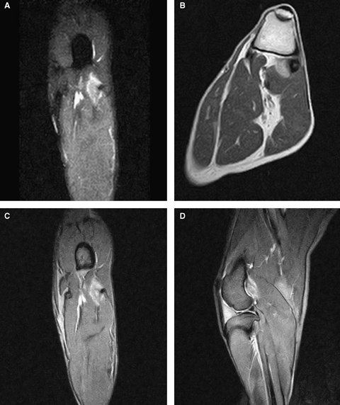
Dog #1: Dorsal STIR (A), transverse T2-weighted (B), dorsal T2*-weighted (C), and sagittal T2*-weighted (D) images of the left stifle of a 7-year-old, neutered female, Border Collie. High signal intensity with a feathery appearance is seen in the lateral head of the gastrocnemius muscle near the sesamoid bone.
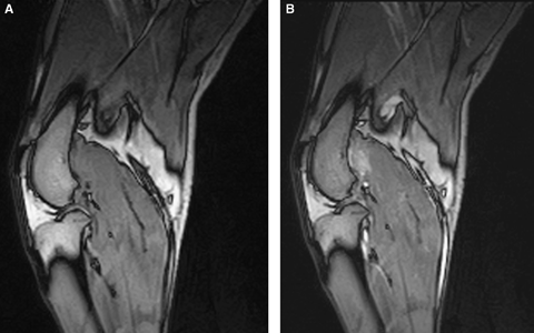
Dog #1: Sagittal T1-weighted images pre- (A) and post- (B) gadolinium administration. There is an iso- to mildly hyperintense signal to the surrounding musculature in T1-weighted sequences in the area of the origin of the lateral head of the gastrocnemius muscle with marked contrast enhancement. Note the normal signal intensity in the femur.
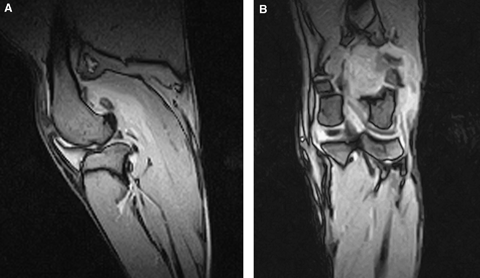
Dog #6: Sagittal (A) and dorsal (B) T2*-weighted images of the left stifle of a 6-year-old, female neutered, Border Collie. This was the most extensive lesion identified. Note the increased volume of the lateral head of the gastrocnemius muscle. Histologically, the lesion was characterized by local fibrosis with a low-grade nonseptic inflammatory reaction, and cartilage and bone proliferation without signs of neoplasia.
All dogs showed abnormality of the lateral sesamoid bone including mineralization in the surrounding tissues observed on radiographs and MR images. The sesamoid bones were in a normal position in all dogs. Contrast enhancement in the center of the sesamoid bone was seen in the two skeletally immature dogs and in the dog with the most extensive lesion. The dog with the most extensive lesion also had increased volume of the lateral head of the gastrocnemius muscle (Fig. 4).
Histologically, the specimen taken during exploratory surgery from mineralized areas of the lateral head of the gastrocnemius muscle was characterized by local fibrosis with a low grade inflammatory, nonseptic reaction, and cartilage and bone proliferation without signs for neoplasia.
Treatment consisted of rest with controlled activity (leash confinement), either alone or in combination with physiotherapy. Except for one dog, all dogs had an excellent outcome and resumed their intended use. The dog with the most extensive lesion, which underwent exploratory surgery (Fig. 4), which was a working dog used in tending sheep and in training herding dogs, had recurrent lameness.
Discussion
On MRI, the signal intensity of normal skeletal muscle is slightly higher than that of water and much lower than fat on T1-weighted images. It is much lower than both fat and water on T2-weighted images.10,11 Generally, increased intracellular or extracellular free water, characteristic for muscle edema, causes T2 prolongation in inversion recovery or fat suppressed T2-weighted sequences.11 On both sequences, edema will have a higher signal intensity than normal muscle, and even subtle areas of edema can be identified.10 If T1 shortening is present, it indicates muscle atrophy, hematoma (Methemoglobin), or fatty infiltration.10,11 The observed contrast enhancement was consistent with increased perfusion or increased permeability of the vessels, for example due to an inflammatory process.
The differential diagnosis for altered SI of skeletal muscle is diverse and includes inflammatory, infectious, autoimmune, neoplastic, and neurologic conditions.11 In this study with the type and appearance of the focal alterations in signal intensity in the lateral head of the gastrocnemius muscle without apparent mass effect (except for dog 6), or loss of muscle volume due to atrophy, most likely represents a myotendinous strain. Differentiation between muscle contusion and myotendinous strain due to excessive tension is difficult based on MRI alone,2 as both are characterized by feathery increased signal intensity in fluid sensitive sequences. In contrast to myotendinous strain, muscle contusions are secondary to direct trauma, leading to fluid accumulation in overlying subcutaneous tissues.11 The differentiation between acute and chronic disorders of the myotendinous junction may be made on MR images, because acute lesions typically involve muscle edema at the myotendinous junction and chronic lesions tend to involve the tendon.12 At the gastrocnemius origin, the myotendinous junction is very broad and a discrete tendon cannot be clearly defined, which makes differentiation between an acute, repetitive, or chronic etiology difficult based on MRI findings. In the present study group the mineralizations at the sesamoid bone and in the surrounding tissue were an indicator for a chronic component.
Myotendinous strains are classified clinically as stretch injury (first degree), partial tear (second degree), and complete tear (third degree) based on absent, mild, or complete loss of muscle function, respectively.2,12 On the basis of the extent of the lesions they can also be classified on MRI.2,10,12 Based on these criteria, the lesions in these dogs were classified as first or second degree.
Myotendinous strains are not commonly recognized in animals and are mostly diagnosed in athletic dogs such as racing greyhounds and field trial dogs.13 Affected animals may be lame or unable to bear weight. Clinical signs of muscle strain depend on the severity and chronicity of the injury. In a mild strain the animal often becomes reluctant to move 12–24 h after strenuous exercise. Severe muscle strains are recognized by swelling and pain of the affected muscle unit.13
In humans, strain injury of the medial head of the gastrocnemius muscle at the middle or proximal leg is termed tennis leg due to its common association with this sport.2 They usually occur at the distal musculotendinous junction,14 but are also observed at the knee2 or in association with an injury of the deeper located plantaris muscle.14 Similarly, lesions of the lateral head of the gastrocnemius occur in humans with posterolateral complex injuries, and are frequently associated with tears of the popliteus tendon, biceps tendon, and plantaris muscle.2
Several factors make a muscle more susceptible to strain, including extension across two joints, long fusiform shape, composition of fast contracting type II fibers, and eccentric action (contraction during elongation).2,12 In eccentric contraction, the muscle fiber is forced to lengthen during contraction, as seen in the gastrocnemius muscle during the initial stance phase.12 The gastrocnemius muscle, as well as the muscles of the hamstring and the biceps brachii muscle have these features and are the most frequently strained in humans.11
In humans, musculotendinous strain of the gastrocnemius muscle is a common racquet sport injury, related to fast acceleration, sudden stops, and turns.14 Given the differences in limb conformation among breeds, inherent biomechanical differences in joint function may be responsible for alterations in loading patterns that contribute to specific injuries or degenerative processes.15 The breed affiliation to Border Collies in our study is striking, and a relation to biomechanical forces or motion pattern may be possible. Border Collies are very active dogs who have a crouched position with a lowered head during herding but also during other activities. Muscles crossing distal limb joints do negative (eccentric) work as they lengthen to absorb energy during weight acceptance in the early stance phase, and they then typically do positive (concentric) work in the late stance phase to provide an active push-off when the foot is caudal to the more proximal joints.15 A crouched posture may increase torque at the tarsus, as the ground reaction force seems to pass close to the knee in many animals, and therefore animals with such a posture might require increased reliance on the gastrocnemius muscle tendon unit.16
Muscle has intrinsic ability to heal by regeneration of myofibrils if the sarcolemmal cells survive and the endomysial connective tissue sheath is not destroyed. Except for the dog with the most extensive lesion, all dogs had an excellent outcome and resumed intended use without recurrence of lameness. The primary treatment for muscle strains is rest. Enforced rest with controlled activity is necessary. If injury is recurrent or severe, longer periods of enforced rest may be necessary. Return to normal function should be expected with most muscle strains.13


