IMAGING DIAGNOSIS—EAR CANAL DISTENSION FOLLOWING EXTERNAL AUDITORY CANAL ATRESIA
Signalment and History
Absence of the normal opening into the left ear canal was noted in a 3-month-old female neutered Red Setter. There were no abnormalities associated with this finding. At 9 months of age, a swelling developed ventral to the left ear pinna. The swelling was progressive, painful, and periaural pruritus was reported. Two months later, the patient was referred for further assessment.
Clinical Findings
The left pinna was normal, but the opening to the external auditory meatus was replaced by haired skin. There was no evidence of a skin lesion at this site, or elsewhere. The cartilaginous ear canal could not be palpated; however, there was a painful swelling in the region of the parotidoauricularis muscle. The right ear was normal. A pin-sized opening in the expected location of the external acoustic meatus was found after the patient was anesthetized.
Imaging
Radiographically, the left external ear canal was not air-filled, and the right was within normal limits. The left tympanic bulla was filled with soft tissue opacity.
Magnetic resonance (MR) images were acquired using a 0.2 T permanent magnet.* T1W (TR=750 ms, TE=26 ms) and T2W (TR=3000 ms, TE=80 ms) images were acquired in dorsal and transverse planes, and T1W images were acquired following administration of gadobenate dimeglumine.†
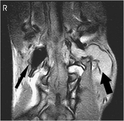
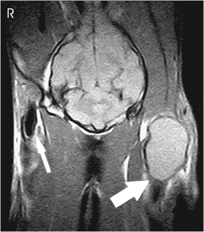
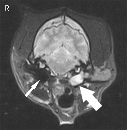
A thick tubular structure, slightly hyperintense to the surrounding muscle with T1W, exited horizontally from the left tympanic bulla, terminating in a uniformly isointense mass ventral to the pinna (1, 2). A band of skin was noted on transverse images where the external opening of the auditory meatus would be expected. The tympanic bulla was filled with material that appeared more hyperintense than the mass lesion with T2W, with a small signal void dorsally (Fig. 3). Neither the mass nor the material in the tympanic bulla enhanced following gadolinium administration.

Dorsal T1W magnetic resonance image at the level of the tympanic bullae. There is material of homogenous intensity filling the left bulla (thick black arrow), and extending out into the distended ear canal. The right bulla is normal (thin black arrow).

Dorsal T1W magnetic resonance image at the level of the ear pinna base. The left ear canal is distended into a mass-like lesion by the same material as the bulla. A skin intensity band (thick white arrow) can be seen at the caudal aspect to the mass; caudal to this is normal air-filled ear canal. The right ear canal (thin white arrow) is normal.

Transverse T2W magnetic resonance image at the level of the tympanic bullae. Mixed intensity material is seen filling the left bulla and proximal ear canal (thick white arrow). The right bulla is normal (thin white arrow).
Diagnosis and Outcome
Considerations for homogenous masses developing from and disrupting the normal ear canal include neoplasia, polyp, granuloma, abscess, hematoma, and solid material trapped in the ear canal including a migrating foreign body or ceruminolith.1 However, with the longstanding atresia of the external auditory meatus, distension caused by accumulation of ceruminous debris was considered most likely. When the mass-like lesion was aspirated through the pinpoint opening, ceruminous debris was obtained.
Owing to the chronicity of the condition, with marked distension of the ear canal, surgery was performed to reconstruct the ear and provide normal drainage. The ear canal opening was enlarged and the ceruminous debris was flushed out. The tympanic membrane was not visible at surgery, and otitis media was confirmed. At 4 weeks, the reconstructed auditory meatus was patent and there was no distension of the ear canal. Assessment of hearing was not performed either pre- or post surgery, although this would have been of interest.
Discussion
Atresia of the ear canal is infrequent in animals.2–4 In this dog, the identification of external auditory canal atresia at a young age makes a congenital or early developmental etiology likely. The ear develops from an infolding of ectodermal cells of the first branchial or pharyngeal cleft, which invaginate and proliferate to form a blind ending tube which extends toward the middle ear.5 The ectodermal cells at the blind end of the first branchial cleft proliferate to form the meatal plug, a solid mass that fills the ear canal. This mass persists into the perinatal period in dogs, opening at 6–14 days.6 Other reports of external ear canal atresia in the dog are similar to ours, with an epidermal-covered band across the external opening of the canal.2–4 Human external auditory canal atresia differs from those examples reported in the dog. In people, abnormalities arising early in gestation have an absent external ear canal; those later in gestation have a stenotic canal with bony abnormalities due to faulty recanalization, which occurs in utero in the human.7 The different presentation of external ear canal atresia in the dog could relate to the increased length of the time the meatal plug is present, continuing into the perinatal period, or differences in canalization between the species.
The incidence of congenital abnormalities of the external ear in dogs is unknown, but in humans is estimated at 1 in 10,000–20,000 live births.8 In humans, unilateral external auditory canal atresia is three to six times more common than bilateral, and the right ear is more commonly affected than the left.9 External auditory canal atresia of suspected congenital or perinatal origin in dogs has all been unilateral; too few patients have been described to evaluate a predilection for the right ear as in humans. There is familial history in 14% of human patients, which could impact on decision-making regards breeding of affected animals.
The computed tomography (CT) findings in external auditory canal atresia in the dog have been described,2 but the MR findings have not. The pressure-related disruption of the ear canal seen in this dog has not been noted previously. Radiographically, ear canal atresia has led to tympanic bulla wall thickening,3 which was not observed in our patient. In this patient, MR imaging was ideal for assessing the congenital abnormality for presurgical planning as the changes were limited to the soft tissues of external ear. If abnormalities in development of the middle or inner ear were suspected, as commonly seen in external auditory canal atresia in humans, CT would have been a useful additional modality.


