Comparison of Computed Tomographic and Standard Radiographic Determination of Tibial Torsion in the Dog
Funded by the Canine Research Grant Program at The Ohio State University.
Presented in poster form at the 31st Annual Conference of the Veterinary Orthopedic Society, Big Sky, MT, February 22–27, 2004, and in abstract form at the 14th Annual American College of Veterinary Surgeons Symposium, Denver, CO, October 6–9, 2004.
Abstract
Objective— To compare the effect of internal tibial rotation on the computed tomographic (CT) and standard radiographic assessment of tibial torsion (TT) in dogs.
Study Design— In vitro study.
Sample Population— Cadaveric canine hind limbs (6 pairs).
Methods— The cranial cruciate ligament was transected, and caudo-cranial radiographic and transverse CT images were obtained with the femur and tibiae in a neutral position, and after 15° internal tibial rotation. Radiographic TT was determined by measuring the distance (d) between the calcaneus and the sulcus of the talus. CT determination of TT was performed using the proximal transcondylar and the distal cranial tibial axes. The distance (d) in the 2 groups and the difference in the CT determination of TT between groups were compared with a hypothetical mean value of 0 mm and 0°, respectively.
Results— The mean distance (d) for the neutral radiographic group was not significantly different from 0 (P=.473); however, for the 15° group it was significantly different (P<.0001). The difference in the CT determination of TT did not differ from 0 (P=.317).
Conclusion— The standard radiographic technique does not discriminate between internal TT and internal rotation of the tibia. Thus, dogs with normal tibial conformation can be depicted by radiography as torsed, whereas dogs with TT may be misinterpreted as normal because of arbitrary positioning.
Clinical Relevance— Lateral displacement of the medial border of the calcaneus on a caudo-cranial radiograph should not be used as the sole arbiter of TT before surgical correction.
INTRODUCTION
TIBIAL TORSION (TT) describes the angular relationship of the proximal articular axis relative to the distal articular axis of the tibia about its longitudinal axis.1 TT beyond the normal, physiologic range because of torsional deformities2,3 or malalignment after fracture repair4,5 has been reported to cause gait alterations,6 as well as stifle joint pathology in humans.7–10
Different methods have been developed for assessment of TT in humans, such as direct goniometric measurement,11 and several radiologic techniques, e.g., fluoroscopy,12 ultrasound,13 computed tomography,14–16 magnetic resonance imaging,17 or computer-assisted quantification of periaxial bone rotation.18 In veterinary medicine, a radiographic method using conventional radiographs to evaluate TT has been reported.19
Disadvantages of conventional radiographic techniques are the projection of a complex three-dimensional structure in a two-dimensional plane resulting in superimposition of the anatomic structures used in tibial axis determination. In addition, the rotational position of the proximal aspect of the tibia is evaluated using femoral landmarks. True caudocranial position of the femur, however, may not necessarily guarantee true caudocranial positioning of the tibia. Suboptimal radiographic positioning, because of the natural recurvation of the femur, or rotation about the stifle joint, which may be enhanced by cranial cruciate ligament (CCL) deficiency, can create a radiographically perceived torsional deformity, or can reduce the apparent magnitude of an actual deformity. Accurate assessment of TT is necessary before corrective osteotomy, such as tibial plateau leveling osteotomy (TPLO), because surgical correction of torsional deformities and inadvertent torsional malalignment are possible with this procedure. Thus, accurate assessment is also required to evaluate the effectiveness of such procedures postoperatively.
Our purpose was to compare the standard radiographic technique with computed tomographic (CT) assessment of TT in the dog, based on the hypothesis that CT assessment was more precise than standard radiographic technique, and would not be affected by minor rotational malpositioning of the tibia.
MATERIALS AND METHODS
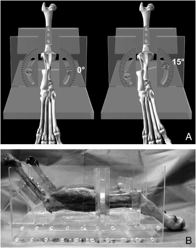
Six paired (n=12) cadaveric hind limbs from mature dogs of grossly normal conformation, visually assessed to be free of orthopedic disease of the stifle, were prepared by removal of all soft tissues proximal to the stifle with careful preservation of the stifle joint capsule and collateral ligaments. The CCL was transected through a lateral arthrotomy to provide a model of a clinically affected CCL deficient stifle. The paired legs were positioned in a custom made holding device (Fig 1A), which rigidly fixed the femur, and allowed quantifiable, independent rotation of the tibia. The angle of the stifle joint was held at 132°; this angle was determined based on the measurement of a number of clinical cases (n=6; mean, 131.9°) during routine caudocranial TPLO radiography. The tibiae were aligned parallel to the base of the holding device (Fig 1B).

(A) Schematic illustration of the limb holding device in a neutral (0°) and a 15° internally rotated position. A custom made 3-hole, 4.5 mm dynamic compression plate and 4.5 mm screws secured with nuts were used to secure the femur in a true caudocranial position. The curvilinear slots in the device allow independent, quantifiable, axial tibial rotation to be performed. Note that in the neutrally positioned leg (left) the medial border of the calcaneus is aligned with the base of the sulcus of the talus. (B) Limbs positioned in the limb-holding device, such that each tibia is aligned parallel to the bottom of the device, and the stifle joint angle is held at 132°.
Imaging
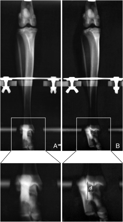
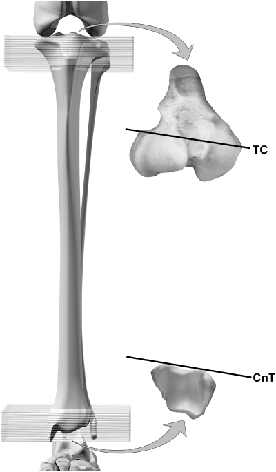
Caudocranial radiographs of both limbs were obtained (Prestige II, GE Medsystems, Milwaukee, WI) and true caudocranial alignment of the femur was confirmed as previously described by Slocum, with the patella centered in the trochlear groove of the femur, and the fabellae bisected by the distal femoral cortices. Each tibia was positioned in a neutral position, such that the medial aspect of the calcaneus was aligned with the base of the sulcus of the talus (Fig 2A). Without altering the position of the limbs within the holding device, transverse, 1 mm thick, contiguous CT (PQS Spiral CT scanner, Picker International, Highland Heights, OH, 130 kV, 125 mA, matrix size 512 × 512, FOV 100, bone algorithm) slices were obtained from the proximal 2 cm of all tibiae including at least 4–6 slices of the distal femur, and from the distal 2 cm of all tibiae including at least 3–4 slices of the proximal aspect of the tarsus (Fig 3). The tibiae were internally rotated 15° about the stifle and fixed in this position within the holding device; this magnitude was chosen because this is the degree of internal rotational instability in the normal, extended canine stifle.20 Caudocranial radiographic images (Fig 2B) and transverse CT images were repeated. The radiographic images were digitized on a flatbed scanner (Epson Perfection®1660 Photo, Epson America, Inc., Long Beach, CA) and measurements were made using commercially available software (Adobe Photoshop®, Version 7.0, Adobe Systems, Inc., San Jose, CA). According to the guidelines of TT assessment for TPLO (Seminar titled: Tibial plateau leveling osteotomy for cranial cruciate ligament repair; Slocum Enterprises, Eugene, OR), the distance (d) between the medial border of the calcaneus at the level of the talocrural joint and the base of the sulcus of the talus was identified and measured (Fig 2). CT determination of TT was based on the angular relationship of proximal and distal axes drawn between reference points. The CT slice within which the specific anatomic landmarks that define each tibial axis could most clearly be identified was chosen for analysis. The proximal transcondylar (TC) axis was defined as the line connecting the caudolateral extent of the extensor sulcus to the prominence at the medial collateral ligament insertion. The distal cranial tibial (CnT) axis was defined as a line parallel to the cranial tibial cortex immediately proximal to the talocrural joint (Fig 3). The axis angle was determined by calculating the angular orientation of these axes with the vertical plane, using commercially available software (eFilm Workstation™, Version 1.8.3, Merge eFilm, Milwaukee, WI). The torsion angle, expressed in degrees, was calculated by determining the difference between the axis angles, as described by Aper et al.21 Deviation from parallelism was described using positive and negative values. For example, a clockwise offset of the distal axis in a left tibia was assigned a negative value.

Caudocranial radiographs of a limb in a neutral position (A) and 15° internal rotation (B). In both radiographs, the fabellae appear bisected by the femoral cortices and the patella is in the center of the trochlear sulcus. In the neutrally positioned limb (A) the medial border of the calcaneus is aligned with the base of the sulcus of the talus. In the internally rotated limb (B) the calcaneus is displaced laterally. Note that in both images the position of the femur is unchanged d, displacement.

Illustration of the tibia (cranial view) showing the area from which the transverse computed tomographic slices were obtained. The transcondylar (TC) axis is the line joining the caudolateral extent of the extensor sulcus with the prominence at the medial collateral ligament insertion. The distal cranial tibial axis (CnT) is a line parallel to the cranial cortex of the tibia immediately proximal to the talocrural joint.
Statistical Analysis
The initial (neutral) and 15° radiographic measurements of the distance d, expressed in mm, and the difference between the initial and 15° CT determination of TT, expressed in degrees, were analyzed using a one-sample t-test (PRISM© Version 4.0a, Graph Pad Software Inc., San Diego, CA), with a hypothetical mean value of 0 mm or 0° respectively. Significance was set at P<.05.
RESULTS
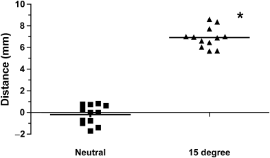
The mean distance (d) for the neutral radiographic group (−0.19±0.90 mm) was not significantly different from the hypothetical mean value of 0 mm (P=.473); however, the mean distance (d) for the 15° radiographic group (6.92±0.94 mm) was significantly different from 0 mm (P<.0001; Fig 4). The mean difference between the initial and 15° CT determination of TT (−0.583±1.93°) did not differ from the hypothetical mean value of 0° (P=.317).

Scatter plot of neutral (0°) and 15° internally rotated radiographic data. Note that a negative value indicates that the calcaneus is medial to the base of the sulcus of the talus. Each data point represents TT expressed in mm (distance “d”) for an individual specimen. The bar indicates the mean value for the group.
DISCUSSION
In human medicine, various CT techniques for determination of TT have been developed, based on identification of reference axes on transverse images of the proximal and distal tibia, femur, or fibula. These techniques differ primarily with regard to the anatomic landmarks and therefore position of the axes. One of the reasons for this may be the rounded form of the human tibial bone and the lack of obvious anatomic ridges.15 A commonly used and reproducible proximal reference axis is a tangent drawn on the dorsal border of the tibia just below the joint space and above the fibular head. However, variations in distal reference point determination exist, including using the lateral malleolus of the fibula for the trans- or bimalleolar axis. This point may not accurately reflect the distal tibial axis, because of the influence of the position of the fibula.22 Jend et al14 developed a method where the distal reference line is formed by joining the center of a circle fitted to the pilon tibiale including the fibular notch, with the midpoint of a line across the fibular notch of the tibia. Waidelich et al23 assess TT with a distal reference axis defined by a line joining the midpoint of an ellipse fitted to the base of the medial malleolus and the midpoint of a line across the incisura fibularis. Nevertheless, nonconformity of axis determination contributes to variability in normal torsion values and indicates that there is no consensus in the most reliable, accurate, or reproducible method or anatomic landmarks.
Canine tibial anatomy offers more pronounced reference points for definition of proximal and distal tibial axes. Aper et al21 demonstrated correlation between direct anatomic measurements, and CT measurement of 2 proximal and 2 distal tibial axes in canine cadaveric tibial bones. Using 2 of these reported axes, TT was measured in this study, and the CT method was compared with the standard radiographic method. Selection of the proximal TC axis and the distal CnT axis was based on the investigators ability to identify the specific anatomic landmarks that define these axes in all of the tibiae studied.
The standard radiographic technique described by Slocum is based on the determination of TT as a linear measurement, whereas the CT method assigns an angular value; therefore these 2 methods cannot be compared directly. For this reason, the results of each method, in a neutral tibial position were compared with the results after 15° internal rotation. Because the real magnitude of TT was unchanged by tibial rotation, significant changes in perceived TT would be artifactual and caused by positioning. The standard radiographic technique relies on the inherent stability of the stifle in extension to minimize tibial rotation. In practice, it is difficult to achieve complete extension of the stifle during positioning for the caudocranial radiographic projection, and for this reason, a stifle joint angle comparable with that obtained clinically was used in our study. Rotational malpositioning of the tibia at the stifle may occur during routine stifle radiography, and because rupture of the CCL results in an increase in range of motion in internal rotation,20 this may increase the likelihood of malpositioning, and result in inaccurate evaluation of TT. The linear measurements obtained with the standard technique demonstrated a significant increase after internal tibial rotation about the stifle. This finding demonstrates that this method is susceptible to rotational artifacts, resulting in inaccurate determination of TT.
In contrast, CT assessment of TT was not affected by 15° internal rotation of the tibia about its long axis. This finding demonstrates that the CT method of TT measurement is unaffected by internal tibial rotation of up to 15°, unlike the standard radiographic method. Interestingly, a study using human cadaveric tibiae demonstrated that angled CT slice orientation, with respect to the long axis of the tibia did not have an effect on torsional assessment.16
A variety of clinical problems have been associated with torsional malformation of the tibia in both human and veterinary patients, such as patellar instability,24–26 stifle osteoarthritis,13 and Osgood–Schlatter's disease.27 The magnitude of TT varies widely between individuals, and more detailed information regarding physiologic versus pathologic magnitudes of TT in veterinary patients is required. Clinical studies focused on breed specific TT values and inter-observer differences are necessary to define developmental or acquired malformation of the tibia. This information would be particularly helpful if corrective tibial osteotomy is to be performed.
The standard radiographic technique is adequate to determine malalignment of limb axes in the frontal and sagittal plane and can be a helpful in screening for TT. However, if accurate assessment of TT is required, CT determination is indicated, as the CT method is unaffected by minor malpositioning.


