REVIEW ARTICLE: Th1/Th2/Th17 and Regulatory T-Cell Paradigm in Pregnancy
Abstract
Citation Saito S, Nakashima A, Shima T, Ito M. Th1/Th2/Th17 and regulatory T-cell paradigm in pregnancy. Am J Reprod Immunol 2010
T-helper (Th) cells play a central role in modulating immune responses. The Th1/Th2 paradigm has now developed into the new Th1/Th2/Th17 paradigm. In addition to effector cells, Th cells are regulated by regulatory T (Treg) cells. Their capacity to produce cytokines is suppressed by immunoregulatory cytokines such as transforming growth factor (TGF)-β and interleukin (IL)-10 or by cell-to-cell interaction. Here, we will review the immunological environment in normal pregnancy and complicated pregnancy, such as implantation failure, abortion, preterm labor, and preeclampsia from the viewpoint of the new Th1/Th2/Th17 and Treg paradigms.
Introduction
T cells play a central role in immunoregulation and immunostimulation. T-helper (Th) cells can be classified into Th1 cells, which produce interleukin (IL)-2 and interferon (IFN) γ and are involved in cellular immunity, and Th2 cells, which produce IL-4, IL-5 and IL-13 and are involved in humoral immunity.1 In the 1980s to 1990s, maternal tolerance toward fetal alloantigens was explained by the predominant Th2-type immunity during pregnancy, which overrules Th1-type immunity, therefore protecting the fetus from maternal Th1-cell attack.2 Indeed, predominant Th1-type immunity has been observed in recurrent spontaneous abortion3,4 and in preeclampsia.5 However, Th2-dominant immunity was also reported in recurrent abortion cases,6,7 and therefore, the Th1/Th2 paradigm is now insufficient to explain the mechanism of why the fetus is not rejected by maternal immune cells. Now, the Th1/Th2 paradigm has been expanded into the Th1/Th2/Th17 and regulatory T (Treg) cells paradigm.8 Th17 cells, which produce the proinflammatory cytokine, IL-17, play important roles for the induction of inflammation.8,9 They have been proposed as a pathogenetic mechanism in autoimmune diseases and acute transplant rejection. In contrast, Treg cells play central roles for immunoregulation and induction of tolerance. Treg cells are now known to inhibit proliferation and cytokine production in both CD4+ and CD8+ T cells, immunoglobulin production by B cells, cytotoxic activity of natural killer (NK) cells, and maturation of dendritic cells (DCs), resulting in induction of tolerance.10,11
This review aims to reexamine the Th1/Th2/Th17 and Treg paradigms in normal pregnancy and complicated cases such as implantation failure, recurrent pregnancy loss, and preterm labor.
Reciprocal developmental pathways between Th1/Th17 subsets and between Th17/Treg subsets
Th1 cells are characterized by the transcription factor T-bet and signal transducer and activator of transcription (STAT) 4, and the production of IL-2, IFN-γ, and tumor necrosis factor (TNF) β. They are involved in the cellular immunity and rejection process. In contrast, mediators of humoral immunity, Th2 cells develop into IL-4-, IL-5-, and IL-13-producing cells by the transcription factor GATA-3 and STAT6. A new lineage of Th cells that selectively produce IL-17 has been proposed. This population, which has been termed Th17, plays a critical role for the induction of inflammation and plays a critical role in the pathogenesis of autoimmune diseases and rejection.8,9 Th17 cells are characterized by unique signaling pathways, receptor-related orphan receptor (ROR) C2 or ROR-α. Th17 cells are involved in the host defense against bacteria, fungi, and viruses. But evidence for the pathogenic role of Th17 in rheumatoid arthritis (RA), psoriasis, multiple sclerosis, and inflammatory bowel disease has been reported.8,9
The function of effector T cells, such as Th1, Th2 and Th17 cells, is regulated by CD4+ CD25+ regulatory T (Treg) cells. CD4+ CD25+ Treg cells are important cells for the maintenance of peripheral tolerance.10,11 The master gene for the differentiation to Treg cells is transcription factor forkhead box P3 (Foxp3).
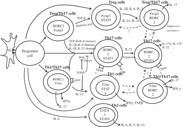
Interestingly, recent data show the reciprocal development pathways between Th17/Th1 subset and Th17/Treg subsets8 (Fig. 1). The progenitor cells differentiate to Th17/Treg intermediate cells, which express both RORC and Foxp3. The differentiation of both Th17 and Treg cells requires transforming growth factor (TGF)-β. When DCs are activated by microorganisms that induce production of pro-inflammatory cytokine IL-6 or IL-1, TGF-β induced differentiation of native T cells diverted away from the induced Treg cells pathway and toward the Th17 cells pathway. The differentiation of human Th17 cells is inhibited by high TGF-β concentrations but requires IL-1β and IL-6.8,9 Prostaglandin E2 (PGE2), which is a mediator of tissue inflammation, induces up-regulation of IL-23R and IL-1R and promotes the differentiation and expansion of Th17 cells. The plasticity of Th17/Th1 lineage has also been reported.8 The existence of IFNγ+ IL-17+-double positive T cells has been reported. Stimulation by IL-12 down-regulates IL-17 production and induces IFNγ production, suggesting Th1 polarization (Fig. 1), but interplay between the Th1 and Th17 lineages has not been fully clarified.

Th1, Th2, Th17, and Treg cells development from CD4+ progenitors. Plasticity of T-cell phenotype between Th1 cells and Th17 cells or Treg cells and Th17 cells is reported.
Interestingly, the conversion of Treg cells to Th17 cells has been reported in humans and mice. IL-6 plays an important role in this process in mice, and IL-1β and IL-2 are the key cytokines in this process in humans.
These data show that Th1/Th2/Th17 and Treg lineages are associated with each other, and they are able to convert to other lineages.
Th1/Th2/Th17 and Treg paradigms in implantation
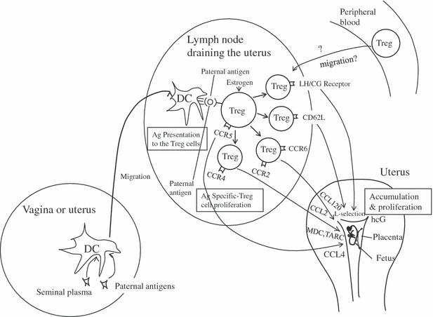
Studies in humans and mice have shown that leukemia inhibitory factor (LIF), IL-1, IL-6, IL-11, vascular endothelial growth factor, placenta growth factor, fibroblast growth factor-2, heparin-binding epidermal growth factor (HB-EGF), and TGF-β play important roles for successful implantation by modulating angiogenesis processes, trophoblast differentiation, or the immune system.12 Recent data show that uterine DCs play a central role for successful implantation.13,14 The number of uterine DCs increases at the implantation period, and depletion of DCs results in severe implantation failure.13,14 DC depletion impairs uterine NK cell maturation, tissue remodeling, and angiogenesis.13,14 Surprisingly, depletion of DCs also causes embryo resorption in syngeneic and T-cell-deficient pregnancy, suggesting that DCs appear to govern uterine receptivity independent of the immunological tolerance.14 Although tolerogenic DCs take a part in inducing materno-fetal tolerance, DCs may play a principal role in implantation. But depletion of Treg cell by anti-CD25 monoclonal antibody on the day or 2.5 days after mating results in severe impairment of implantation in allogeneic mice, but this was not observed in syngeneic mice, suggesting that Treg cells are essential for inducing immunological tolerance.15,16 Treg cells are already increased in the lymph nodes draining the uterus 2 or 3 days after mating, suggesting that Treg cells accumulate into the lymph nodes draining the uterus before the implantation.13,14 Robertson et al. reported seminal plasma, but not sperm, plays an essential role for the induction of paternal antigen-specific tolerance.19 The maternal immune system prepares for the semiallograftic embryo to come into the uterus. Alvihare et al. reported that expansion of Treg cells in the lymph nodes draining uterus was also observed in allogeneic mice and syngeneic mice.17 They proposed that pregnancy hormones such as estrogen might induce Treg cells numbers and Arruvito et al.18 showed that Treg cells increased during the follicular phase of the menstrual cycle, suggesting that estrogen plays a part for the expansion of Treg. But our recent data showed paternal antigen-specific Treg cells proliferate in the lymph nodes draining the uterus 3 days after coitus. When BALB/c female mice were mated to DBA/2 male mice, fetuses express DBA/2-derived Mls Ia antigen on the cell surface. As Mls Ia antigen is recognized by T-cell receptor Vβ6, CD4+ CD25+ Foxp3+ Vβ6+ cells are paternal antigen-specific Treg cells. Ki67 is a marker for cell-proliferation; therefore, Ki67+ CD4+ CD25+ Foxp3+ Vβ6+ cells are proliferating fetal antigen-specific Treg cells. Interestingly, Ki67+ Vβ6+ Treg cells increase in the uterine draining lymph node on 3.5 days post-coitus in BALB/c x DBA/2 mating but not in BALB/c x BALB/c mating, suggesting that paternal antigen-specific Treg cells proliferate in the uterine draining lymph node before the implantation.19 After implantation, on day 5.5 post-coitus, Ki67+ paternal antigen-specific Treg cells increase in the uterus in BALB/c × DBA/2 mating.20 These findings demonstrate that paternal antigen-specific Treg cell in draining lymph nodes quickly move to the pregnant uterus and proliferate in the uterus, resulting in the induction of paternal antigen-specific tolerance at the early stage of pregnancy (Fig. 2).20 The ligands such as CCR2, CCR4, CCR5, CCR6, and CD62L are known as molecules for Treg cell migration. CCL2, CCL22, CCL17, CCL4, CCL20, or L-selection in the uterus might selectively accumulate Treg cells from peripheral tissues.21 Recent data demonstrate that some Treg cells express LH/CG receptor, and human chorionic gonadotropin produced by human trophoblast has an ability to attract Treg cells to the uterus.22 Further studies are needed to clarify whether paternal antigen-specific Treg cells express these chemokine receptors or LH/CG receptors on their surface. Th1/Th2/Th17 balance at implantation in such Treg cell-deficient or depleted mice has not been reported, and further studies are needed.

A model for the paternal antigens-specific Treg cells expansion, proliferation, and mobilization from vagina to pregnant uterus. As a first step, DCs uptake paternal antigen, and these DCs migrate to lymph nodes draining the uterus. DCs present paternal antigens to Treg cells, and Treg cells proliferate before the implantation. These paternal antigen-specific Treg cells migrate to the pregnant uterus by chemokine and hcG-induced chemoattractant mechanisms.
In humans, augmented Th1-type immunity or suppressed Th1-type immunity in endometrium is observed in repeated implantation failure.23 Deceased number or function of Treg cells might be a cause of augmented Th1-type immunity in such cases. Jasper et al.24 reported that primary unexplained infertility is associated with reduced expression of Foxp3 mRNA in endometrial tissue. It can be speculated that decreased Treg cells might induce implantation failure, resulting in unexplained infertility.
Th1/Th2/Th17 and Treg paradigms in normal pregnancy
Many studies have reported a predominant Th2-type immunity and suppressed Th1-type immunity during pregnancy.2,3,25 This tendency is more clear at the feto–maternal interface (Table 1). Both Th2 cells and T cytotoxic (Tc) 2 cells accumulate in the decidua basalis,26,27 and uterine DC cells can differentiate naïve T cells to Th2 cells.28 Therefore, both Th2 cell migration and Th2 cell differentiation induce Th2-type immunity at the feto–maternal interface. But the systemic immune system does not change so much.25,26 Moreover, IL-4, IL-5, IL-9, and IL-13 knockout mice show normal pregnancy in allogeneic pregnancy,28 suggesting that predominant Th2-type immunity might not be essential for successful pregnancy. Administration of excess amount of Th1-type cytokine such as IL-2 or IFNγ induces abortion in mice, and stimulation of toll-like receptor (TLR) induces Th1-type cytokine production, resulting in abortion.29 But IFNγ also plays an important role for vascular remodeling at the early stage of murine pregnancy.30 Thus, Th1-type immunity is well controlled to avoid overstimulation of Th1-type immunity. Treg cells might play a part in this process.
| Normal pregnancy | Abortion | Depletion of Th1, Th2,Th17 or Treg cells | |||
|---|---|---|---|---|---|
| Peripheral blood | Uterus | Peripheral blood | Uterus | ||
| Th1 cells | ↘ | ↓ | ↗→ | ↑→ | Abortion is not observed. |
| Th2 cells | ↗ | ↑ | → | ↓→↑(conflict data) | Abortion is not observed. |
| Th17 cells | →↘ | ↗ | →↗ | → (missed abortion)↑ (inevitable abortion)↑ (recurrent abortion: inevitable abortion) | There is no data, but IL-17 null mice are fertile. |
| Treg cells | ↑ | ↑↑ | → | → | Abortion and implantation failure are observed in allogeneic pregnancy. |
- →: no change, ↗: slightly elevate, ↑: elevate, ↑↑: markedly elevate, ↘: slightly decrease, ↓: decrease.
IL-17 plays an important role for the pathophysiology in RA.8,9 The symptoms of RA usually improve during pregnancy,2 suggesting that Th17 cells might be decreased during pregnancy (Table 1).
Two recent articles show that the frequency of circulating Th17 cells to CD4+ T cells is very low (0.64–1.4%) in healthy subjects.31,32 In our study, the frequency of Th17 cells to CD4+ T cells during all stages of pregnancy period was similar to that in non-pregnant women,32 but Santner-Nanan et al.33 reported that the frequency of Th17 cells in the third trimester of pregnancy was significantly lower compared to that of non-pregnant women. Further studies are needed to determine circulating Th17 cell levels during pregnancy. The main producer of IL-17 in both peripheral blood and deciduas is CD4+ T cells, and IL-17-expressing CD8+ T cells are rare (approximately 0.1%).32 CD14+ monocytes, CD56dim NK cells, and CD56bright NK cells do not express IL-17.34
Previous studies demonstrate that elevation of IL-17 is observed in acute renal rejection,35 suggesting that increased Th17 cells in pregnancy decidua might be disadvantageous for the maintenance of pregnancy.33
Unexpectedly, the frequency of Th17 cells in the decidua is significantly higher compared to that in peripheral blood. The uterine cavity is not completely sterile, and therefore, Th17 cells might play a role to induce protective immune response against extracellular microbes. Recent reports show that IL-17 increases progesterone secretion by JEG-3 human choriocarcinoma cells and induces the invasive capacity of JEG cells.36 These data suggest IL-17 may be useful for a successful pregnancy.
Inflammation is necessary for successful implantation, but excessive inflammation can cause embryo resorption. Treg cells might regulate excessive inflammation in the uterus at the implantation period. Indeed, endometrial (decidual) Treg cells increase in mice and humans,16,18,37–39 and Treg cell number in allogeneic pregnant mice is much higher compared to that in syngeneic pregnancy (Table 1).39 And these Treg cells in the decidua selectively inhibit the mixed lymphocytes reaction (MLR) to umbilical mononuclear cells, suggesting selective migration of fetal antigen-specific Treg cells to the decidua.40 In humans, extravillous trophoblasts express polymorphic histocompatibility antigen, HLA-C, which can elicit an allogeneic T-cell-response. Tilburgs et al.41 recently reported that pregnancy with a HLA-C-mismatched child induces an increased percentage of activated T cells in decidual tissue. Interestingly, HLA-C-mismatched pregnancies exhibit significantly increased suppressive capacity in one-way MLR reaction to umbilical mononuclear cells, suggesting decidual Treg cells inhibit HLA-C-recognized-T-cell attack. These findings show that Treg cells also play an important role for regulating fetal rejection by maternal immune cells in human pregnancy.
Th1/Th2/Th17 and Treg paradigms in abortion
Predominant Th1-type immunity is observed in abortion, but predominant Th2-type immunity is also reported in recurrent pregnancy loss.2–4,6,7 Therefore, adequate balance for Th1/Th2 immunity, i.e., slightly shifted to Th2-type immunity, may be suitable for the maintenance of pregnancy. Overstimulation of Th1 immunity or Th2 immunity might be harmful for successful pregnancy. A pro-inflammatory cytokine IL-17 induces the expression of many mediators of inflammation. Recent data show an increased prevalence of Th17 cells in peripheral blood and decidua in unexplained recurrent spontaneous abortion patients.42,43 The master transcription factor for Th17 cells, RORc and IL-23, which play a crucial role in Th17 cells expansion, is also increased in the decidual tissue in recurrent spontaneous abortion cases.43 An inverse relationship between the numbers of Th17 cells and Treg cells is observed in peripheral blood and decidua of unexplained recurrent spontaneous abortion cases.43 IL-6 is a key cytokine that blocks the development of Treg cells and induces the differentiation of Th17 cells. The serum IL-6 and soluble IL-6 receptor (sIL-6R) levels are increased. On the other hand, the trans-signaling inhibitor, soluble gp130 (sgp130) level, is decreased in recurrent spontaneous cases.44 After paternal lymphocytes alloimmunization, sIL-6R levels are decreased and sgp130 levels are increased, and Treg cell number is increased.44 These findings suggest IL-6 trans-signaling plays a very important role for regulating Th17/Treg balance.
We have already reported that Th17 cells increased in decidua of inevitable abortion, i.e., progression stage of abortion.42 And the number of IL-17+ cells is well correlated with that of neutrophils, suggesting that IL-17 plays an important role in neutrophil infiltration. But Th17 cells did not change in the decidua of missed abortion cases that did not show vaginal bleeding, uterine cramping or cervical deletion.42 Importantly, all the abortion cases in Wang’s study were inevitable abortion.43 These findings suggest that increased Th17 cells are a consequence of fetal loss, but not a cause of fetal loss. After embryonic death, increased IL-1 or IL-6 production and decreased TGF-β production might cause increased Th17 cells and decreased Treg cells in the uterus.
Our group first reported the decreased numbers of peripheral and decidual Treg cells in spontaneous abortion cases.37 Yang et al.45 reported that the proportion of Treg cells in both decidua and peripheral blood in unexplained recurrent spontaneous abortion was significantly lower than those in control women. And the immunosuppressive activity of Treg cells in repeated miscarriage cases have reduced suppressive capacity compared with normal fertile women.18 Persistent TLR stimulation or IL-6 suppression of Treg function have been reported,46,47 and therefore these reduced Treg functions in recurrent spontaneous abortion might be caused by chronic inflammation. In a mouse model, when BALB/c CD25− lymphocytes are injected into BALB/c nu/nu T-cell-deficient mice, they did not have CD4+ CD25+ Treg cells. These Treg-deficient mice showed abortion in allogeneic pregnancy, but this was not observed in syngeneic pregnancy.17 But it has not been clarified how greatly Treg cells induce abortion in allogeneic pregnancy. We have injected various amounts of anti-CD25 monoclonal antibody on 4.5 and 7.5 days after mating and found that an over 60% decrease of Treg cells could induce abortion in allogeneic pregnancy.48 CBA/J female mice mated with DBA/2J male mice is a good model for abortion. Interestingly, Zenclussens et al.16 reported fewer Treg cells associated with elevated Th1-type immunity in CBA/J × DBA/2J mating. Adoptive transfer of CD4+ CD25+ cells from normal pregnant mice prevented fetal loss, but adoptive transfer of CD4+ CD25+ T cells from non-pregnant mice had no effect for protecting fetal loss. Transfer of CD4+ CD25+ T cells from pregnant mice on day 4 of pregnancy did not prevent abortion.16 Interestingly, transfer of CD4+ CD25+ Treg cells to abortion-prone mice induced the expression of LIF and TGF-β, but the expression of Th1-type cytokines such as IFNγ and TNFαwas unchanged, and Th1/Th2 ratio did not change.49 These findings show that the protective effect against abortion by Treg cells might not be caused by the correction of abnormal Th1/Th2 balance.
Th1/Th2/Th17 and Treg paradigms in preterm labor
Preterm labor is associated with subclinical infection, and intrauterine inflammatory response causes preterm labor and delivery. Therefore, the secretion of pro-inflammatory cytokines such as IL-1, IL-6, IL-8, and TNFα is increased in amniotic fluid, decidual tissue, and chronic tissue. Although the secretion of Th1-type cytokines, i.e., IL-2, IFNγ, and TNFα, which mediate inflammatory reactions, is up-regulated in preterm labor,50 the secretion of Th2-type cytokines, i.e., IL-4 and IL-6, is also elevated.51
TGF-β might be a key cytokine that regulates Th17/Treg lineage. As IL-17 is a key cytokine that induces inflammation, IL-17 might have some roles in pathophysiology of preterm labor. Our groups recently found that Th17 cell number is increased in the chorioamniotic membrane of preterm delivery cases with chorioamnionitis (CAM).34 Amniotic fluid IL-17 levels in severe CAM (stage III) preterm delivery cases were significantly higher than those in CAM-negative preterm delivery cases. Amniotic fluid IL-17 levels were positively correlated with IL-8 levels. Interestingly, although IL-17 did not enhance IL-8 secretion by amniotic mesenchymal cells, TNFα-induced IL-8 secretion was enhanced by IL-17 in a dose-dependent manner. Amniotic mesenchymal cells express IL-17 receptor, and therefore, IL-17 signaling pathway might up-regulate TNFα-induced IL-8 secretion by amniotic mesenchymal cells. Indeed, the IKK inhibitor and MAPK inhibitor significantly inhibited IL-17 with TNFα-induced IL-8 secretion in amniotic mesenchymal cells.34 These findings show that Th17 cells promote inflammation at the feto–maternal interface in preterm delivery.
Th1/Th2/Th17 and Treg paradigms in preeclampsia
In the early days of reproductive immunology when the Th1/Th2 paradigm was first proposed, Th1/Th2 balance in preeclampsia was examined by measuring cytokines or detecting intracellular cytokines by flow cytometry.5 It has been clarified that predominant Th1-type immunity is present in preeclampsia.5 A later study demonstrated that the production of Th1-type cytokine, IFN-γ, is also enhanced in NK cells.52 Secreted IL-12p70 and IL-18, which differentiate Th1 cells from the progenitor cells, are increased in preeclampsia.5,52 Predominant Th1-type immunity may suppress the tolerance system, resulting in preeclampsia.
As a next stage, our interest shifted to the Treg cells in preeclampsia, because Treg cells were proposed as major contributors to the maintenance of tolerance during pregnancy.17,37,38 Recent reports described decreased numbers of Treg cells in preeclampsia,53,54 although two other articles found stable Treg cells in preeclampsia.55,56 However, the sample number was too small in one study,56 and the other study55 evaluated only total CD4+ CD25+ as Treg cells. The frequency of a CD4+ CD25high cell population was not described. In humans, Treg cells are CD4+ CD25high cells, and CD4+ CD25dim cells have no ability for immunoregulation.
Recently, Santner-Nanan et al.33 reported very interesting findings. They studied the frequency of CD4+ CD25high, CD4+ CD127lowCD25+, and CD4+ Foxp3+ cells in preeclampsia. Treg cells are involved in all of these three populations. The frequencies of the three populations in preeclampsia were significantly lower than those in normal pregnancy, and the immunosuppressive function of Treg cells did not change in preeclampsia. These findings strongly support that inadequate tolerance because of small numbers of Treg cells may be present in preeclampsia.
Recently, the developmental and functional links between induced Treg (iTreg) cells and Th17 cells have been reported.8,9 Th17 cells and iTreg cells share a requirement for TGF-β, and high TGF-β concentrations induce Treg cells. Terminal differentiation of Th17 cells requires IL-1β and TGF-β.57 An imbalance between Treg cells and Th17 cells has been proposed as a pathogenic mechanism in several human diseases. Santner-Nanan et al. recently reported an increased population of peripheral blood Treg cells and a decreased population of peripheral Th17 cells in normal pregnancy. In contrast, a decreased population of Treg cells and an increased population of Th17 cells compared to non-pregnancy levels in preeclampsia have been reported.33
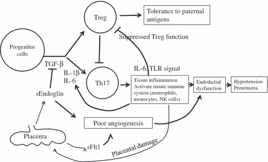
Preeclampsia is associated with exaggerated systemic inflammatory changes58 and poor angiogenesis because of increased levels of soluble Flt-1 and soluble endoglin59 (Fig. 3). Very interestingly, soluble endoglin is an inhibitor of TGF-β; therefore, TGF-β signaling is suppressed in preeclampsia. Furthermore, IL-1β and IL-6, which induce Th17 cell differentiation, are produced by monocytes in preeclampsia. These factors may easily differentiate Th17 cells, which induce exaggerated inflammation, resulting decreased numbers of Treg cells. Increased Th17 cells may induce exaggerated systemic inflammatory changes and vascular endothelial dysfunction. Furthermore, chronic inflammation may impair Treg function.47 The chronic inflammation theory, endothelial dysfunction theory, poor angiogenesis theory, and immune maladaption theory are interrelated, and the imbalanced differentiation of Treg cells and Th17 cells may explain the pathophysiology of preeclampsia.

The interrelationship between inflammation, poor angiogenesis, endothelial damage, and imbalance of Th17 and Treg differentiation.
Conclusions
The new paradigm, i.e., the Th1/Th2/Th17 and Treg paradigms, has been proposed, and emerging literature provides many new findings in reproductive biology and immunology. The balance and correlation between Th1 cells, Th2 cells, Th17 cells, and Treg cells should be discussed to understand reproductive immunology, and it is important to find new therapies for implantation failure, recurrent pregnancy loss, preterm labor, and preeclampsia.


