Increased plasmin-α2-antiplasmin levels indicate activation of the fibrinolytic system in systemic amyloidoses
Systemic amyloidosis is marked by accumulation of amyloid deposits in various tissues throughout the body. In most cases, impairment of organ function by these deposits leads to morbid and complex pathological conditions. Primary systemic amyloidosis (AL) is caused by accumulation of monoclonal immunoglobulin light-chains (LC), which are overproduced by plasma cells [1]. AL is the most prevalent form of systemic amyloidosis. Secondary systemic amyloidosis (AA) can be recognized by the accumulation of serum amyloid A protein, a process that is thought to be a hazardous side-effect of chronic inflammation [2]. Thirdly, hereditary forms of systemic amyloidosis exist that are caused by mutations in transthyretin (ATTR; [3]) or, more rarely, by lysozyme, fibrinogen or other proteins [4–8], causing these proteins to accumulate into amyloid deposits. Patients suffering from systemic amyloidosis are at risk for developing bleeding complications, which may have life-threatening consequences [9–17]. This problem has not yet been fully elucidated, but can partially be explained by decreased coagulation activity [17–22].
Amyloid is defined by the presence of deposits of fibrillar protein aggregates in tissue, which can be stained by dyes such as Congo red. Amyloids of different origin have common structural characteristics, most notably a unique quarternary fold, termed the cross-β structure, with a 4.7 Å inter-sheet regularity. We have identified tissue-type plasminogen activator (tPA), an activator of fibrinolysis, as a multiligand receptor for amyloid cross-β structure in vitro [23]. Plasmin is a serine protease that mediates proteolysis of many substrates, including fibrin. Increased plasmin activity has previously been implicated in bleeding [24–30]. Accumulating protein deposits in systemic amyloidosis patients share the structural characteristics that activate plasmin formation and their amyloidogenic progenitors are present in plasma [31,32]. The levels of these circulating proteins are predictive for the clinical outcome of the diseased patients.
From this perspective, we hypothesized that the observed bleeding tendency of amyloidosis patients may be attributed at least in part to constitutive fibrinolysis. To test this hypothesis, we have determined the plasmin-α2-antiplasmin (PAP) complex levels in systemic amyloidosis patients and compared them with age- and sex-matched healthy controls.
Systemic amyloidosis patients were recruited at Groningen University Medical Center and Utrecht University Medical Center as described earlier [33–35]. Selection was based on positive Congo red biopsies and number and location of sites with amyloid depositions. Thirty-one patients with systemic amyloidosis were included in the study. Five patients suffered from hereditary TTR amyloidosis, nine from AA and seventeen from AL. Blood was drawn by venipuncture and collected in 6-mL Vacutainer tubes (BD, Franklin Lakes, NJ, USA, #388410) with a final concentration of 0.32% sodium citrate. Plasmas were isolated by 10 min centrifugation at 2700 × g at room temperature. Samples were snap-frozen and temporarily stored at –20 °C before transport to the laboratory and storage at –80 °C. Plasma concentrations of PAP complexes were determined in duplicate within 5 months using a commercially available ELISA (Technoclone GmbH, Vienna, Austria, Cat. No. TC11060), according to the manufacturer’s instructions. Data were drawn and analyzed in graphpad prism 4 for Windows; statistical analysis of differences between individual groups was performed by two-tailed non-parametric t-tests (Mann–Whitney). Overall differences between more than two groups were compared by one-way anova (Kruskal–Wallis).
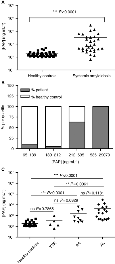
Plasma levels of PAP complexes were significantly elevated in patients with systemic amyloidosis, compared with healthy control subjects, as shown in Fig. 1A. Elevated PAP levels were found in 71.0% of the patients suffering from systemic amyloidosis, compared with 2.5% of control subjects (elevated PAP levels are defined as being above the sum of the mean value of 40 healthy controls and 3 SD). This is indicative for a shift in the hemostatic balance towards a fibrinolytic state in systemic amyloidosis patients. Division of patients and controls into quartiles is illustrated in Fig. 1B. The first and second quartile together comprises 33 healthy controls (82.5%) and only three systemic amyloidosis patients (9.7%). The third quartile comprises seven control subjects (17.5%) and 11 patients (35.5%). The highest quartile consists only of systemic amyloidosis patients (54.8%). Thus, systemic amyloidosis is often accompanied by an increase in PAP levels.

Activation of the fibrinolytic system in systemic amyloidosis. (A) Plasma plasmin-α2-antiplasmin (PAP) levels were determined by ELISA in 31 patients with systemic amyloidosis (38.7% male, average age 51.8 ± 10.5) and compared with those of 40 healthy controls (37.5% male, average age 49.4 ± 7.3). Data were analyzed by two-tailed non-parametric t-test and a highly significant difference was found between both groups, indicating the profibrinolytic state of the patient population. (B) Division in quartiles demonstrates that all subjects with high fibrinolytic activity are systemic amyloidosis patients. (C) Subanalysis of our patient population showed a significant difference in PAP levels between the three forms of amyloidosis, as determined by one-way anova (***P < 0.0001). Individual two-tailed non-parametric t-tests revealed that high PAP levels were prominent in AA and AL amyloidosis, but not in TTR amyloidosis, as compared to controls.
Next, the question arose of whether the hyperfibrinolytic state seen in systemic amyloidosis was preferentially or equally divided over the three forms of systemic amyloidosis. A two-tailed one-way anova indicated a P-value < 0.0001, indicating significant differences between groups. Individual non-parametric t-tests showed that only AA and AL patients had strongly elevated PAP levels compared with healthy controls, whereas ATTR patients showed no increased PAP levels (Fig. 1C). This suggests that a hyperfibrinolytic state is mainly present in AA and AL patients, with no significant difference between these two groups, although on average AL patients have slightly higher PAP levels compared with AA patients. While PAP levels in ATTR patients are significantly lower than in AL patients, similar differences between AA and ATTR patients did not reach statistical significance, probably because of lack of power.
Generation of plasmin has been associated with inflammation [36]. CRP is clinically used as a marker for inflammation. Twenty-four out of 31 patients had been routinely tested. We analyzed those values with the same statistical methods used for analyses of PAP complex levels. We found that CRP was significantly elevated in AA, but not in AL or ATTR (not shown). No correlation between PAP and CRP levels could be demonstrated in our patient population, suggesting that elevated PAP levels are not directly related to inflammation in our patients. The elevated CRP levels in AA can be explained by the chronic inflammation that leads to AA amyloidosis [2,31,37]. In addition, albumin levels, routinely measured in 26 out of 31 patients, were equal between ATTR, AA and AL amyloidosis (not shown). This suggests that determination of PAP levels in our study was not influenced by variation in general protein metabolism, which could have been caused by lowered liver function or nephropathy.
Systemic amyloidosis has a poorly understood bleeding phenotype. Previous data have indicated that this bleeding tendency can, in part, be attributed to decreased coagulation. Both lowered levels or acquired deficiencies of certain coagulation factors (i.e. FX, FV and von Willebrand factor) are reported as being responsible [17,20–22]. In particular, recruitment of FX to amyloid deposits has been considered causative. This explanation, based on activity measurements alone, has been challenged [19]. In a large population of AL patients decreased amounts of active FXa were found, rather than lowered levels of FX. Others have hypothesized that decreased activation of FX might be due to the loss of tissue factor from blood vessels to amyloid deposits and/or inactivation by soluble protein aggregates [38]. Loss of tissue factor may also affect the stability of the clot in another way. Recent data show that tissue factor, via activation of FXI, is involved in stabilizing fibrin. FXIa enhances fibrin generation but also strengthens the clot because it inhibits fibrinolysis through the generation of thrombin-activatable fibrinolysis inhibitor [39]. Taken together it is evident that bleeding induced by amyloidosis can be ascribed to decreased coagulation.
We found here that 71.0% of systemic amyloidosis patients have elevated PAP levels, as compared with 2.5% in control subjects, which is indicative for elevated plasmin generation and fibrinolytic activity in systemic amyloidosis. Elevated PAP levels were most pronounced in AA and AL patients, but not in ATTR patients. Our findings indicate that systemic amyloidosis can be accompanied by constitutive plasmin formation, which is reflected in the circulating PAP complexes. Given the previous notion that tPA is activated by misfolded protein aggregates in general, we presume that the fibrinolytic system in these patients is activated directly by the amyloidogenic misfolded proteins. Consequently, this is a physiological response in order to remove these potentially harmful protein structures to alleviate the symptoms.
In certain AA and AL patients, this response of the fibrinolytic system may become pathological and lead or contribute to bleeding. This hypothesis is illustrated by our finding that one AL patient with very high PAP levels in our study (13 455 ng mL−1; sample taken in June 2004), was treated and reported for having fibrinolysis-related bleeding symptoms in September 2000 [10]. This would suggest a long-term constitutive hyperfibrinolytic state in amyloidosis patients. This patient had widespread vascular amyloid deposits (H.M. Lokhorst, personal communication), which might explain the systemic character of the observed bleeding symptoms. Elevated fibrinolytic activity has been reported as being of importance in individual cases of systemic amyloidosis patients suffering from bleeding episodes [10,40]. Interestingly, hemorrhaging has also been described in ATTR patients, which can be ascribed to ruptures of the cranial microvasculature caused by localized amyloid deposits in vessel walls [11–13,41]. Similarly, reports state that amyloid deposits in small vessels can cause splenic rupture in AA [42]. Apparently, mechanic vessel rupture by localized accumulating amyloid causes bleeding. However, the bleeding tendency seen in most patients is not only related to vessel rupture by amyloid [14–16,19] and lacks a well-defined cause.
Current studies point towards prefibrillar amyloidogenic proteins as in vitro activators of fibrinolysis (our unpublished data). The differences in the fibrinolytic status of patients with specific amyloidoses (i.e. ATTR, AA, AL) may be explained by the differences in the presence of these activating species in vivo. Indeed, a generally less severe bleeding tendency is observed in ATTR patients than in AA and AL patients (B. P. C. Hazenberg, pers. comm.). Further studies are required to identify the exact molecular characteristics of amyloidogenic proteins that are able to stimulate fibrinolysis. This may broaden our perspective on the role of the fibrinolytic system. Recent developments have already pointed to more roles for plasmin than mere involvement in fibrinolysis. Most notably, it has been described that plasmin formation mediates inflammatory responses in a number of conditions, for example atherosclerosis [36]. Perhaps, plasmin formation in general, aids in the removal of misfolded protein aggregates, a response that may turn harmful under pathological conditions, such as systemic amyloidoses. Further investigations are required to validate these hypotheses.
In conclusion, we suggest hyperfibrinolysis as a causative factor for bleeding events in certain forms of systemic amyloidosis. Screening for hyperfibrinolysis in idiopathic amyloidosis-related bleeding may therefore prove useful. Amelioration of symptoms may be achieved by administration of antifibrinolytics, such as tranexamic acid or epsilon amino caproic acid, which has already been reported in individual cases [10,43]. We propose that amyloid deposits inflict physical damage and impairment on vessels and organs and, at the same time, sequester and activate fibrinolysis, resulting in hemorrhage. A better understanding of this mechanism may offer new targets and more effective treatment of systemic amyloidosis and/or amyloidosis-related hemorrhaging.
Acknowledgements
We thank J. Bijzet for his assistance in collecting blood samples and B. N. Bouma for critically reading the manuscript and valuable advice. The work of C. Maas was funded by a grant kindly provided by the Dutch Thrombosis Foundation.
Disclosure of Conflict of Interests
B. Bouma and M. F. B. G. Gebbink are employed and shareholders of Crosbeta Bioscience VC, a company developing diagnostics and therapeutics for protein misfolding diseases. The other authors state that they have no conflict of interest.


