Stabilization of Juxta-Physeal Distal Tibial and Fibular Fractures in a Juvenile Tiger Using a Hybrid Circular–Linear External Fixator
Presented in part at the 36th Annual Conference of the Veterinary Orthopaedic Society, Steamboat Springs, CO, March 2009.
Abstract
Objective
To report stabilization of closed, comminuted distal metaphyseal transverse fractures of the left tibia and fibula in a tiger using a hybrid circular–linear external skeletal fixator.
Study Design
Clinical report.
Animal
Juvenile tiger (15 months, 90 kg).
Methods
From imaging studies, the tiger had comminuted distal metaphyseal transverse fractures of the left tibia and fibula, with mild caudolateral displacement and moderate compression. Multiple fissures extended from the fractures through the distal metaphyses, extending toward, but not involving the distal tibial and fibular physes. A hybrid circular–linear external skeletal fixator was applied by closed reduction, to stabilize the fractures.
Results
The fractures healed and the fixator was removed 5 weeks after stabilization. Limb length and alignment were similar to the normal contralateral limb at hospital discharge, 8 weeks after surgery. Two weeks later, the tiger had fractures of the right tibia and fibula and was euthanatized. Necropsy confirmed pathologic fractures ascribed to copper deficiency.
Conclusion
Closed application of the hybrid construct provided sufficient stability to allow this 90 kg tiger's juxta-articular fractures to heal with minimal complications and without disrupting growth from the adjacent physes.
Juxta-physeal fractures present challenges in planning and executing a successful fracture repair; these challenges are magnified in a large, nondomesticated animal such as a tiger. We report successful use of a hybrid circular–linear external fixator for stabilization of juxta-physeal fractures of the distal tibia and fibula in a juvenile tiger. Unfortunately, the tiger subsequently sustained fractures of the contralateral tibia and fibula and was euthanatized. Necropsy revealed that the tiger had osteoporotic bone disease, which was ascribed to copper deficiency.1
CLINICAL REPORT
A 15-month-old intact male white Bengal tiger was admitted to the University of Florida for stabilization of left tibial and fibular fractures sustained 24 hours previously, after falling from a ledge. The tiger was in excellent general health and had been fed a diet of raw meat and whole chickens, with Oasis™ FT (Apperon Inc., Marshalltown, IA) supplement added to the meat. At admission, the tiger weighed 90 kg, was bright, alert, and calm. Physical examination findings, as permitted, were normal except that the tiger had an obvious left hind limb lameness. On orthopedic examination after sedation with buprenorphine (0.01 mg/kg orally), pain, crepitus, and torsional instability were elicited on manipulation of the left distal crus. Substantial soft tissue swelling was present at the level of, and distal to, the palpable crural instability.
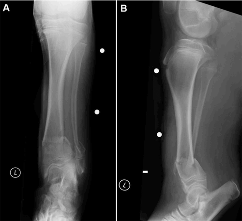
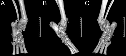
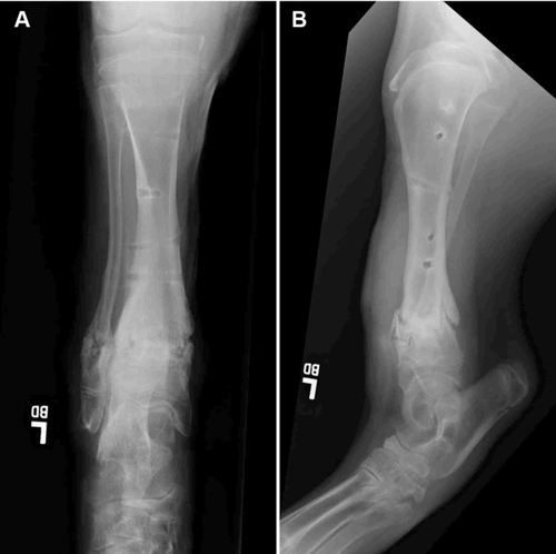
The tiger was premedicated with buprenorphine (0.01 mg/kg orally) and anesthesia induced with ketamine (3.6 mg/kg intramuscularly [IM]), medetomidine (30 μg/kg IM), and midazolam (0.03 mg/kg IM) administered using a dart. The tiger was intubated and maintained with isoflurane (1.5%) in oxygen for radiographs and computed tomography (CT) of the left crus. There were closed, comminuted, transverse fractures of the distal left tibial and fibular metaphyses with mild caudolateral displacement (Figs 1). Physeal involvement was not apparent. Radiographs of the contralateral crus were taken to assist in preoperative planning. A helical CT scan of both pelvic limbs was performed from the stifle to the distal metatarsus using an 8-slice multidetector row CT unit (Toshiba Acquilion 8, Toshiba America Medical Systems, Tustin, CA). Axial images were reconstructed with a slice thickness of 2 mm; sagittal and dorsal plane reformatted images were also created with a slice thickness of 2 mm. All images were reconstructed using bone and soft tissue algorithms and were reviewed on a PACS workstation (Kodak Directview Version 5.1, Carestream Health, Woodbridge, CT) in bone and soft tissue windows. From the CT study, there were comminuted distal metaphyseal transverse fractures of the left tibia and fibula, with mild caudolateral displacement and moderate compression. Multiple fissures extended from the fracture through the distal metaphyses, extending toward, but not involving the distal physes (Fig 2). The bone density and comminution of the fractures were not suggestive of a pathologic fracture. A plantar-lateral fiberglass splint (3M™ Vetcast™ Plus Veterinary Casting Tape, 3M Medical Division, St Paul, MN) was applied to the left pelvic limb and the tiger was recovered from anesthesia until a treatment plan was developed.
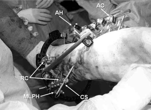
Two days later, the tiger was premedicated with medetomidine (28 μg/kg IM) and ketamine (2.3 mg/kg IM) with a dart. After induction of anesthesia, the tiger was intubated and maintained with isoflurane (1.5%) in oxygen. The tiger was positioned in dorsal recumbency, and the left pelvic limb was clipped and aseptically prepared for surgery. Under fluoroscopic guidance (Premier system model no. 60000, Fluoroscan Imaging Systems Inc., A Hologic Company, Bedford, MA), a 1.6-mm Kirschner wire (IMEX™ Veterinary, Inc., Longview, TX) was inserted from lateral-to-medial, through the skin and subcutis on the cranial aspect of the crus, immediately proximal to the distal tibial and fibular physes. This Kirschner wire was used as a landmark identifying the distal physes for insertion of fixation wires. A 1.8-mm olive wire (Ilizarov stainless steel bayonet point wire with stopper, Smith & Nephew Orthopaedics, Memphis, TN) was inserted from medial-to-lateral, through the distal tibial metaphyseal segment, immediately proximal to the physis, and secured to a complete circular ring (Taylor Spatial Frame 180 mm full ring, Smith & Nephew Orthopaedics) component with 2 fixation bolts (Ilizarov slotted wire fixation bolts, Smith & Nephew Orthopaedics). The Kirschner wire identifying the distal physes was removed. Next, a 1.6-mm Kirschner wire (Smooth fixation wire, IMEX™ Veterinary, Inc.) was placed through a fixation bolt attached at a cranial position on the ring into the metaphyseal segment to temporarily stabilize the ring during placement of subsequent olive wires. Two additional 1.8-mm olive wires were placed from craniomedial-to-caudolateral and craniolateral-to-caudomedial, and were captured with fixation bolts. Flat washers (Ilizarov spacing washers, Smith & Nephew Orthopaedics) were used when needed to elevate the point of attachment of the wires away from the surface of the ring. The end of 1 wire was secured craniolaterally with a fixation bolt placed in a 2-hole Rancho cube (Rancho system cube, 2 holes, Smith & Nephew Orthopaedics), while the end of another wire was secured caudolaterally with a fixation bolt placed through a male hinge (male low profile hinge, Smith & Nephew Orthopaedics). All wires were tensioned sequentially to 110 kg using a calibrated tensioning device (Ilizarov dynamic wire tensioner, Smith & Nephew Orthopaedics). The temporary cranial Kirschner wire in the distal tibial segment was removed. A straight carbon fiber connecting bar (Jet-X bar, Smith & Nephew Orthopaedics) was attached to the ring along the medial surface of the crus using an articulating hinge (Jet-X bar-to-ring clamp, Smith & Nephew Orthopaedics). A 5-mm fluted, partially threaded, positive-profile half-pin splintage pin (Jet-X short half-pin, Smith & Nephew Orthopaedics) was placed in the proximomedial tibial metaphysis and loosely secured to the connecting bar with an articulated clamp (Jet-X bar-to-pin clamp, Smith & Nephew Orthopaedics). The fracture was distracted by applying traction to the ring and aligned manually as the hinge and clamps were tightened. Acceptable alignment of the fracture was confirmed fluoroscopically. A centrally threaded positive-profile full-pin splintage pin (Jet-X traction pin, Smith & Nephew Orthopaedics) was placed from medial-to-lateral in the distal diaphysis of the proximal tibial segment. The full-pin was secured to the connecting bar on the medial side of the crus with an articulated clamp. A 5-hole Rancho cube (Rancho system cube, 5 holes, Smith & Nephew Orthopaedics) and centering sleeve (Rancho system 5-mm centering sleeve, Smith & Nephew Orthopaedics) were used to secure the full-pin to the ring laterally. Three additional half-pin splintage pins were placed on the connecting bar into the tibial diaphysis. These pins were each secured to the connecting bar with articulated clamps. A single half-pin splintage pin was placed cranially into the proximal tibial diaphysis and secured to the ring at a medial position using an articulated clamp, connecting bar, and articulating hinge (Fig 3).
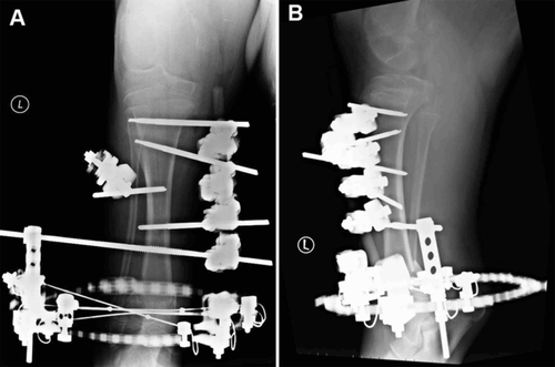
Postoperative radiographs showed good alignment and apposition of the fractures, with a possible fissure fracture propagating between the central fixation pins (Fig 4). Sterile gauze squares were placed over the pin and wire insertion wounds, and the interior of the fixator was packed with sterile sponges. A modified Robert Jones bandage incorporating the paw was applied. Buprenorphine (0.01 mg/kg orally every 4–6 hours for 4 days) then tramadol (2 mg/kg, orally every 12 hours for 6 weeks) and meloxicam (0.05 mg/kg orally once daily for 3 weeks) were administered for postoperative pain management. Acepromazine (1 mg/kg orally as required) and diazepam (0.25 mg/kg orally twice daily for 1 week) were administered for sedation. Cephalexin (30 mg/kg orally twice daily for 6 weeks) was administered for antibacterial prophylaxis. Calcium citrate (30 mg/kg orally once daily) was given in the diet, and the tiger was provided access to direct sunlight during the convalescent period. The tiger was restricted to cage confinement and remained calm, comfortable, and playful during hospitalization.
Approximately every 3rd day, the bandage was changed and the pin- and wire-skin interfaces cleaned under sedation with medetomidine (20 μg/kg subcutaneously) given by hand injection. The tiger was tractable and minimally responsive to stimulation after this low dosage of medetomidine in conjunction with the residual sedation from the pain management regimen. After the bandage was removed, the fixation element-skin interfaces were cleaned using 0.05% chlorhexidine solution. The medetomidine was not reversed after the bandage changes. The cranial half-pin was removed 3 weeks after surgery because the pin impinged on the cranial tibial muscle, resulting in discomfort and pin-tract drainage. One of the olive wires was also removed because of associated purulent drainage.
Radiographs taken 5 weeks after surgery revealed remodeling of the cortices and progressive mineralization of the surrounding bridging fracture callus. The fixator was removed and the limb was reradiographed (Fig 5). Radiographs of the contralateral tibia were taken and measurements of tibial length were made on the mediolateral radiographs from the intercondylar eminences to the center of the distal articular surface of the tibial cochlear. Length was comparable between the left (254 mm) and right (255 mm) tibiae. The distance from the center of the most distal fixation wire to the distal tibial physis increased from 7.5 mm immediately postoperatively to 12.5 mm at the time of fixator removal. The tiger was discharged 8 weeks after surgery and had only a subtle weight-bearing left pelvic limb lameness when walking. No grossly evident angular or torsional deformities were present in the left crus.
Thirteen days after discharge, the tiger was admitted to Texas A&M University's Veterinary Medical Teaching Hospital for evaluation of an acute nonweight-bearing right pelvic limb lameness of 24 hours duration. The lumbar spine, pelvis, pelvic limbs, left thoracic limb, and thorax were radiographed. There were incomplete folding fractures of the right distal tibial and fibular diaphyses. The previous surgically repaired left tibial and fibular fractures were healed. On thoracic radiographs, there were multiple, healed rib fractures. Decreased bone density was apparent on the inner cortical surfaces of the metaphyses of multiple long bones. Based upon the presence of multiple fractures in osteopenic bones, it was concluded that the tiger had pathologic fractures secondary to a nutritional or metabolic bone disease. The tiger was euthanatized and necropsy confirmed that the fractures were pathologic. The tiger had osteoporosis that was ascribed to copper deficiency.1
DISCUSSION
Hybrid fixators are being used with increased frequency to stabilize fractures in dogs and cats.2-4 Primary attributes of hybrid fixators include the system's modularity, allowing for multidimensional flexibility in construct design and fixation. Hybrid fixators also use small diameter wires as fixation elements that are secured to ring components. The wires are placed at divergent angles, thus allowing stabilization of short juxta-articular fracture segments while minimizing disruption of adjacent soft tissues, preventing nerve injury, reducing operative time, and improving patient tolerance.5 These attributes were particularly advantageous in stabilizing the juxta-physeal fractures in this skeletally immature tiger.
The location and configuration of the fractures presented additional concerns with respect to selection of fixation devices. Consideration was given to using traditional AO techniques using a plate and screws to stabilize this fracture. This approach would have required considerable soft tissue dissection and disruption of the periosteal blood supply. Despite having an apparently large distal tibial fracture segment (measuring ∼40 mm in length), achieving rigid internal fixation would have been difficult with available veterinary orthopedic implants because of extensive fissuring. We evaluated a number of fracture-specific bone plates designed for stabilization of juxta-articular fractures in people; however, these implants are only manufactured as 3.5-mm plates, which would have been biomechanically inadequate as the sole means of fracture fixation. Procuring a custom-made plate or plates was not considered reasonable because of the inevitable delay in treatment while manufacturing the implants.
There were multiple fissures in this fracture, extending toward the distal tibial physis. Fissures can propagate during and after implant application, increasing the complexity of the fracture, which can result in fixation failure. Preoperative CT images were valuable because they substantiated the surgeon's impression that anatomic reconstruction of this tiger's tibia was not feasible because of fracture impaction and fissuring. The olive wires, used as fixation elements on the ring component, immobilized and compressed the fissures, thereby preventing fissure propagation.6, 7 The olive wires were placed at divergent angles on the ring component, providing multiplanar stability.7, 8 Application of the hybrid fixator by closed reduction minimized disruption of the periosseous soft tissues, thus preserving periosteal blood supply. Further, use of tensioned wires for ring fixation elements allowed for axial micro-motion of the secured fracture segment, which can stimulate callus formation.9, 10
We have moved away from using traditional circular fixator constructs11, 12 to predominantly using hybrid circular–linear fixator constructs2, 4, 13, 14 for static applications in dogs and cats. Hybrid constructs are simple to apply and associated with less postoperative morbidity.2-4, 13, 14 Unfortunately, the components of the system (IMEX™ Veterinary, Inc.) we normally use in dogs and cats were not large enough to use in this tiger. We were able to obtain access to the Smith & Nephew Orthopaedics Hybrid External Fixator System. The construct we used in this tiger consisted of a single full-ring with 3 divergent olive wires stabilizing the distal fracture segment and biplanar hybrid connecting bars with positive-profile, end-threaded half-pins and a centrally threaded positive-profile full-pin stabilizing the proximal fracture segment. The fixator components and fixation pins and wires used to stabilize this tiger's fracture cost ∼$8500, and thus, the routine use of this system in animals is unlikely.
Reports describing the use of hybrid constructs for fracture stabilization in dogs and cats document good to excellent limb use during convalesence.2-4, 13, 14 This assumption cannot be directly extrapolated to nondomestic species such as a tiger, because of patient curiosity and the propensity for mutilation of unfamiliar adornments. With cage confinement and administration of analgesics and sedatives, this tiger readily tolerated the fixator. The tiger's compliance was attributed to the light weight of the components and stability provided by the hybrid fixator system. Safe corridors for placement of fixation pins have been defined in dogs to minimize postoperative morbidity associated with impingement of soft tissues by fixation pins.15 Using these corridors, this tiger required only infrequent tranquilization and examination for bandage changes, fixator modification, or pin- and wire-tract cleaning. The cranial fixation pin was removed 3 weeks postoperatively because of pin-tract drainage ascribed to impingement of the pin on the cranial tibial muscle. One olive wire was also removed at this time because of purulent drainage. The discharge resolved after removal of these implants. The fixator was removed when the fracture was bridged with confluent callus and cortical remodeling was evident radiographically.
The distal tibial physis is responsible for ∼50% of limb length in dogs.16 Similar information is not available for tigers, but we were cognizant to avoid placing fixation elements through or distal to the distal tibial and fibular physes in this tiger, to avoid restricting further physeal growth. We were able to provide stable fracture fixation, while preserving the distal tibial and fibular physes, which allowed for continued longitudinal bone growth. Subjectively, both pelvic limbs had identical conformation, and serial radiographs confirmed that crural length remained similar bilaterally, with continued growth from the left distal tibial and fibular physes.
Although this tiger's bone density and cortical thickness appeared normal radiographically at initial admission, the fixation pins and wires were noted to easily penetrate the bone during surgery. Although we are always suspicious of nutritional bone diseases in juvenile captive nondomestic felines, the ease of pin placement was our first indication that this tiger might have underlying bone pathology. Results of pre- and postoperative serum biochemical profiles performed during the tiger's hospitalization at the University of Florida did not reveal any objective evidence of calcium or phosphorous abnormalities in this tiger, and subjective assessment of sequential radiographs and CT supported adequate bone density and normal bone healing.
The history indicated that since weaning, this immature tiger had been fed a diet that was composed predominantly of meat and whole chickens, with mineral supplementation. Other tiger cubs in the collection had previously been raised on similar diets without apparent orthopedic problems; however, because the supplement was manually top dressed, the exact amount of calcium and other trace minerals consumed would have been difficult to document or control in an individual animal. The tiger was given therapeutic doses of calcium and routinely exposed to sunlight after surgery as prophylaxis to the development of nutritional hyperparathyroidism. Despite these measures, necropsy revealed that the tiger suffered from an underlying osteoporotic bone disease ascribed to copper deficiency.1


