Iatrogenic Sciatic Nerve Injury in Eighteen Dogs and Nine Cats (1997–2006)
Abstract
Objective— To report clinical features associated with iatrogenic peripheral nerve injury in dogs and cats admitted (1997–2006) to a referral teaching hospital.
Study Design— Retrospective study.
Animals— Dogs (n=18), 9 cats.
Methods— Patients had acute signs of monoparesis attributable to sciatic nerve dysfunction that developed after treatment. Neurologic examination and electrodiagnostic testing were performed. Surgical therapy was used for nerve entrapment and delayed reconstructive surgery used in other cases.
Results— Of 27 nerve injuries, 25 resulted from surgery (18 with treatment of pelvic injuries). Iliosacral luxation repair resulted in tibial (4 cats) and peroneal (3 dogs) nerve dysfunction. Other causes were intramedullary pinning of femoral fractures (3), other orthopedic surgery (cemented hip prosthesis [2] and tibial plateau-leveling osteotomy [1]), and perineal herniorrhaphy [1]. Nerve injury occurred after intramuscular injection (1 cat, 1 dog). Immediate surgical treatment was removal of intramedullary nails, extruded cement, or entrapping suture. Delayed nerve transplantation was performed in 2 dogs. Within 1 year, 13 patients recovered completely, clinical improvement occurred in 7, and there was no improvement in 7. Five of the 7 dogs that did not recover had acetabular or ilium fracture.
Conclusion— Iatrogenic sciatic nerve injury occurred most commonly during treatment of pelvic orthopedic diseases and had a poor prognosis. Clinical variation in sciatic nerve dysfunction in dogs and cats can be explained by species anatomic differences.
Clinical Relevance— Iatrogenic sciatic nerve injury leads to severely debilitating locomotor dysfunction with an uncertain prognosis for full-functional recovery.
INTRODUCTION
ALTHOUGH PERIPHERAL nerve injury has been described in dogs and cats, there are few reports of iatrogenic nerve injury. The sciatic nerve, the largest peripheral nerve, is protected relatively well from external injury because of its deep location close to bone and heavy muscle mass.1–3 When considering pelvic fracture as a possible cause of sciatic nerve injury, iatrogenic lesions inflicted during surgical treatment have seldom been clearly differentiated from traumatic injuries because, in most reported cases, there has been no preoperative neurologic examination.4–5 Its deep location, close proximity to the pelvic bone, and limited visibility make the sciatic nerve vulnerable to damage during surgical exploration of the wing of the ilium, acetabulum, sacroiliac joint, and caudal proximal femoral region.3–7 Sciatic nerve injury has also been reported as a complication of the surgical correction of perineal hernia in dogs.8 Furthermore, the caudal thigh musculature is a common site for intramuscular injections in dogs and cats and sciatic nerve damage can result directly from needle injury, the agent used, or later by scarring involving the nerve.9
Our purpose was to report the occurrence of iatrogenic peripheral nerve injury in dogs and cats. Only lesions of the sciatic nerve or its branches ramifications were observed. We describe the pathophysiology of the injury based on the surgical procedure initially performed and on the results of subsequent clinical and electrophysiologic examination.
MATERIALS AND METHODS
Animals
Between 1997 and 2006, 103 patients (72 dogs, 31 cats) were admitted with monoparesis associated with a peripheral nerve lesion. Of these, 27 (18 dogs, 9 cats) patients had iatrogenic nerve injury and are the subjects of this report. To be included, patients had to be neurologically normal before treatment and, generally, clinical signs had to appear immediately after treatment.
In the 18 patients (12 dogs, 6 cats) with pelvic fractures, neurologic examination was performed before surgery to rule out any trauma-induced nerve lesions. In these patients, including non-ambulatory patients, no neurologic abnormalities were detected, and all preoperative clinical signs were directly related to the orthopedic problem, indicating that subsequent nerve injury was associated with pelvic fracture repair. In the remaining cases the paresis was clearly associated with the treatment patients had received.
Neurologic Findings
Patients with sciatic nerve paralysis presented with a dropped-hock and knuckling of the digits. The limb could support weight because the quadriceps muscles were functioning. Sensory analgesia was detected on the lateral, dorsal, and plantar surfaces of the lower limb. The cranial tibial and gastrocnemius tendon reflexes were decreased or absent, as was the withdrawal reflex. Patients with peroneal nerve paralysis had hyperextension of the tarsus and knuckling of the digits. Cutaneous analgesia was identified on the dorsal aspect of the paw and cranial aspect of the leg; a cranial tibial reflex was absent. Patients with tibial nerve injury had a dropped hock and overextended digits with analgesia of the caudal aspect of the limb and plantar aspect of the paw. This clinical classification is used in Table 1 and throughout the text of this report.
| InitialProblem | Patient | Clinical Signs | NeurophysiologicExamination | Neurolocalization | Treatment | Follow-Up | Outcome |
|---|---|---|---|---|---|---|---|
| Acetabular fracture/plating | |||||||
| 1 | 2 y M Pekingese, 8 kg | Sciatic nerve | Tibial nerve+Peroneal nerve++ | Tibial+peroneal nerve | Physiotherapy | 6 m | Fair |
| 2 | 2.5 y M Irish, Setter 28 kg | Sciatic nerve | Tibial nerve++Peroneal nerve++ | Tibial+peroneal nerve | Physiotherapy | 1 y | Poor |
| 3 | 13 m M Husky, 19 kg | Sciatic nerve | Tibial nerve+Peroneal nerve+ | Tibial+peroneal nerve | Physiotherapy | 8 m | Good |
| 4 | 2 y F Belgian Shepherd, 21 kg | Peroneal nerve | Tibial nerve+Peroneal nerve++ | Tibial+peroneal nerve | Physiotherapy | 6 w | Poor |
| 5 | 4 y F Mixed-breed, 32 kg | Peroneal nerve | Tibial nerve−Peroneal nerve++ | Peroneal nerve | Physiotherapy | 1.5 y | Good |
| Ilial fracture/plating | |||||||
| 6 | 2 y F Golden Retriever, 28 kg | Sciatic nerve | Tibial nerve+Peroneal nerve+ | Tibial+peroneal nerve | Physiotherapy | 2 w | Poor |
| 7 | 1.5 y F Labrador Retriever, 28 kg | Sciatic nerve | Tibial nerve++Peroneal nerve++ | Tibial+peroneal nerve | Physiotherapy | 1 y | Poor |
| 8 | 2 y M Golden Retriever, 33 kg | Sciatic nerve | Tibial nerve++Peroneal nerve++ | Tibial+peroneal nerve | Physiotherapy | 1.5 y | Poor |
| 9 | 1 y M Mixed-breed, 26 kg | Peroneal nerve | Tibial nerve−Peroneal nerve+ | Peroneal nerve | Physiotherapy | 6 w | Fair |
| Iliosacral luxation/lag screw | |||||||
| 10 | 4 y M Miniature Pinscher, 5 kg | Peroneal nerve | Tibial nerve−Peroneal nerve+ | Peroneal nerve | Physiotherapy | 6 w | Good |
| 11 | 3 y M Irish Setter, 28 kg | Peroneal nerve | Tibial nerve+Peroneal nerve++ | Tibial+peroneal nerve | Physiotherapy | 6 m | Good |
| 12 | 6 y F Poodle, 9 kg | Sciatic nerve | Tibial nerve−Peroneal nerve+ | Peroneal nerve | Physiotherapy | 4 w | Good |
| Dysplasia/THR | |||||||
| 13 | 5 y M German Shepherd, 36 kg | Sciatic nerve | Tibial nerve−Peroneal nerve++ | Peroneal nerve | Cement removalNeurotization | 15 m | Fair |
| 14 | 3 y F Mixed-breed, 33 kg | Sciatic nerve | Tibial nerve++Peroneal nerve++ | Tibial+peroneal nerve | Cement removal | 8 m | Poor |
| Perineal hernia/Suture | |||||||
| 15 | 10 y M Teckel, 9 kg | Sciatic nerve | Not performed | Tibial+peroneal nerve | Suture removal | 4 w | Good |
| Femur fracture/intramedullary pin | |||||||
| 16 | 11 m M Dobermann, 24 kg | Peroneal nerve | Not performed | Peroneal nerve | Pin removalFracture plating | 6 w | Good |
| Cruciate ligament rupture/TPLO | |||||||
| 17 | 7 y M Labrador Retriever, 26 kg | Peroneal nerve | Tibial nerve−Peroneal nerve+ | Peroneal nerve | External neurolysis | 3 m | Good |
| Intramuscular injection | |||||||
| 18 | 1 y F American Staffordshire, 22 kg | Sciatic nerve | Tibial nerve++Peroneal nerve++ | Tibial+peroneal nerve | Nerve transplantation | 2 y | Fair |
- w, weeks; m, months; y, years; −, no electrophysiological abnormalities; +, slight findings of denervation (isolated fibrillations, positive sharp wave, NCV normal or decreased); ++, severe findings of denervation (continuous fibrillations, NCV severely decreased or absent); poor, no clinical improvement, permanent sciatic, or peroneal nerve paralysis; fair, clinical improvement but remaining neurologic deficits; good, complete healing or situation allowing good locomotor function; THR, total hip replacement; TPLO, tibia plateau leveling osteotomy.
Electrophysiology
After a delay of 5–10 days, all patients had electrophysiologic investigation. General anesthesia was induced with midazolam (0.2 mg/kg intravenously [IV]) or medetomidine (5 μg/kg IV) together with methadone (0.2 mg/kg IV) and propofol (4 mg/kg IV). After orotracheal intubation, anesthesia was maintained with isoflurane and oxygen. Body temperature was maintained >37°C.
A digital electromyography (EMG) system (Nicolet Viking Quest portable system, VIASYS Healthcare, Kleinosthheim, Germany) was used for EMG and nerve conduction recordings. Stainless-steel 0.45 mm × 40 mm concentric needle electrodes were used for recordings from all muscles of the affected limb. Coccygeal and gluteal musculature were also examined. Insertional and spontaneous EMG activity (fibrillation potentials and positive sharp waves) was recorded on anesthetized animals.
Motor nerve conduction was assessed10,11 in peroneal and tibial nerves of both hind limbs. Compound muscle action potentials were evaluated for amplitude and duration; motor nerve conduction velocity (MNCV) and sensory nerve conduction velocity (SNCV) were measured. Evaluation of F-waves and spinal somatosensory-evoked potentials were not performed.
Physiotherapy
An individual physiotherapy plan was established for each patient. Adequate pain management with opioids and/or non-steroidal drugs allowed an early start with rehabilitation. Initial procedures consisted of local massage 2–3 times/day. The massage technique was not used in the region of pelvic fracture during the initial 2 weeks. To reduce muscle atrophy, passive exercises were performed and, after a couple of days, active exercise was started and gradually increased. Passive exercise consisted of bicycling movements, stretching and simulation of the flexor reflex. Assisted active exercises (standing with and without assistance, standing in combination with passive movement of the contralateral limb) were progressively replaced by short but frequent assisted walking. Patients were regularly walked on a leash 3–5 times/day. In 15 dogs active exercise also included hydrotherapy (assisted swimming or walking on an underwater treadmill) 1–2 times/day depending on the clinical improvement and general condition of the dog.
Treatment
Implementation of a conservative or surgical treatment plan was based primarily on the cause of the nerve injury. In patients with paw knuckling, a protective shoe or preferably an ankle–foot orthotic12 was used to prevent toe injury during locomotion in the recovery phase. For patients with no clinical improvement, these devices were also used to prevent further toe injury. Surgical therapy consisted of immediate or early removal of the implant that caused the nerve injury and/or delayed reconstructive or palliative surgical procedures on the nerve. Fibrous entrapment of the nerve was treated by external neurolysis (microdissection of the nerve from surrounding fibrous tissue). Peroneal neurotization13 (anastomosis between a healthy donor nerve proximally and the distal part of the injured nerve) using the tibial nerve as donor and the peroneal nerve as recipient was used in patients with an irreversible isolated very proximal peroneal nerve lesion. An autologous nerve graft was used in patients where complete destruction of a nerve segment near its target muscles had occurred.
Follow-Up
All patients, except those showing rapid resolution of neurologic signs and early return to normal function, had repeated neurologic examination at 10 days (suture removal), 4 weeks, 3 months, and then every 3 months during the first year after surgery. In some patients where no clinical resolution was observed, a second electrophysiologic examination was performed for comparison with the initial electrodiagnostic findings to determine if re-innervation was proceeding and/or if nerve conduction was measurable.
RESULTS
Abnormal EMG findings were detected in all patients (Tables 1 and 2). Insertional activity included trains of positive waves and isolated fibrillation potentials in 4 dogs (dogs 3, 10, 12, 17) and 6 cats (cats 2–7) that overall had a good functional recovery. Dogs 6 and 9 with ilial fractures and cat 8 had similar electromyographic patterns but without return of normal functional activity. In these 13 patients, the nerve responded well to electrical stimulation and NCV was normal (MNCV: 60–70 m/s [normal >50 m/s]; SNCV:∼60 m/s [normal >60 m/s] or reduced [<50 m/s]).
| InitialProblem | Patient | Clinical Signs | NeurophysiologicExamination | Neurolocalization | Treatment | Follow-Up | Outcome |
|---|---|---|---|---|---|---|---|
| Acetabular fracture/plating | |||||||
| 1 | 6 y M European shorthair | Sciatic nerve | Tibial nerve+Peroneal nerve++ | Tibial nervePeroneal nerve | Physiotherapy | 6 m | Fair |
| 2 | 2 y F European shorthair | Sciatic nerve | Tibial nerve+Peroneal nerve+ | Tibial nervePeroneal nerve | Physiotherapy | 1 y | Good |
| Iliosacral luxation/lag screw | |||||||
| 3 | 3 y F Somalian cat | Tibial nerve | Tibial nerve+Peroneal nerve− | Tibial nerve | Physiotherapy | 2 w | Good |
| 4 | 2 y F European shorthair | Tibial nerve | Tibial nerve+Peroneal nerve+ | Tibial nervePeroneal nerve | Physiotherapy | 4 w | Good |
| 5 | 4 y M European shorthair | Tibial nerve | Tibial nerve+Peroneal nerve− | Tibial nerve | Physiotherapy | 3 m | Good |
| 6 | 4 y M European shorthair | Tibial nerve | Tibial nerve+Peroneal nerve− | Tibial nerve | Physiotherapy | 6 m | Good |
| Femur fracture/intramedullary pin | |||||||
| 7 | 1.5 y M European shorthair | Peroneal nerve | Tibial nerve−Peroneal nerve+ | Peroneal nerve | Pin removalExternal neurolysis | 6 w | Good |
| 8 | 2 y F European shorthair | Peroneal nerve | Tibial nerve+Peroneal nerve+ | Tibial nervePeroneal nerve | Pin removal | 4 w | Fair |
| Intramuscular injection | |||||||
| 9 | 7 y M European shorthair | Sciatic nerve | Tibial nerve++Peroneal nerve++ | Tibial nervePeroneal nerve | NoneEuthanasia | ||
- w, weeks; m, months; y, years; −, no electrophysiological abnormalities; +, slight findings of denervation (isolated fibrillations, positive sharp wave, NCV normal or decreased); ++, severe findings of denervation (continuous fibrillations, NCV severely decreased or absent); poor, no clinical improvement, permanent sciatic or peroneal nerve paralysis; fair, clinical improvement but remaining neurologic deficits; good, complete healing or situation allowing good locomotor function.
Continuous trains of fibrillation potentials associated with a loss of nerve conduction were observed in 8 dogs (1, 2, 4, 7, 8, 13, 14, 18) and 2 cats (1, 9) that did not recover or only incompletely recovered. Dogs 5 and 11 with the same electrophysiological patterns returned to a normal functional state. In 3 dogs (7, 8, 14), a second electrophysiological examination was performed after 3 months. Denervation patterns and absence of conduction were observed.
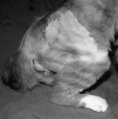
All nerve injuries associated with surgical treatment of pelvic fractures (12 dogs, 6 cats) were treated conservatively with physiotherapy. Sciatic nerve paralysis after plating long oblique ilium fractures only occurred in 4 large-breed dogs (≥25 kg) and had poor recovery. Acetabular fracture repair with bone plating induced sciatic nerve injury (peroneal and tibial components) in 6 patients (4 dogs, 2 cats), and in dog 5 it induced an isolated peroneal nerve lesion: 5 of these patients (3 dogs, 2 cats) functionally recovered (2 dogs and 1 cat returned to normal activity within 1.5 years) whereas 2 dogs (1, 2) and 1 cat (1) still had debilitating deficits (loss of strength, knuckling of paw) after 6 months. Repair of iliosacral joint luxations resulted in sciatic or peroneal nerve injury in 3 dogs and in tibial nerve palsy in 4 cats (Fig 1). These deficits were transient in all 7 patients and resolved completely within 6 months.

Tibial nerve paralysis in a cat after surgical repair of iliosacral joint luxation.
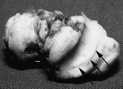
Patients with implant-associated sciatic nerve injury were treated by immediate implant removal (extruded cement in 2 dogs with total hip replacement [THR], intramedullary [IM] pin in 1 dog and 2 cats with femoral fracture, and an entrapping suture in 1 dog with perineal hernia). Caudal extrusion of cement during cup implantation in THR damaged the sciatic nerve and the peroneal nerve in an almost irreversible way in 2 dogs. The sciatic nerve was exposed by an approach to the caudal aspect of the hip joint with tenotomy of the tensor fasciae latae and superficial gluteal muscles. The nerve was partly encircled by a cauliflower-shaped mass of cement near the sciatic notch and had protruded through a perforation of the ischial part of the acetabulum. The nerve was carefully dissected from the cement mass and had some scarring and narrowing where it had been in contact with the cement mass, which was chiseled off the ilium (Fig 2). Cement removal did not result in the resolution of neurologic deficits. Delayed palliative reconstructive surgery after 6 weeks (peroneal neurotization) improved the clinical outcome in 1 dog (13) with peroneal paralysis.

Cement mass, (from total hip replacement) chiseled from the ischial bone. Arrows show the groove in which the sciatic nerve was embedded.
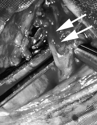
Migrating or misplaced IM femoral implants induced peroneal nerve paresis in 2 cats and 1 dog. Pain and proprioceptive deficits without withdrawal reflex deficits were neurologically observed. In dog 16 and cat 8, collapse at the fracture site caused the IM pin to partially spear the sciatic nerve (Fig 3). Cat 7 was admitted with chronic lameness that developed 4 months after femoral fracture treatment. In dog 16 and cat 7, complete recovery occurred within 6 weeks. In cat 8, where the IM pin had speared the sciatic nerve, there was no improvement 4 weeks after surgery.

Surgical exploration of the sciatic nerve in a cat after intramedullary pinning of femoral fracture. The end of the pin speared the sciatic nerve and induced severe axonotmesis.
Entrapment of the sciatic nerve by a suture around the sacrotuberal ligament during perineal herniorrhaphy in 1 dog caused an immediate sciatic paralysis that resolved within 4 weeks after early suture removal by a standard caudal approach to the hip joint.
Injury to the sciatic nerve occurred after intramuscular injection in the caudal thigh in 1 dog and 1 cat. After implantation of an autologous contralateral tibial nerve autograft, the dog recovered to a good functional state but strength deficits remained visible when walking slowly. The owners of the cat elected euthanasia. On necropsy, sciatic nerve necrosis induced by intramuscular injection was confirmed.
In 1 dog, perineural fibrosis that developed within 6 weeks after tibial plateau leveling osteotomy entrapped the peroneal nerve caudal to the fibular head. The diagnosis was histologically confirmed. Lameness and slight knuckling of the paw completely resolved within 3 months after external peroneal neurolysis.
DISCUSSION
Patients with iatrogenic injuries represented 26.2% (27/103) of the patients with peripheral nerve injuries admitted to our clinic; only the sciatic nerve was affected. This may be owing to the nerve's deep location and its limited visibility during surgical procedures in this region.3 This lesion was more often observed in dogs (18 cases) than in cats (9 cases). Anatomic differences (greater size, more fatty tissue in dogs) and the variety of pelvic surgical procedures performed in dogs may explain this finding. Males were slightly overrepresented (11 dogs, 6 cats) and most patients were young (mean age, 3.5 years; range, 11 months to 10 years). No breed predilection was observed but medium to large breed dogs (14 dogs) were more frequently affected which may reflect a more difficult surgical approach but being overweight was not a consistent risk factor.
The sciatic is an important nerve for locomotion and weight bearing and its functional deficit is debilitating. Even with physiotherapy, only some animals will be able to compensate for sciatic nerve damage by flipping the paw and walking on the plantar surface after many months.2,3 Injury to less-essential nerves, e.g. median nerve injury occurring during elbow arthrotomy, results in slight lameness that can be confused with the original orthopedic condition. Consequently, such lesions may remain undiagnosed and thus the incidence of iatrogenic peripheral nerve injury may be substantially underestimated. Gilmore3 reported that the most common cause of sciatic nerve injury is trauma associated with ilial body fractures. Intrapelvic damage to the nerve is very difficult to recognize because of its anatomic location and in this report no attempt was made to visualize the sciatic nerve when stabilizing pelvic fractures. Furthermore, absence of a preoperative neurologic examination suggests that the incidence of iatrogenic sciatic nerve injury could have been underestimated.3
Inability to adequately visualize the nerve during pelvic surgical procedures is probably the most common cause of iatrogenic sciatic nerve injury3–5 and may explain why the site of injury was located proximal to the great trochanter in most patients (24/27) and why pelvic orthopedic surgery was the primary cause of injury (12 dogs, 6 cats) in our patient population. Excessive manipulation or elongation of the nerve during acetabular—or ilial-fracture fragment manipulation will lead to neurapraxia (transient physiologic conduction block of nerve transmission that will typically resolve in 3–6 weeks1). Three patients (2 dogs, 1 cat) that regained sciatic function probably had neurapraxia or axonotmesis (disruption of axons with maintenance of intact endoneurial tubules1).To prevent this complication, external neurolysis could be used before fracture reduction to allow greater mobility of the nerve during manipulation.14 If the sciatic nerve is not carefully elevated from the ilium during exposure of the fracture fragments, the nerve may also become entrapped either in the fracture gap itself or within the jaws of the reduction forceps, leading to an irreversible crushing injury. This may have occurred in 5 dogs (5/9) that did not recover functionality. Nerve regeneration, even if only axonotmesis is present, is impaired by endoneurial fibrous tissue, leading to poor functional results.14
With iliosacral joint luxation, elongation (during manipulation) or compression (under the sacral wing with the tip of the elevator used during repositioning) of the sciatic nerve probably led to reversible deficits in all 7 patients. Incorrectly placed implants could also lead to an isolated injury of the S1-root but this complication was not observed in our patients.
The tibial nerve was affected in cats and in the peroneal nerve in dogs. Normally the peroneal nerve appears more susceptible to injury than the tibial nerve in both human and animal patients. One explanation is that the peroneal component is composed of fewer and larger funiculi with less connective tissue support than the tibial component.1 Nerves or nerve segments composed of large and tightly packed funiculi are more vulnerable to mechanical injury than those in which the bundles are smaller or more widely separated by a greater amount of connective tissue.1 In the former, the forces acting on the nerve are concentrated on the funiculi, which form the main content. In the latter, they are broken up and dispersed by the connective tissue packing and the funiculi, being more loosely arranged, are more easily displaced within the nerve thereby reducing the effects of deforming forces. In cats, the tibial nerve is more developed than in dogs,1 and lies more laterally than the peroneal nerve, under the sacral wing, which may make it more vulnerable to iatrogenic injury and explain our clinical and neurophysiologic findings.
During THR, when methylmethacrylate is used to cement the acetabular component, keyholes may be made in the ischium and ilium to improve fixation. If one of the holes perforates the bone, cement herniation can occur and is often visible on postoperative radiographs but is generally considered benign. However, if the extrusion occurs in the immediate vicinity of the sciatic nerve, compression and “burning” associated with the exothermic reaction during hardening may occur.15 Removing the cement to relieve compression did not lead to resolution of the neurologic deficits in either of our patients. It seems likely that thermal damage within the nerve results in fibrous tissue replacement, thereby impairing the sprouting of regenerating nerves.
Three cases were associated with the use of IM pins to stabilize a femoral fracture. The most consistent neurologic deficit noted in these 3 patients was loss of proprioception. When IM pins are incorrectly inserted, the main concern is that the sciatic nerve will be impaled by the pin; however, the more common finding is that the pin lies in close proximity to the nerve and transmits pressure to the nerve through the surrounding soft tissues during leg motion. Fibrous reactive tissue scar can also form around the dorsal aspect of the pin and can entrap and gradually constrict the nerve. This pathogenesis is supported by the delayed onset of clinical signs in most reported cases.6,7 In 3 of our patients, the nerve pathway was explored and the cause was either direct contact with the IM pin or development of fibrous scar tissue surrounding the nerve. Early surgical intervention to remove the implants and external neurolysis at the fibrous constriction resulted in partial or complete functional recovery in these 3 cases. Individual species incidence rates indicated that cats are nearly twice as likely as dogs to develop this disease.
The cat femur is straighter and does not have the slight degree of curvature observed in the dog femur. This may possibly affect IM pin positioning resulting in medial pin placement or excessive pin length, both of which are recognized causal factors for iatrogenic sciatic nerve damage.16
Damage to the sciatic nerve may also occur during perineal herniorrhaphy if sutures placed around the sacrotuberous ligament penetrate or entrap the nerve. Severe pain and non-weight-bearing lameness are the main clinical signs. An emergency caudolateral approach to the hip joint is indicated for investigation and suture removal. Although recovery was quick in our patient it may take several weeks to months, and may not be complete.17
More peripheral lesions are rare. Intramuscular injections into or near the sciatic nerve may result in variable clinical signs with the prognosis for recovery dependent on the nature of the injected material and the degree of axonal damage. Both the drug and the vehicle may cause tissue damage.9 Two of our patients did not regain normal functional use of the affected limb. Treatment of cranial cruciate ligament rupture by tibial plateau leveling osteotomy led to delayed progressive dysfunction of the peroneal nerve through entrapment in fibrous tissue in 1 dog. The cause of this unusual complication was most probably a technical error made while performing the osteotomy, which lead to a soft-tissue injury that progressed to perineural fibrosis.
The success of peripheral nerve regeneration after iatrogenic injury is largely determined by the degree of disruption to the supporting elements of the nerve. The prognosis for recovery of a completely transected nerve, even if re-anastomosed, is uncertain. Iatrogenic sciatic nerve crushing lesions associated with the treatment of pelvic fractures had the worst prognosis in our patients. The proximal location of most lesions (in 24 patients proximal to the great trochanter) results in a longer and incomplete regeneration process that negatively influences the prognosis. In such cases, neurophysiologic findings are important in assessing the degree of damage, the possible need for surgical intervention, and the subsequent prognosis.18


