Cerebellar volume and proton magnetic resonance spectroscopy at term, and neurodevelopment at 2 years of age in preterm infants
Abstract
Aim To assess the relation between cerebellar volume and spectroscopy at term equivalent age, and neurodevelopment at 24 months corrected age in preterm infants.
Methods Magnetic resonance imaging of the brain was performed around term equivalent age in 112 preterm infants (mean gestational age 28wks 3d [SD 1wk 5d]; birthweight 1129g [SD 324g]). Cerebellar volume (60 males, 52 females), and proton magnetic resonance spectroscopy (1H-MRS) of the cerebellum in a subgroup of 58 infants were assessed in relation to cognitive, fine motor, and gross motor scores on the Bayley Scales of Infant and Toddler Development-III. Different neonatal variables and maternal education were regarded possible confounders.
Results Cerebellar volume was significantly associated with postmenstrual age at time of magnetic resonance imaging. Cerebellar volume corrected for postmenstrual age was significantly and positively associated with cognition. Cognitive scores related significantly with N-acetylaspartate/choline (NAA/Cho) ratio obtained from cerebellar 1H-MRS in 53 infants. Correction for neonatal and maternal variables did not change these results. Cerebellar variables were not related to motor performance.
Interpretation In preterm infants, both cerebellar volume and cerebellar NAA/Cho ratio at term equivalent age were positively associated with cognition; however, no relation was found with motor outcome at 2 years of age. These findings support the importance of the cerebellum in cognitive development in preterm infants.
Abbreviations
-
- 1H-MRS
-
- Proton magnetic resonance spectroscopy
-
- BSITD-III
-
- Bayley Scales of Infant and Toddler Development-III
-
- IVH
-
- Intraventricular haemorrhage
-
- NAA/Cho
-
- N-acetylaspartate/choline
What this paper adds
- •
Cerebellar volume of the preterm infant, assessed at term equivalent age, is positively associated with developmental outcome at 2 years corrected age.
- •
Cerebellar N-acetylaspartate/choline ratio acquired with 1H-MRS at term equivalent age is positively related to cognition at 2 years corrected age.
- •
The cerebellar development influences cognitive performance in preterm infants.
Preterm infants are known to be at risk for impaired neurodevelopment.1 Many studies have focused on cerebral injury and altered development in relation to neurological outcome.2–4 New insights have recognized that the cerebellum plays a role in higher functioning, such as motor learning, memory and cognition, and in behavior.5,6
Disrupted cerebellar development in the first 12 weeks after birth has been reported following extreme preterm birth.7 The exponential growth of the cerebellum between 28 to 42 weeks of gestation may explain an increased vulnerability of the cerebellum during late gestation.8,9 While extensive cerebellar injury can be visualized with dedicated cranial ultrasound through the mastoid window, smaller punctate lesions are best detected with magnetic resonance imaging (MRI). Cerebellar haemorrhagic lesions are related to long-term neurodevelopmental disabilities and the underrecognized role for the cerebellum regarding cognition, learning, and behavior has been revealed.10 Several studies have now demonstrated a relation between cerebellar volume and neurodevelopment assessed from early childhood into adolesence.11–13 Reduced cerebellar volumes are usually seen in the context of associated supratentorial lesions.11,14
Proton magnetic resonance spectroscopy (1H-MRS) can be used to assess brain integrity and maturation with age.15 A decreased cerebral N-acetylaspartate/choline (NAA/Cho) ratio has been shown to predict abnormal neuromotor development in full-term infants with perinatal asphyxia.16 In addition, a two-fold increase of cerebellar NAA/Cho is seen between the brain of the preterm and term infants, and between term age and adulthood.17 To the best of our knowledge, no studies have been performed in preterm infants relating changes in cerebellar 1H-MRS with neurodevelopmental outcome.
We hypothesized that cerebellar volume and cerebellar NAA/Cho ratio at term equivalent age are associated with neurodevelopment at 24 months corrected age in preterm infants after correction for neonatal variables and maternal education. Cerebellar 1H-MRS was performed in a subgroup of the cohort.
Methods
Neonates born at less than 31 weeks’ gestation who reached term equivalent age between January 2007 and June 2008 were recruited for a prospective preterm cohort study performed in the Wilhelmina Children’s Hospital in Utrecht, the Netherlands. Nine neonates who were either born outside our referral district, had dysmorphic features, or had a congenital infection were excluded. Of the 167 consecutively admitted neonates, 19 died in the neonatal period, parents of 15 neonates declined to participate, 15 neonates were examined on a 1.5 Tesla system, and six were lost to follow-up. The study cohort therefore consisted of 112 infants. Neonatal details are represented in Table I. MRI of the brain was acquired around term equivalent age, with a mean age of 41 weeks and 5 days (range 39–45wks). Written informed parental consent was obtained for all infants. This study was approved by the Medical Ethics Committee of the University Medical Centre Utrecht.
| Total (n=112) | |
|---|---|
| Gestational age, mean (SD) | 28wks 4d (1wk 5d) |
| Birthweight, mean (SD) | 1129g (324) |
| Small for gestational age (weight <10th percentile) | 3 (2,7) |
| Male, n (%) | 60 (53.6) |
| Birthset: singleton/twins, n (%) | 83 (74.1)/29 (25.9) |
| PPROM, n (%) | 24 (21.4) |
| Sufficient antenatal steroids, n (%) | 86 (76.8) |
| Race, n (%) | |
| Caucasian | 88 (78.6) |
| Other | 18 (16.1) |
| Mixed | 6 (5.4) |
| Apgar score at 5min, median (range) | 9 (1–10) |
| Late-onset sepsis with positive blood culture, n (%)a | 51 (45.5) |
| Days of ventilation, median (range) | 5 (0–40) |
| Intraventricular hemorrhage (IVH), n (%) | |
| No IVH | 74 (66.1) |
| IVH I | 8 (7.1) |
| IVH II | 19 (17.0) |
| IVH III | 8 (7.2) |
| IVH IV | 3 (2.7) |
| PVL grade 1 on cranial ultrasound, n (%) | 59 (52.7) |
| Maternal education, n (%)b | |
| Low | 28 (25.0) |
| Middle | 47 (42.0) |
| High | 35 (31.3) |
| Postmenstrual age at MRI, mean (SD) | 41wks 5d (1wk 1d) |
| Cerebellar variablesc | |
| Volume, median (IQR) | 24.3 (3.7) ml |
| 1H-MRS TE 35 NAA/Cho, median (IQR) | 0.663 (0.181) |
| 1H-MRS TE 144 NAA/Cho, median (IQR) | 0.333 (0.092) |
| 1H-MRS TE 144 Lac/NAA, median (IQR) | −0.160 (0.174) |
| 1H-MRS TE 144 Lac/Cho, median (IQR) | −0.050 (0.057) |
- aForty-two of these late-onset sepsis was caused by coagulase-negative staphylococci. bMaternal education of two mothers is missing. cCerebellar volume: results presented of 112 infants; cerebellar 1H-MRS TE 35: results presented of 47 infants; cerebellar 1H-MRS TE144: results presented of 53 infants. PPROM, preterm prolonged rupture of membranes; sufficient antenatal steroids, two doses of steroids administered 24 hours before labour; PVL, periventricular leukomalacia; IQR, interquartile range; 1H-MRS TE 35, proton magnetic resonance spectroscopy with echo time of 35ms (short echo time); 1H-MRS TE 144, proton magnetic resonance spectroscopy with echo time of 144ms (long echo time); NAA/Cho, N-acetylaspartate/choline; Lac/NAA, Lactate/N-acetylaspartate ratio; Lac/Cho, lactate/choline ratio.
Magnetic resonance imaging
Magnetic resonance investigations were performed on a 3.0 Tesla MR system (Achieva, Philips Medical Systems, Best, the Netherlands) using a Sense head coil. The infants were sedated with 50 to 60 mg/kg oral chloralhydrate. Heart rate, transcutaneous oxygen saturation and respiratory rate were monitored during scanning. For hearing protection Minimuffs (Natus Medical Incorporated, San Carlos, CA, USA) were used. A neonatologist was present throughout the examination.
The protocol contained conventional sagittal T1-weighted (repetition time [TR]=886ms; echo time [TE]=15ms; scan time=3.03min; slice thickness=3.0mm), axial 3D T1-weighted (TR=9.4ms; TE=4.6ms; scan time=3.44min; slice thickness=2.0mm) and axial T2-weighted imaging (TR=6293ms; TE=120ms; scan time=5.40min; slice thickness=2.0mm). From May 2007 1H-MRS was added to the scanning protocol. In 58 infants cerebellar 1H-MRS was acquired with short and/or long echo time (TR=2000ms; TE=35ms [short TE]/144ms [long TE]; PRESS [point-resolved spectroscopy]; NSA [number of signal averages]=96; automatic water suppression).
Magnetic resonance imaging was evaluated independently by two experienced neonatologists (LSdV, MJNLB) and blinded to the results of the neurodevelopmental assessment. In case of disagreement, a third reader (FG) was consulted. The white matter signal intensity, size of the subarachnoid space, presence of white matter cysts, size of the ventricles, and thickness of the corpus callosum were scored as (1) normal, (2) mildly abnormal, and (3) moderately/severely abnormal (adjusted from Woodward et al.4; Table SI, supporting material online). The white matter score is the sum of these subscores (range 5–15) and was applied as an indicator for white matter injury.
Cerebellar variables
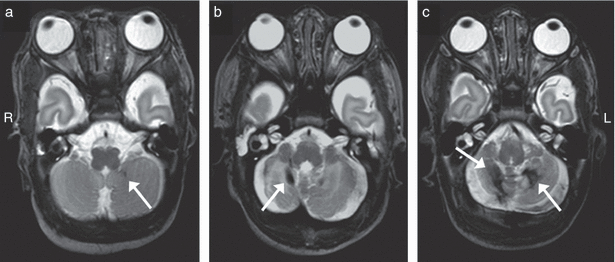
The number of cerebellar lesions was recorded on conventional MRI (Fig. 1). These lesions had a decreased signal intensity on the T2-weighted and an increased intensity on the T1-weighted sequence, suggestive of haemorrhage.

Examples of cerebellar lesions. In most cases of cerebellar lesions (n=17), (a) punctate lesions were found, (b) except in one child who showed signs of a larger hemorrhage in one hemisphere as well as a punctate lesion in the contralateral hemisphere, (c) and one child with a large bilateral cerebellar hemorrhages. The lesions are indicated by white arrows. R, right side; L, left side.
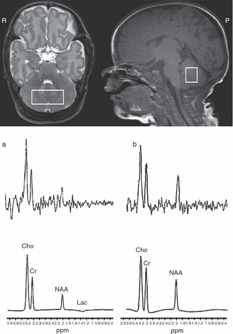
Cerebellar 1H-MRS was acquired with a short and/or long TE. Forty-four infants had spectra of good quality with both echo times, three only with short TE, and nine only with long TE. Two infants were excluded due to poor quality of both spectra and in 54 infants no spectroscopy was performed because of changes in the protocol and/or time constraints. A region of interest was positioned to be as large as possible (minimum size: 1.0cm3; Fig. 2). NAA (2.02ppm), Cho (3.25ppm), Creatine (Cr, 3.02ppm), and if present the lactate peak (Lac; 1.33ppm) were fitted by software of the Philips MR system. The peak area ratios were calculated of NAA/Cho (both TE), Lac/NAA, and Lac/Cho (TE 144ms).

Cerebellar proton magnetic resonance spectroscopy. Planning position of the region volume of interest in the cerebellum in the axial and sagittal plane, from which magnetic resonance spectroscopy data are obtained (top images: axial and sagittal view respectively). Examples of spectra acquired at long echo time (144ms), showing in (a) a low N-acetylaspartate/choline (NAA/Cho) ratio and (b) a normal NAA/Cho ratio. Top: crude spectra; bottom: fitted spectra. R, right side; P, posterior; Cr, creatine; Lac, lactate; ppm, parts per million.
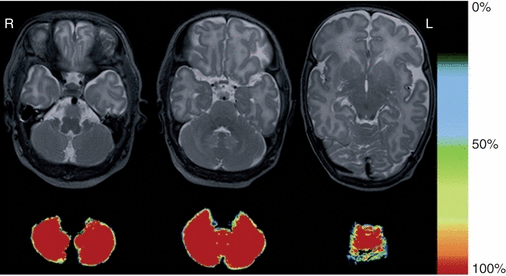
The cerebellar volume was measured with an in-house developed, fully automatic, probabilistic brain segmentation method.18 Voxels were classified by k-Nearest Neighbour classification. Voxel features were the signal intensities and the x-, y-, and z-coordinates on the axial T1- and T2-weighted images. To eliminate over classified gray matter tissue, an atlas was composed from co-registering manual cerebellum segmentations of a set of seven so-called training patients (i.e. patients without any MRI abnormalities from this cohort; Fig. 3). Validation of the method was performed using the manual segmentation of the seven training infants. Manual segmentation of the cerebellum was used as criterion standard. The Dice similarity index for the cerebellum was 0.93. The accurateness of all segmentations was visually confirmed independently by two researchers (BJMvK and MJNLB). Because of large haemorrhages in the cerebellum of two infants, the automatic segmentation was inadequate and manual editing was needed.

Example of cerebellar segmentation at different levels. The color bar indicates the probability (0–100%) that a voxel is cerebellar tissue. For example, the red voxels are 100% cerebellar tissue, whereas 25% of the volume of light blue voxels contribute to the cerebellar volume. R, right side; L, left side.
Neurodevelopmental outcome
At 2 years corrected age (mean 24.2mo [SD 0.6]), the children were assessed with the Bayley Scales of Infant and Toddler Development-III (BSITD-III) by a single developmental specialist (ICvH) who was blinded to the MRI findings.19 Only the cognitive and fine and gross motor subtests were used. The language subtest was not performed due to the limited time a child can concentrate on the different tasks during one session. Both scaled scores of these subtests as well as the cognitive and total motor composite scores were calculated corrected for preterm birth (mean in a normative population: 10 [SD 3] and 100 [SD 15] respectively).
Other factors included in the analysis
An intraventricular haemorrhage (IVH) and maternal education were included as possible confounders. An IVH was graded as normal/mild (no IVH or IVH I–II) or moderate/severe (IVH III–IV).20 Education was classified as low, middle, and high, according to the CBS (Statistics Netherlands, The Hague; http://www.cbs.nl/en-GB/menu/home/default.htm).
Data analyses and statistics
SPSS version 15.0 (SPSS Inc., Chicago, IL, USA) was utilized for all analyses. The distribution of the different variables were analysed for normality. Linear regression was used to assess the relation between cerebellar volume or cerebellar NAA/Cho ratio and neurodevelopmental outcome. The neonatal variables of gestational age, birthweight z-score, sex, white matter score and IVH, and maternal education were included in the analysis as possible confounders. As the absolute cerebellar volume showed a significant relation with postmenstrual age, the cerebellar volume/postmenstrual age ratio was used in the analyses. Only the cerebellar NAA/Cho ratio (n=53) was used in the multivariable analyses using a general linear model as the quality of this spectra was better than the spectra acquired with TE 35ms and in the univariable analyses the relation between the NAA/Cho ratio at TE 35ms and BSITD-III was not significant. A p<0.05 was considered statistically significant.
Results
MRI findings
In this cohort, 11 infants (9.8%) showed normal MRI, 87 (77.7%) mildly abnormal white matter, and 14 (12.5%) moderately abnormal white matter. No infants showed severe white matter injury. During the neonatal period, 38 infants were diagnosed with an IVH on cranial ultrasound examinations (Table I). Eight neonates developed post-haemorrhagic ventricular dilatation requiring intervention. Fifteen infants had small (<1cm) punctate haemorrhages in the cerebellum. Two infants had a larger haemorrhage (one bilateral, one unilateral) associated with volume loss in the affected cerebellar hemisphere (Fig. 1). Infants with a moderate/severe IVH were significantly more likely to show cerebellar lesions than infants with no/mild IVH (4/17 vs 7/95 respectively; p=0.039). The infant with a larger unilateral cerebellar haemorrhage had an IVH grade III and the infant with bilateral haemorrhages also had an IVH with an ipsilateral small venous infarction. No association was found between cerebellar lesions and white matter injury.
Cerebellar 1H-MRS
In this study, 56 infants were included with quantifiable MR spectra with TE 35 and/or TE 144ms (Fig. 2). In the two infants with larger cerebellar haemorrhages no cerebellar 1H-MRS could be performed due to the large amount of blood. There was a significant positive relation between gestational age and NAA/Cho, Lac/Cho and Lac/NAA (TE 144ms; p=0.010; p<0.001, and p<0.001 respectively). No other statistically significant relations were found between the metabolite ratios and gestational age, postmenstrual age and neither with white matter injury or cerebellar lesions.
Cerebellar volume
The cerebellar volume was normally distributed. The median (interquartile range) of the cerebellar volume in the total study cohort was 24.3 (3.7) ml. Gestational age showed a relation with cerebellar volume: infants with a lower gestational age tended to have a smaller cerebellum. However, this was mainly a result of the two infants with a low gestational age and a larger unilateral or bilateral cerebellar haemorrhage. The association between gestational age and cerebellar volume disappeared after exclusion of these two infants (gestational age 26wks 2d: cerebellar volume=14.1ml and gestational age 25wks: cerebellar volume=7.0ml respectively). No overall volume differences were found between the other fifteen infants with small cerebellar haemorrhages and those without cerebellar lesions (n=15: 24.2 [6.1] ml, n=95: 24.5 [3.6] ml respectively). Cerebellar volume was positively related with postmenstrual age at the time of the scan (p<0.001). No relation was demonstrated between cerebellar volume and the different metabolite ratios at TE 35 and 144ms, white matter injury or IVH grade. These associations did not change after exclusion of the two infants with larger cerebellar haemorrhages.
Neurodevelopmental outcome and relationship to MRI findings
The median (range) cognitive scaled score and composite score were 11 (4–19; 8.9% of infants scored ≤−1SD) and 105 (70–145; 8.9% of infants scored ≤−1SD) respectively. The median (range) fine motor scaled score was 13 (5–19; 2.7% of infants scored ≤−1SD) and gross motor scaled score 9 (6–15; 8.0% of infants scored ≤−1SD) resulting in a total motor composite score of 107 (73–142; 2.7% of infants scored ≤−1SD). Mann–Whitney U analysis showed no differences in the BSITD-III scores between infants with (n=17) and without (n=95) cerebellar lesions. Kruskal-Wallis analysis showed that cognitive scores were significantly different between children of a mother with a low, middle, or high educational level (p=0.001; median 9, 10, and 12 respectively).
Cerebellar volume/postmenstrual age and the cerebellar NAA/Cho ratio acquired with TE 144ms showed a significant positive relation with cognition (p=0.018 and p=0.007 respectively). These values remained significant after correction for multiple comparisons. A trend was seen between cerebellar volume/postmenstrual age and fine motor performance and between the cerebellar NAA/Cho ratio acquired at TE 144ms and gross motor skills (p=0.076 and p=0.095 respectively; Table SII, supporting material online).
With multivariable analyses using a general linear model and the neonatal variables gestational age, birthweight z-score, sex, white matter score, IVH, and maternal education as possible confounders, the positive relation between both cerebellar volume and NAA/Cho ratio, and cognitive scores remained statistically significant (cerebellar volume β=8.6, p=0.009, R2-model=0.23 and NAA/Cho ratio β=11.7, p=0.036, R2-model=0.28 respectively). In addition to the neonatal parameters and maternal education, no association could be demonstrated between cerebellar variables and motor function. Exclusion of the two infants with large cerebellar haemorrhages did not alter the results of the multivariable analyses.
Discussion
This study assessed the relation between cerebellar volume and cerebellar spectroscopy at term equivalent age of preterm infants and neurodevelopment at 24 months corrected age. An association could be demonstrated between cognitive scores on the BSITD-III and both cerebellar volume/postmenstrual age and cerebellar NAA/Cho ratio at TE 144ms after correction for neonatal and maternal confounders. Cerebellar volume showed a positive relation with postmenstrual age. The cerebellar variables did not reveal an association with motor scores.
Prediction of developmental abilities in preterm infants is challenging. This study implies that cerebellar volume and NAA/Cho ratio at term equivalent age are associated with developmental outcome at 2 years corrected age. This highlights their potential role as biomarkers for prognostic purposes. The cerebellar volume obtained in this cohort was in the range of data reported previously,21 although the volumes were larger compared to data reported by Shah et al.11 It was hypothesized this could be due to differences in segmentation techniques and the infants in our cohort were older at birth. Others have shown that cerebellar volume in preterm infants assessed at term equivalent age is reduced compared to full-term controls and this reduction is more common in the presence of supratentorial lesions.8,11,14 We were, however, unable to show an association with an IVH or white matter injury. This could be due to the smaller number of infants with a severe IVH or severe white matter injury, a higher mean gestational age in our cohort and earlier intervention of post-haemorrhagic ventricular dilatation.22 Our findings are in agreement with Limperopoulos et al.,8 who also found reduced cerebellar volumes in preterm infants with normal MRI findings. This does suggest that only visual analysis of the MRI is not sufficient to detect subtle cerebellar changes. Both direct destructive injury and indirect events like ischemia, infection, or deficits in trans-synaptic communication between cerebellum and cerebrum could cause cerebellar disease.9 This hypothesis is supported by one study showing reduced grey and white matter volumes on the contralateral cerebral hemisphere in infants with unilateral cerebellar injury.23
We were able to demonstrate a significant relation between cerebellar volume at term equivalent age and developmental outcome at 2 years corrected age in preterm infants after correction for white matter injury. This is even more remarkable since our cohort consisted of relatively healthy preterm born infants. Shah et al.11 found no relation with IVH and a weak association between cerebellar volume and outcome that disappeared after correction for white matter injury. We employed a different version of the Bayley Scales (BSITD-III vs Bayley Scales of Infant Development, Second Edition). Additionally, they used a semi-automatic segmentation method with manually outlining of the cerebellum and we used a fully automatic segmentation method, which may have led to more objective measurements. Other studies reporting cerebellar volumes assessed at an age range from 24 months into adolescence did reveal an association between cerebellar volume and cognition.12,13 Parker et al.24 reported a decline in cerebellar volume between adolescence and young adulthood in adolescents who were born preterm, and they hypothesized that altered cerebellar development in children born preterm persists until adolescence.
In our study, a reduced cerebellar volume was significantly associated with cognition; however, the association with motor performance did not reach statistical significance. Limperopoulos et al. assessed in both preterm infants and full-term infants the relation between cerebellar lesions and neurodevelopment.10,25 An impaired motor function was reported in infants with large cerebellar haemorrhages resulting in cerebellar hemispheric and/or vermis atrophy compared to preterm controls without cerebellar lesions or to term infants with small punctate cerebellar lesions respectively. In our study, fifteen infants displayed small punctate cerebellar lesions only recognized with MRI, one further infant had a large unilateral and another a large bilateral haemorrhage, both recognized with cranial ultrasound. The cognitive and total motor outcome of the first child was well within the normal range, the second child had composite scores of 70 and 88 respectively.
Using 1H-MRS cerebellar metabolism can be demonstrated in vivo. The NAA/Cho ratio increases in cerebrum and cerebellum of healthy preterm infants between 30 and 44 weeks’ gestation.17 NAA is considered to increase with neuronal development and to be synthesized by proliferating oligodendrocyte progenitor cells in the developing white matter.26 A positive relation was found between cerebellar NAA/Cho ratio and both gestational age and cognition, indicating differences in neuronal density and function that could be linked to maturation and neurodevelopment. A positive trend was found between cerebellar NAA/Cho ratio and postmenstrual age, however the increase in NAA/Cho in this time-frame was not significant. It was considered that we were unable to demonstrate this association due to the narrow time-frame in which the infants were scanned. The increase in cerebellar volume in this short period was more extensive and therefore we were able to show a positive relation between postmenstrual age and cerebellar volume. In this study, cerebellar volume and cerebellar NAA/Cho ratios at term equivalent age were used as ‘readout’ for the events in the neonatal period and it was assessed whether these features could be used as biomarkers for neurodevelopment. Further studies will be needed to determine the role of different potential risk factors for changes in cerebellar development.
In this population, there was no association between gestational age or birthweight and cognition. The mean gestational age of this cohort was almost 29 weeks (25–31wks) and only five infants had a birthweight less than 10th percentile. Only seven children scored ≤−1SD on the cognitive scale. Apparently, the population in our region is relatively affluent and benefits from adequate (prenatal) health care. This could contribute to the small number of infants with a poor score on the BSITD-III compared to studies performed in other countries. It has recently been suggested that developmental delay is underestimated on the BSITD-III.27 However, the mean of the Griffiths’ Developmental Assessment scale at 15 months corrected age in this study cohort was 102 with only three infants scoring below 85 (data not shown). None of the infants was diagnosed with cerebral palsy at the corrected age of 2 years, however one child was diagnosed to have mild cerebral palsy at the age of 3.5 years. This is in agreement with the decreasing incidence of cerebral palsy in infants admitted to our unit as recently presented by van Haastert et al.28
This study has several limitations that need to be addressed. The language scale was not assessed due to time constraints. Comparison with the Mental Developmental Index of the Bayley Scales of Infant Development, Second Edition is therefore not possible. However, the cognitive outcome of the BSITD-III is possibly a better reflection of the cognition in non-Caucasian infants, as language difficulties do not affect this score. Next, findings of this study are restricted to preterm infants, without comparison to healthy full-term controls. Finally, cerebellar volume was assessed at one time point. We hypothesize that sequential imaging soon after birth and at term equivalent age will provide additional information regarding cerebellar growth, allowing a more accurate prediction of adverse developmental outcome.
In conclusion, cerebellar volume and NAA/Cho ratio assessed in preterm infants at term equivalent age were associated with cognition at 2 years corrected age, but no relation was found with motor function. Our results suggest that the cerebellum should be taken into account, when trying to understand subsequent neurodevelopmental outcome of preterm infants.
Acknowledgements
The authors thank Dr Cuno S P M Uiterwaal for his support in the statistical analysis. The authors are grateful to the families who took part in the study, Niels Blanken and the other MR technicians of the MR institute for their expertise and enthusiastic help, and we thank our colleagues in the Neonatal Intensive Care Unit of the Wilhelmina’s Children’s Hospital in Utrecht, the Netherlands. This research was funded by The Netherlands Organization for Health Research and Development, project 94527022. There was no involvement of the funder in study design, data collection, data analysis, manuscript preparation, and/or publication decisions.


