CASE REPORT: Iridociliary melanoma with secondary lens luxation: distinctive findings in a long-horned cowfish (Lactoria cornuta)
Abstract
This report describes a long-horned cowfish, which was diagnosed with buphthalmia and lens sub-luxation in the right eye, conditions that progressed to complete anterior lens luxation and secondary keratoconus. Three months after the initial evaluation, a pigmented mass was observed protruding from the vitreous. An enucleation was performed under general anesthesia. Ocular histopathology revealed an iridociliary melanoma. Reports of intraocular melanomas are extremely rare in fish. To the authors’ knowledge, this is the first report of an iridociliary melanoma that led to buphthalmia, lens luxation, and keratoconus in a fish. Histological findings of lens luxation are also demonstrated. Due to the presence of a complex suspensory apparatus involving the teleost lens, this report speculates that lens luxation is a more devastating disease process in teleosts than in mammals.
Introduction
Cowfish are small box-like fish (also called boxfishes or trunkfishes) from the order Tetraodontiformes, family Ostraciidi.1 They are often found around coral reefs of the tropical Indo-Pacific Ocean.1–3 Closely related to the pufferfish, members of the order Tetraodontiformes have many peculiar features and one of them is body anatomy.4,5 They utilize their unique body structure for patterns of behavior and defensive mechanisms.5 Their scales form hexagonal bony plates that are fused into a solid, triangular carapace. Only the eyes, mouth, nostrils, fins, anus, and tail protrude and are exposed through this ‘armor’.1,2,5 Yet, they exhibit high stability and high maneuverability.6 Other protective features in species of the genus Lactoria include two pairs of parallel long horns, one located dorso-anteriorly and the other ventro-posteriorly, thus pointing forward and backward, respectively.1,4,5 Adults are shy and solitary7 whereas juveniles often form small groups.8 When threatened or harassed, they can exude a powerful toxin (ostracitoxin), which is lethal to organisms that are present in a confined space, including itself.5
Ocular disorders are common in fish, either as a primary disease process or secondary to a systemic disease.9 However, the literature on ocular diseases of fish is still scarce, especially regarding animals from the family Ostraciidi. Additionally, ocular tumors are infrequently described.10 Whether this is because they actually rarely occur or are not routinely diagnosed is unknown. Thus, the purpose of this report is to describe a long-horned cowfish that was diagnosed as having an iridociliary melanoma, which led to buphthalmia, secondary lens luxation, and keratoconus. Additionally, findings pertaining to lens luxation in fish are discussed.
Case report
A 3-year-old male cowfish with a history of a progressive enlargement and protrusion of the right eye (OD) was evaluated by the Colorado State University Ophthalmology Service. The examination was not detailed as it took place at the Landry’s Downtown Aquarium in Denver, where equipment was limited. The fish could swim normally and navigated well around obstacles in the fish tank. After gross observation, it was placed into a 2.5-gallon hospital tank and sedated with 50 ppm of tricaine methanesulfonate (MS222 – Finquel; Argent Chemical Laboratories, Redmond, WA), which was placed in buffered water. Throughout the ocular examination, the fish was constantly moved in and out of the water, for periods not longer than the time the examiner (EG) could hold his own breath. Ophthalmic examination was performed with a Finoff transilluminator (Welch Allyn, Skaneateles Falls, NY, USA) and slit-lamp biomicroscope (SL-15; Kowa, Torrance, CA, USA). Intraocular pressures were taken with a TonoVet® (on the P setting) (Jorgensen Laboratories, Loveland, CO, USA). Additionally, both indirect (20D lens; Volk Optical, Mentor, OH, USA) and direct ophthalmoscopy (Panoptic, Welch Allyn, Skaneateles Falls, NY, USA) were performed. The left eye (OS) was normal (Fig. 1b). Moderate buphthalmos was present OD (Fig. 1a). The cornea was normal and a prominent annular ligament was present in both eyes (OU), surrounding the peripheral iris and extending into the cornea (Fig. 2). Biomicroscopic examination showed no flare. There was a punctate, central, anterior capsular cataract and a slight anterior lens sub-luxation (Fig. 2). Intraocular pressures were normal at 5 OD and 6 OS. Limited fundic examination was within normal limits OU. Despite the fact that air bubbles were not detected in the ocular examination, an ocular ultrasound was recommended to rule out the presence of retrobulbar gas bubbles. The difficulty in transporting the fish to the university precluded this examination. This recommendation was based on the fact that other fish in the same facility were reported to have had ocular gas bubble disease. However, there was no history of gas bubble disease in any fish in the tank where the cowfish was housed. Ocular ultrasound measurements would also have been ideal for the documentation and monitoring of buphthalmos in this case, as would caliper measurements of the corneal diameter; this was not performed because of the lack of equipment and due to the fact that the right eye clearly appeared larger than the left. No treatment was prescribed. Close monitoring for progression of the buphthalmia was recommended and a 6-month recheck examination was scheduled.
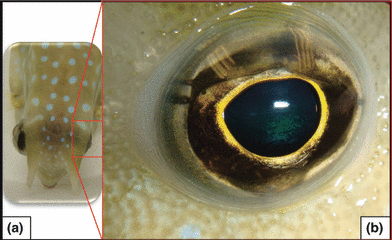
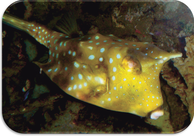
(a) Appearance of the cowfish upon initial presentation. Note the presence of moderate buphthalmos OD. (b) The left eye was normal.
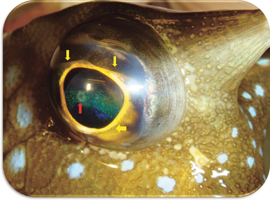
Ocular findings upon initial presentation OD. Moderate buphthalmos was present as well as a central anterior capsular cataract (red arrow). Note the presence of a pronounced annular ligament (yellow arrows), normally found in this species. The skin color change in contrast to Fig. 1 can be found in some fish during stress.
During the following 3 months, the aquarists noticed buoyancy changes and worsening of the eye condition in the cowfish. This consisted of progression of the buphthalmia and gradual cataract formation along with displacement of the lens into the anterior chamber. At that time, a recheck examination was carried out. Visual function of the eye was difficult to assess due to the buoyancy problems. The fish was sedated using the same protocol as in the first ocular evaluation. Examination findings OD included severe buphthalmia, focal corneal edema, keratoconus, mild flare, complete anterior lens luxation, and immature cataract formation (Fig. 3). The intraocular pressures were 2 mmHg OD and 7mmHg OS. Additionally, a large, dark-brown mass was seen protruding toward the pupil from the ventral portion of the posterior segment, which precluded examination of the fundus. Due to the poor prognosis for vision and profound ocular changes, an enucleation was recommended. As the fish was already under sedation, the decision was made to proceed with enucleation immediately. General anesthesia was induced by placing the fish in a 10-gallon tank of water and bringing the level of tricaine methanesulfonate (MS222) to 100 ppm. The stage of anesthesia was maintained by means of a closed anesthetic system. Intraoral intubation was used to provide anesthetic flow through the gills by ram ventilation, thereby allowing the fish to be surfaced out of the water for the surgical procedure. A sub-conjunctival technique was used for the eye removal, as previously described.11 No gas bubbles were detected in the retrobulbar space peri-operatively. The orbital space was left open, due to the inability to suture the remaining defect. After the removal of the globe, the orbital space was inspected and no signs of invasion of the tumor could be detected. The orbit was flushed with dilute betadine prior to re-immersion in the water. The animal recovered uneventfully and immediately regained its normal buoyancy. Postoperative medical therapy consisted of a constant bath of oxytetracycline (50 mg/kg Q24h for 3 days) because an injectable antibiotic could not be used due to the body armor in this fish. The fish remained in a hospital tank for 2 weeks, isolated from any other fish due to the potential for ostracitoxin release.
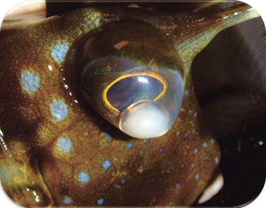
Ocular examination 3 months after initial evaluation. There is worsening of the buphthalmos OD, complete anterior lens luxation and immature cataract, focal corneal edema, as well as secondary keratoconus.
The enucleated eye was fixed in 10% formalin and processed routinely for histological examination. Gross examination of the sectioned eye revealed an anteriorly luxated lens and the presence of a dark mass at the level of the posterior chamber. Sections were stained with hematoxylin and eosin. Histologically, the mass seemed to circumferentially arise from the ciliary body. On low power, the pronounced annular ligament surrounding the anterior peripheral iris could be easily visualized (Fig. 4a). A locally extensive, moderately defined, nodular mass was partially effacing the ciliary body with extension into the retractor lentis muscle as well as into the base of the iris (Fig. 4b). The mass was composed of large melanocytic cells with abundant intracytoplasmic melanin pigment obscuring nuclear detail (Fig. 4c). Diffuse pathologic retinal and choroidal detachment were also noted. No histological signs of gas bubble disease or intraocular hemorrhage were present.
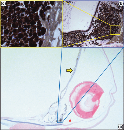
(a) Sub-gross image of the ventral part of the sectioned eye (OD) demonstrating the lens luxation and presence of diffuse retinochoroidal detachment (arrow). The annular ligament can be seen in the anterior chamber, cranial to the iridocorneal angle at the level of the iridocorneal angle (red star). (b) Section of the uveal melanoma showing partial invasion of the iris root (yellow square) (H&E, ×100). (c) Large round melanocytic cells with abundant intracytoplasmic melanin pigment obscuring nuclear detail (H&E, ×400).
Approximately 9 months after surgery, the fish was doing well and the skin was almost closing the orbital space (Fig. 5). Up to the time this case report was written, there was no evidence of tumor growth in the orbit or elsewhere.
The appearance of the surgical site 8 months postoperatively. The peri-orbital skin is almost closing the remaining wound, by secondary intention.
Discussion
The etiology of most spontaneous neoplasms in fish has not been elucidated.10 The majority of the reported neoplasms in fish involve discrete, focal, benign growths that generally behave less aggressively than neoplasms in mammals.12 These were true of the ocular neoplasm in the fish of this report. Histologically, tumors in fish do not differ significantly from their mammalian counterparts. This allows for the classification of fish tumors on much the same criteria as mammalian tumors.13 For example, the major criteria of malignancy in animal melanomas are their mitotic indices and their location.14 Mitotic figures were not apparent in this mass suggesting that it was a well-differentiated, likely benign, melanoma. However, the exact biological behavior of ocular melanomas in fish is not known, so histological grade alone cannot be used as a criterion of malignancy for this case. Although we were unable to determine if the melanoma in this fish was primary or secondary, the fact that the fish was alive and doing well 9 months postoperatively suggests that the tumor was primary rather than metastatic. The solitary nature of this mass would also support this hypothesis. This agrees with the findings previously reported that fish tumors rarely exhibit metastatic behavior, whether or not phenotypic characteristics of malignancy are present.10
Spontaneous ocular tumors that have been reported in fish include fibroma, smooth muscle carcinoma and neurofibroma,15 orbital myxosarcoma,16 retinoblastoma,17 ciliary body meduloepithelioma,18 retrobulbar hemangioma,19 and lymphosarcoma.20 According to a study of animal models of uveal melanomas, these tumors can be spontaneously found in fish as well as dogs, cats, horses, rats, mice, and birds.21 However, we were able to find only one report of an intraocular melanoma in a fish.22 Primary pigmented intraocular tumors have also been found in cattle, sheep, and rabbits.23
Lens luxation is an uncommon finding in fish. One study described lens sub-luxation, luxation, and buphthalmos in rock-fish; all these abnormalities were secondary to the presence of multiple intraocular gas bubbles.24 Distinctive gross and histological features of ocular gas bubble disease have been described.24,25 In the present case, a complete ocular examination and histological evaluation ruled out the presence of intraocular gas bubble disease.
The mass that was detected ophthalmoscopically in the ventral aspect of the eye could have been occluding drainage of aqueous humor. In teleosts, the production of aqueous humor occurs at the dorsal ciliary epithelium and drains through the ventral portion of the eye, into the ventral canalicular network and vitreo-retinal vessels.26 The location of the ventral mass may have significantly impaired aqueous outflow in this case. This could have contributed to the pathogenesis of buphthalmia, despite the fact that the intraocular pressure was normal to low at the time of enucleation. A likely contributing factor to the apparent progression of the buphthalmia was lens luxation. This led to the formation of keratoconus, which was exacerbated by the large size and weight of the piscine lens.
Buphthalmia is not an uncommon ocular finding in fish;9,10,22,24,25 however, lens luxation is a rare clinical sign. A potential reason for this may be related to the anatomy of the suspensory apparatus of the teleost lens, which is more complex than that of mammals. An electron microscopy (EM) study in 11 species of bony fish revealed that all species had five suspensory ligaments surrounding the equatorial plane of the lens. These ligaments prevent unwanted lateral and rotational lenticular movements.27 Furthermore, EM detected the presence of two additional posterior ligaments in one fish species (dorsal and ventral posterior ligaments). These ligaments were attached from the posterior pole of the lens to the falciform ligament.27 The presence of multiple lens ligaments may indicate that more force is required to cause displacement of the lens. Additionally, a more severe intraocular disease process is likely present in eyes of fish with sub-luxated or luxated lenses. Moreover, the consequences of lens luxation seem far more devastating in teleosts than in mammals. This would help explain clinical and histological findings in this report, in that the complete anterior lens luxation was accompanied by diffuse retinal and choroidal detachment.
It is likely that the incipient central cataract in the cowfish of this report was due to a corneal–lens contact secondary to the lens sub-luxation. The authors speculate that, as the tumor growth progressed in the posterior chamber (as noted on histopathology to be ultimately affecting the entire equator of the globe), more ligaments were disrupted subsequently leading to a complete lens luxation which, in turn, caused the retinochoroidal detachment. Such theory would be particularly true, especially if the presence of the dorsal and ventral posterior lens ligaments could be demonstrated in the cowfish as well; however, this have not been evaluated yet. To the authors’ knowledge, this is the first report of a spontaneous iridociliary melanoma leading to buphthalmia, anterior lens luxation, and keratoconus in a fish.


