Indium-111 labeled vitamin B12 imaging of a ciliary adenoma with concurrent grade 2 soft tissue sarcoma of the leg in a Labrador Retriever
Abstract
An 11-year-old, male castrated, Labrador Retriever was evaluated for the presence of rapidly growing concurrent leg and intraocular masses. Metastasis was noted in the chest at time of initial presentation. Indium-111 labeled vitamin B12 imaging was performed, and there was significant uptake by both primary tumors and the lung metastases. Enucleation and amputation were performed for palliative relief. The leg mass was a grade 2 soft tissue sarcoma and the ocular mass a ciliary adenoma. The dog remained symptom-free for approximately 10 weeks before developing signs of respiratory distress. He was euthanized 12 weeks after initial presentation, and there was diffuse infiltration of the lungs with metastatic sarcoma. Indium-111 labeled vitamin B12 imaging identified a ciliary adenoma in this case and may provide a useful differentiation technique for evaluation of intraocular and retrobulbar masses if it can be demonstrated that there is differential uptake between inflammatory or infectious conditions and neoplasia.
INTRODUCTION
Ciliary body adenomas are common primary ocular neoplasms in dogs. They occur most often in middle-aged dogs, and an over-representation of Labrador Retrievers has been noted.1 Adenomas tend to be slow-growing tumors with a low potential for metastasis.1 Soft tissue sarcomas tend to be rapidly growing, locally invasive neoplasms with a low incidence of metastasis. Aggressive surgical treatment is recommended to prevent local recurrence.2 This report describes the case of a dog with a ciliary adenoma and a primary appendicular soft tissue sarcoma.
The purpose of this study is to describe the changes in the biological behavior of a benign neoplasm upon development of an independent malignant neoplasm. We also wish to introduce a unique noninvasive imaging modality, indium-111 labeled vitamin B12 single photon emission computed tomography (SPECT) scanning. This technique appears to have great potential in both human and veterinary oncoscintigraphy due to its ability to identify small neoplastic masses and its potential to differentiate between neoplastic and inflammatory mass lesions.
CASE REPORT
An 11-year-old, male castrated, Labrador Retriever was presented to Colorado State University Veterinary Teaching Hospital (CSUVTH) for evaluation of an ocular mass and development of a fast-growing mass on the carpus. The dog was current on vaccinations and had no history of travel to regions where systemic mycoses are endemic. The mass in the right eye had been noted by the owner 4 years before presentation. The owners elected to monitor the mass for progression of disease because it had not seemed to change in character or size. The mass remained unchanged at repeat examinations for 4 years until 2 weeks prior to presentation at the referring veterinarian (RDVM). At that time, the dog had developed a second mass on the palmar aspect of the left carpus, which appeared to be growing rapidly. The owners noted a sudden increase in the size of the ocular mass concurrent with development of the carpal mass.
The RDVM noted an intraocular mass in the right eye and a 2-cm mass on the palmar aspect of the left carpus on examination. The soft tissue mass on the palmar aspect of the carpus was determined on radiographs not to be invading bone. Fine needle aspirate of the mass was nondiagnostic. A 3-cm mass and two smaller soft tissue masses were visible in the right cranial lung field on chest radiographs taken by the RDVM. In a patient with a known malignancy and a possible second malignancy, the most likely rule-out is metastatic disease. Given the dog's history and geographic location, fungal disease was considered less likely. No attempt was made to collect a sample from the lung masses for histological examination. The dog presented to CSUVTH 1 week after examination at the RDVM for further evaluation of the masses in the eye and on the leg.
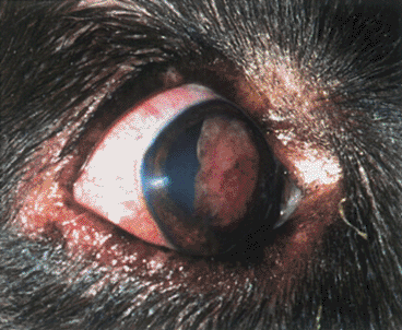
Physical examination showed that there was an 8-mm nonpigmented, exophytic mass protruding through the pupil of the right eye into the anterior chamber (Fig. 1). It appeared to arise from the nasal portion of the ciliary body. Adhesions between the iris and the mass were noted, as was mild anterior synechiae where the iris had been displaced by the pressure from the mass. Approximately 60% of the pupillary aperture was occluded by the mass. The medial aspect of the lens was displaced caudally. No abnormalities were identified in the conjunctiva, cornea, sclera, aqueous humor or retina. Aqueous flare was not identified by biomicroscopy. Intraocular pressures were 17 and 16 mmHg OD and OS. A bulging, firm, nonmovable soft tissue mass (3 × 3 × 2 cm) was identified on the palmar aspect of the carpus just distal to the carpal pad. No other abnormalities were identified on physical examination.

Photograph of the right eye of an 11-year-old, castrated male Labrador Retriever with an intraocular adenoma. Note the pink mass protruding from behind the iris and spreading across the pupil.
An elevation in serum alkaline phosphatase (217 IU/L, reference range 18–160 IU/L) was the only abnormality on serum biochemical profile. There was mild enlargement of the adrenal glands on abdominal ultrasound. The ocular mass was well visualized on ultrasound of the eye. It was displacing the lens caudally and the medial aspect of the iris rostrally. The base of the mass was confirmed to be located in the nasal portion of the ciliary body.
Fine needle aspirate of the carpal mass was repeated and was inconclusive. Incisional biopsy of the carpal mass was performed for staging purposes; the histopathologic findings were consistent with a soft tissue sarcoma. Further differentiation and grading of the mass were not possible. An indium-111 labeled vitamin B12 SPECT scan was performed as part of a funded research project, for inclusion in which the dog had to have a known malignancy. The purpose of the project was to study the biological activity of indium-111 labeled vitamin B12 in a variety of tumors.
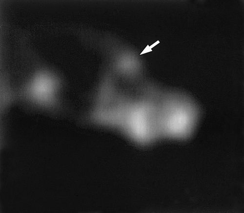
Indium-111 labeled vitamin B12 (37 MBq) was injected intravenously and general inhalation anesthesia was induced and maintained for 2 hours. SPECT images were acquired of the head and neck, as well as the thorax (GE Millennium VG camera). Focal increased uptake of the radiopharmaceutical was noted in the right eye, as well as adjacent to the left carpus and three areas of uptake were noted in the cranial right lung lobe (Fig. 2). The areas of uptake in the lung tissue were consistent with the location of nodules on chest radiographs.

Sagittal plane SPECT image of the right side of the skull showing increased indium-111 Vitamin B12 uptake at the level of the right orbit, compatible with the ciliary adenoma (arrow). Uptake of the radiopharmaceutical in the areas of the parotid and sublingual salivary glands and the most rostral aspect of the nose is normal.
The size of the carpal mass and degree of surface involvement precluded complete surgical excision. Although a combination of surgery and radiation therapy was discussed, reconstructive surgery with full-thickness grafts would have been necessary to close the defect. Given the aggressive and rapid growth of the mass, it was considered unlikely that the graft would be healed in time for radiation to retard recurrence of the mass. Preoperative radiation was considered inappropriate given the speed of tumor growth and level of patient discomfort. Given the presence of presumptive pulmonary metastasis and pain, palliative amputation was recommended for the carpal mass with enucleation due to impending iridocorneal angle obstruction in the eye. The owner was not ready to proceed with surgery at this time. The dog was released on morphine extended-release tablets for analgesia (1 mg/kg PO, BID PRN).
The dog represented 2 weeks later for enucleation and amputation. The ciliary body mass had increased to 12-mm in size and occluded the outflow tract. Intraocular pressures were not measured prior to enucleation. The carpal mass had doubled in size and was causing significant discomfort in spite of analgesia. Transconjunctival enucleation and amputation were uneventful.
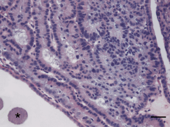
The leg mass was a subcutaneous flattened nodule that was easily dissected away from the underlying tendons, periosteum, and was distinct from the joint capsule. It was composed of spindloid cells forming sheets and palisading cords with multifocal necrosis. Cells had anisokaryosis with megalokaryosis and 0–2 mitotic figures per ×400 field. The mass was interpreted as a grade 2 soft tissue sarcoma. The ocular mass was a discrete nodule arising from the ciliary body. It was composed of epithelioid cells forming tightly packed cords and tubular structures aligning on moderate fibrovascular stroma. The epithelioid cells had modest cytoplasm that was rarely pigmented. They had central to paracentral round nuclei with mild anisokaryosis and rare scattered mitoses. The ocular mass histologically was consistent with a ciliary adenoma (Fig. 3).

Eye; ciliary adenoma. There is a multilobular mass composed of tightly packed, epithelial lined tubular structures supported by modest fibrovascular stroma. Asteroid hyalosis is within the adjacent vitreous (asterisk). H bar = 50 µm.
The dog recovered well from surgery and was discharged 3 days after surgery. The dog quickly returned to normal activity level. Recheck chest radiographs were taken 6 weeks postoperatively after a brief episode of exercise intolerance on a trip at high altitude and compared with previous radiographs. Multiple lung masses were noted, the largest of which appeared to be 5 cm in diameter. The owners elected to continue to monitor the dog for respiratory signs and limit his exertion at high altitude. Twelve weeks post presentation, the dog was euthanatized for progressive respiratory distress.
On necropsy, multifocal, 0.5–5 cm umbilicated masses were randomly scattered throughout the lungs. The masses had multifocal necrosis with hemorrhage and were composed of densely packed fusiform cells. These histological findings were similar to those of the carpal mass suggesting pulmonary metastases of the carpal sarcoma. There was no evidence of metastasis from the ciliary adenoma.
DISCUSSION
Ciliary body neoplasms are the second most common primary intraocular neoplasm in the dog. Increased incidence has been described in a number of breeds, including German Shepherds, Cocker Spaniels, Golden Retrievers and Labrador Retrievers.1,3 Typically, ciliary adenomas are pigmented or nonpigmented masses arising from the retroiridal region, whereas adenocarcinomas tend to be pink masses located in or extending into the anterior chamber and attaching to the iris root.4 Despite the typical growth patterns, differentiation between ciliary body adenomas and adenocarcinomas can be challenging. Anterior chamber fluid analysis is thought to have poor diagnostic yield in cases in which free cells are not observed during ophthalmic examination and, for this reason, it was not performed in this dog.3 Reported abnormalities that occur secondary to ciliary adenomas and adenocarcinomas include glaucoma, preiridal fibrovascular membranes, retinal separation, asteroid hyalosis, and intraocular hemorrhage.1,3 Both ciliary adenomas and adenocarcinomas tend to be slow-growing masses, and adenocarcinomas metastasize uncommonly and late in the disease process.1 The authors are not aware of any association in the literature between ciliary body neoplasms and primary soft tissue sarcomas.
Soft tissue sarcomas are locally aggressive tumors of mesenchymal origin. They tend to be pseudoencapsulated, separating easily from the surrounding tissues and leaving behind microscopic disease. Recurrence is common, especially when aggressive surgical excision is not pursued. Metastasis occurs in 10–30% of cases depending on grade. Soft tissue sarcomas respond poorly to chemotherapy although adjunctive radiation therapy may offer benefit in cases of incomplete resection. Given their growth characteristics and overlapping immunohistochemical marker uptake, tissue of origin can be very difficult to determine. Aggressive surgical excision with > 3 cm margins in all directions is the mainstay of treatment.2
Radiolabeled vitamin B12 whole-body scanning is a new diagnostic tool currently being evaluated in both veterinary and human oncology. Vitamin B12 is utilized by mitochondria in metabolically active cells and so there is uptake by all cells. The hypermetabolic rate of neoplastic cells leads to significantly upregulated uptake and usage. Whole body scans of animals injected with indium-111 labeled vitamin B12 will have foci of radioactivity at sites of neoplastic activity. Currently, B12 scanning has been used to identify sarcomas, melanomas, hematopoeitic tumors, carcinomas and metastases of similar neoplasms.5
The marked uptake of B12 demonstrated by the ocular mass in this case was unexpected given its small size and may have clinical application. Previously, differentiation of neoplastic and non-neoplastic processes in cases involving mass lesions hidden by nontransparent corneas, mass lesions in the posterior aspect of the globe and retrobulbar mass lesions has been diagnostically challenging. Prognosis, diagnostic plans, and therapeutic plans may be significantly affected by identification of a mass as neoplastic, inflammatory or infectious. If it can be demonstrated that indium B12 scanning is able to differentiate between inflammatory and neoplastic processes, it may become an important adjunctive diagnostic tool. There is, however, a concern that the high sensitivity of this test will not allow differentiation between malignant and benign neoplasms. Further study is currently underway to evaluate this concern.
Another interesting aspect of this case is the sudden change in biological behavior of the intraocular mass after the development of a second primary neoplasm. Multiple studies have demonstrated immunosuppressive effects of sarcomas, including effects on a variety of cytokines, serum immune complexes and alterations in signal transduction and apoptosis of T cells and natural killer (NK) cells.6,7 Reciprocal changes in levels of interferon-γ and tumor necrosis factor α with interleukin (IL)-6 may lead to downregulation of antitumor cytokines.8 Late stages of tumor growth may lead to generalized T-cell dysfunction, or decreased antineoplastic activity specifically, as a result of decreased T-cell responsiveness vs. decreased antigen presentation activity.8 Given studies showing decreasing antitumor T-cell responsiveness with increasing tumor burden, development of the soft tissue sarcoma may have decompensated this dog's ability to suppress growth of the ciliary adenoma, which had remained quiescent for several years, by affecting one or more immunomodulatory pathways.


