The Management of Anal Fissure: ACPGBI Position Statement
Introduction
Anal fissure is a linear ulcer in the squamous epithelium of the anal canal located just distal to the dentate line. It is usually located in the posterior midline but occurs anteriorly in a fifth or more or patients. It typically causes pain during defaecation which may last for 1–2 h afterwards [1]. The most consistent finding on physical examination is spasm of the anal canal due to hypertonia of the internal anal sphincter. It has been postulated that this may either be due to or be the result of ischaemia [2]. All management options aim to reduce anal tone. They include general measures such as dietary fibre supplements, adequate fluid intake, and topical analgesics, medical treatments such as glyceryl trinitrate(GTN) ointment, calcium channel blockers (eg diltiazem cream) and botulinum toxin. Surgery includes lateral sphincterotomy, advancement flap procedures and fissurectomy. This position statement recommends evidence-based practice associated with these treatment options.
Methodology
Searches of the Cochrane Database, Pub Med MEDLINE and EM-BASE were performed using keywords relevant to each section of this position statement. They were limited to English language articles. Additional publications were retrieved from the references cited in articles identified from the primary search of the literature. All evidence was classified according to an accepted hierarchy of evidence and recommendations graded A–C on the basis of the level of associated evidence and/or noted as Good Practice and/or part of NICE/SIGN recommendation or Rapid Technology Appraisal (Table 1).
| Level of evidence | Grade of evidence | ||
|---|---|---|---|
| I | Evidence obtained from a single randomized controlled trial or from a systematic review or meta-analysis of randomized controlled trials | A | Evidence of type I or consistent findings from multiple studies of type IIa, IIb or III |
| IIa | Evidence obtained from at least one well-designed controlled study without randomization | B | Evidience of type IIa, IIb or III and generally consistent findings |
| IIb | Evidence obtained from at least one other well-designed quasi-experimental study | C | Evidence of type IIa IIb or III but inconsistent findings |
| III | Evidence obtained from well-designed non-experimental descriptive studies, such as comparative studies, correlation studies and case studies | D | Little or no systematic evidence |
| IV | Evidence obtained from expert committee reports or opinions and/or clinical experiences of respected authorities, case reports | GP | Recommended good practice based on the clinical experience of the expert group and other professionals* |
- Adapted from Eccles M, Mason J1 and NHS Executive. Clinical Guidelines: Using Clinical Guidelines to improve patient care within the NHS. London: 1996.
- *Previous experience and the literature in this area suggests that given the relative lack of evidence for many health care procedures, expert opinion and professional consensus are likely to be an important part of this process.
Aetiology
Fissures associated with internal anal sphincter hypertonia are probably ischaemic in nature (Level IIb, Grade B).
The aetiology of the typical fissure is not clear. Trauma from passing a large or hard stool is a common initiator [3], but many traumatic fissures heal and others do not. Resting anal pressure is higher in patients with an anal fissure [4]. Ambulatory manometry has shown persisting high anal resting tone interpreted as due to hypertonia of the internal anal sphincter with poor spontaneous relaxation in patients with a chronic fissure [5]. In a study examining the influence of ischaemia, it was found that the higher the sphincter pressure, the lower the anodermal bloodflow. This was most pronounced posteriorly where most fissures occur and was followed by a return of normal bloodflow after sphincterotomy [6]. It was postulated that the pain caused by anal fissure was because of ischaemic ulceration, perhaps due to sphincter spasm reducing the blood flow in vessels penetrating the internal anal sphincter [7] Although this has never been proved with certainty, it remains the most commonly supported theory.
The aetiology of fissure formation in females who have had a vaginal delivery whether complicated or assisted or in patients with a rectocele may be different. Scar formation may be associated with ischaemia and poor healing, but in addition resting sphincter pressure is low [8].
If the fissure is not situated in the midline or if it is multiple or painless, the association with other pathologies should be considered. These include Crohn’s disease, ulcerative colitis, HIV and associated secondary infections, tuberculosis, syphilis, and neoplasia including leukaemia or carcinoma.
Diagnosis
Diagnosis is made from the history and examination (Level IV, Grade GP).
Anal Fissure is common. It occurs mostly between the second and fourth decades of life with an equal distribution between men and women with a lifetime incidence of 11.1% [9]. The diagnosis is usually suspected on the history alone. The symptoms include anal pain during and after defaecation which may last several hours. Bleeding is common and tends to be bright red and is often seen on the toilet paper. The patient may complain of periodic episodes indicating chronicity.
In most patients physical examination by inspection on gentle traction of the buttocks will show the fissure. A sentinel tag at the distal pole of the fissure, a hypertrophied anal papilla at its proximal extent and the appearance of the circular fibres of the internal sphincter muscle in its base indicate that the fissure is chronic. The majority of fissures are in the midline posteriorly, 8% occur both posteriorly and anteriorly [10,11].
Digital rectal examination or endoscopy should be not be carried out in most patients at the time of the initial consultation owing to the likelihood of causing pain. If the fissure is seen on inspection then treatment can be initiated. If it is not apparent then an examination under anaesthetic should be advised to make the diagnosis and also to exclude anorectal sepsis which is associated with fissure or may be present in its own right.
The presence of diseases associated with fissure as listed above should be suspected if the patient reports general symptoms of weight loss or weakness or abdominal symptoms referable to the gastrointestinal track. A family history of inflammatory bowel disease or a morphologically unusual fissure away from the midline should be regarded with suspicion. At the time of the initial consultation it is possible to pass a paediatric proctoscope to determine whether the rectal mucosa is inflamed. This is usually pain free.
Treatment
Conservative
Conservative treatment will heal a proportion of acute anal fissures (Level I, Grade A).
Conservative treatment includes increasing liquid intake, stool softeners and topical analgesics. In a prospective trial dietary bran supplements (5 g three times a day) and warm sitz baths were superior with fewer recurrences than topically applied local anaesthetic or hydrocortisone cream [12]. Recurrence rates were reduced from 68–16% at 1 year following continued conservative management [13].
Medical Therapies
Relaxation of internal anal sphincter tone is achieved by the reduction of intracellular calcium in the smooth muscle cells thereby reducing muscle tone. This can be achieved by nitric oxide donation using GTN or by direct intracellular calcium depletion using calcium channel blockers (diltiazem or nifedipine). Irreversible acetylcholine neuromuscular blockade using botulinum toxin also reduces resting tone.
Glyceryl trinitrate
Topical GTN heals anal fissure better than placebo, irrespective of dose but is associated with headache in around 25% of patients (Level 1, Grade A).
Glycerine trinitrate is a vasodilator and causes relaxation of smooth muscle. When applied topically to the anus two to three times daily, the internal sphincter is relaxed and the fissure heals significantly better than placebo. Healing occurs in only 60% of patients in the short-term, with recurrence rates of around one-third over 18 months. Patients with recurrence may respond to further GTN, but a proportion will require sphincterotomy [14]. The dose of GTN (0.2% or 0.4%) does not influence the efficacy but increases the incidence of side effects, particularly headache which occurs in about a quarter of patients [15,16]. Commercially available GTN ointment (‘Rectogesic’ 0.4%) is often more easily available than 0.2% GTN ointment, [17] and [18]. Meta-analysis has shown that topical GTN twice daily is effective although the placebo response is around 30% (Fig. 1).

Topical glyceryl trinitrate: fissure healing. (Courtesy of Professor Nelson[19].)
Calcium channel blockers
Topical diltiazem has similar efficacy to GTN but with fewer side effects and should be recommended as first line treatment in the management of anal fissure. Patients should be warned about pruritis ani (Level 1, Grade A).
Calcium channel blockers, such as diltiazem and nifedipine improve fissure healing by inhibiting calcium ion entry through voltage-sensitive areas of vascular smooth muscle causing muscle relaxation and vascular dilatation. In a randomized comparison of topical diltiazem 2% with topical GTN 0.2% applied twice daily there was no difference in healing rates [20]. Diltiazem is, however, rarely associated with headache, and only occasionally associated with pruritis ani [21]. Diltiazem 2% and GTN 0.2% are unlicensed so individual drug and therapeutics hospital guidelines will dictate availability. Oral nifedipine has been shown to give good healing rates, but is associated with greater systemic side effects than the topical preparation [22]; a similar finding was seen with oral diltiazem [23].
Botulinum toxin
Botulinum toxin is associated with a similar rate of healing of anal fissure as GTN but is more expensive. It may be used for a fissure resistant to topical GTN or diltiazem. The technique, dose and site of injection do not affect the rate of healing (Level 1, Grade A).
Contraction of the internal sphincter is mediated by sympathetic neuronal activation. Botulinum toxin irreversibly binds to presynaptic nerve terminals preventing acetylcholine release and thereby stopping neural transmission. Botulinum toxin thus induces a relative hypotonia, reducing resting anal canal pressure. This effect lasts for 2–3 months until acetylcholine reaccumulates in the nerve terminals [24].
There are many different published techniques for injecting botulinum toxin. The dose has varied from 10–100 u (mean 23 u based on 20 trials) with a mean healing rate of 75.6% and a range of 44–100% irrespective of the technique. Most frequently the injection has been carried out on either side of the fissure into the internal sphincter. Botulinum toxin has been shown to be as effective than GTN in the primary healing of fissure [25,26], and appears to be as effective for a fissure resistant to GTN [27]. There is no difference in healing rates between the different commercially available products including Botox or Dysport [28]. Botox costs approximately £200/100 u. Grouping of patients on the same operating list and follow-up at the same outpatient clinic improves cost effectiveness, as one vial can be used to treat four patients.
Suggested recommendation for medical management
An acute fissure should initially be treated with increased intake of oral fluids, fibre, stool softeners and analgesics combined with the local application of diltiazem 2% cream. A chronic fissure should be managed with diltiazem 2% topically twice daily for 6–8 weeks where such a prolonged treatment schedule is clinically more acceptable. Failure or recurrence after the application of a topical preparation should be treated with botulinum toxin 20–25 u in two divided doses injected into the internal sphincter on either side of the fissure. Failed medical management or recurrence warrants anorectal physiological testing in females, or in males having had previous anal surgery.
Surgery
The aim of surgery is to reduce resting anal canal tone due to the internal anal sphincter thereby increasing blood supply to the anoderm to improve healing. Surgical options include lateral sphincterotomy, fissurectomy and advancement flap procedures. In the past anal dilatation, posterior sphincterotomy have been used, but there is little evidence to support their continued use. In patients with a low resting pressure an anal advancement flap is a logical option.
Lateral sphincterotomy
Lateral sphincterotomy heals more anal fissures with lower recurrence than medical management but is associated with a significantly higher rate of incontinence to flatus. It should be reserved for patients who fail medical treatment (Level 1, Grade A).
Lateral sphincterotomy has been shown to be more effective than medical management (Fig. 2). One study reported an 85% cure rate, with 5% showing persistence and 10% recurrence. There was however a significant continence disturbance with 30% of patients having difficulty controlling flatus, 20% soiling, and 3–10% having episodes of leakage which appeared to depend on whether a closed or open lateral sphincterotomy had been carried out. Overall there was a 90% patient satisfaction rate [29]. A meta-analysis of the four randomized controlled trials assessing open vs closed lateral sphincterotomy found no significant difference, but there was a trend to greater healing and greater flatus incontinence in the open group [29]. By limiting the sphincterotomy to the length of the fissure, healing rates are not reduced but the frequency of incontinence is lessened [31].

Glyceryl trinitrate vs sphincterotomy: fissure healing. (Courtesy of Professor Nelson [30]).
Based on the evidence, the optimal surgical technique should involve the use of an anal retractor to identify the intersphincteric groove followed by an incision over the groove at 3 o’clock with blunt dissection of the internal sphincter away from the mucosa. The internal sphincter is then divided to the length of the fissure, but for no more than half the length of the sphincter. No difference in healing or complications has been found whether the anal skin incision is closed or not.
Fissurectomy
Fissurectomy with or without posterior sphincterotomy has been found to be useful when the fissure is associated with a fistula [32], but posterior sphincterotomy has lost favour as it may cause a ‘keyhole deformity’ resulting in mucous leakage in up to a third of patients [33]. Fissurectomy includes excision of the fibrotic edge of the fissure, curettage of its base, and excision of the sentinel pile and/or anal papilla if present. When used in association with botulinum toxin in the treatment of a chemically resistant fissure, it appears to enhance healing while, avoiding the risk of a sphincterotomy [34].
Anal dilatation
Anal dilatation heals fewer fissures and is associated with higher rates of incontinence than lateral sphincterotomy and is normally not indicated in the management of anal fissures (Level 1, Grade A).
Manual dilatation of the anus does not appear to heal anal fissure although it may lead to significant symptomatic relief. However, when uncontrolled, digital anal stretch can cause sphincter disruption and incontinence [35]. Different techniques of anal dilatation have been tried. These include the use of an anal dilator as outpatient treatment [36], dilatation in conjunction with sphincterotomy [37] and gentle dilatation under total neuromuscular blockade [38]. The last of these reported a retrospective review which showed minimal incontinence. In a meta-analysis anal dilatation caused significantly more incontinence and healed fewer fissures than sphincterotomy [30].
Anal advancement flap
An anal advancement flap is effective in healing an anal fissure and is followed by minor complications only. It should be recommended in patients with a low resting anal pressure (Level 1, Grade A). Various flaps have been described but a rotational or V–Y flap may reduce complications (Level III, Grade B).
An island flap in which a circumcised area of perianal skin is advanced proximally to cover the fissure has been shown to be effective in healing with no incidence of incontinence [39,40]. An alternative to this is a V–Y advancement flap or a rotational flap, which are both associated with lower rates of donor site wound complications, reported to be as high as 60% [41].
‘Ideal’ management recommendations
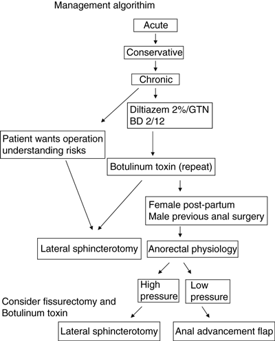
Lateral sphincterotomy should be used when medical management fails in men or women with normal to high resting tone. An alternative may be fissurectomy and botulinum toxin. In patients with low anal resting tone an anal advancement flap is a preferable option (see management algorithm).
Anal fissure in children
Fissure in children should be treated conservatively initially. If it fails to respond, local GTN or calcium channel blockers should be tried. Lateral sphincterotomy or fissurectomy should be reserved for those failing to heal with medical treatment (Level IIa, Grade B).
Most fissures occur in children aged between 6 and 24 months usually as a result of a mechanical tear. If chronicity develops, associated underlying pathologies should be ruled out as in adults. Diagnosis is by the history and examination. Stool negativism i.e. the deliberate reluctance to defaecate is not uncommon. It is important to palpate the abdomen for signs of faecal loading. An acute fissure usually heals in 10–14 days with conservative management, including dietary modification and osmotic laxatives [42]. If the fissure persists for 6–8 weeks chemical sphincterotomy should be considered. GTN 0.2% topically twice daily has been shown to be effective in treating children [43]. There is little information on diltiazem or botulinum toxin treatment in children.
Surgery is rarely indicated. Anal dilatation in the management of constipation and faecal soiling has not been found to be beneficial and is associated with a high rate of recurrence [44]. For an indolent fissure resistant to healing, fissurectomy and lateral sphincterotomy have been found to be beneficial [45,46]. The surgical technique is the same as for adults.
For further full text reference information, please refer to the recently published systematic review by Bhardwaj R and Parker MC [47].
Conflicts of Interest
None declared