Congenital tracheal stenosis managed with external chest compression exhalation technique for acute airway obstruction relief
Case report
Sir—We report a 10-month-old female child with a history of congenital tracheal stenosis (CTS) and right lung agenesis. She was born at 27 week and then transferred to us for cardiac evaluation at 2 months of age. During tracheal intubation for MRI, a 3.0 uncuffed endotracheal tube (ETT) did not pass through the glottis. A 2.5 uncuffed ETT was placed but could not be advanced more than 1 cm beyond the cords. A direct laryngobronchoscopy (DLB) was performed that showed severe tracheal stenosis. She remained in the intensive care unit (ICU) intubated with 2.5 uncuffed ETT on synchronized intermittent mechanical ventilation with a rate of 12 breaths per min. At 4.5 months of age, a tracheostomy was performed and a 3.0 cuffless tracheostomy tube was inserted.
After tracheostomy, she began having daily episodes of oxygen desaturation requiring multiple interventions. Albuterol, budesonide and ipratropium via inhalation were started. She continued retaining CO2 with episodes of bradycardia associated with acute airway obstruction. This was felt to be secondary to air trapping and pulmonary hyperinflation by a ball-valve mechanism. These episodes resulted in cardiopulmonary arrest three times which responded to cardiopulmonary resuscitation. Heliox 60/40 temporarily improved her ventilation; however, after 3 days, she returned to pre–heliox status. At that time, the ICU physicians decided to use external chest compression (ECC) to facilitate exhalation of the trapped air. This was accomplished by bag-trach ventilation followed by gentle manual compression of her left hemithorax at a slow rate during expiration. The trachea was suctioned using an 8 French suction catheter. Vecuronium, midazolam and fentanyl intravenous were required. The patient had no subsequent cardiac arrests and continued to thrive until 8 month of age when she became ready for tracheal reconstruction.
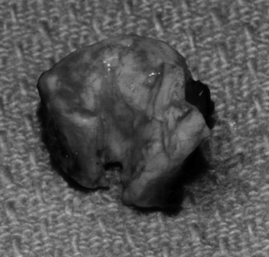
On the day of the surgery, the trachea was intubated with a 4.0 uncuffed ETT that was placed 1 cm proximal to the stenosis with closure of the stoma. Following sternotomy, the patient was placed on cardiopulmonary bypass (CPB). During bypass, flexible bronchoscopy was performed and the proximal and distal parts of the tracheal rings were identified by the surgeon. It was noted that the stenosis length was about 2 cm extending approximately to 0.5 cm above the carina. The external diameter of the stenosis was normal, but there was hyperplasia of the tracheal ring with about 1.5 mm orifice (Figure 1). The mid-portion was transected and the trachea was split vertically at the inferior and superior portions until normal trachea was seen. The trachea was then resected at these two points and reapproximated. A pericardial patch was used to cover the anterior split. Naso-endotracheal intubation was performed with the tip of the ETT placed just above the carina.

Gross specimen of the resected segment of the tracheal rings.
A week after the surgery, tracheal extubation was tried but failed. She subsequently developed transient pulmonary hypertension that was treated with intravenous sildenafil. One week post-reconstruction, a DLB showed moderate tracheomalacia. She was placed back on heliox. Four weeks post-reconstruction, extubation was attempted that failed and a DLB showed severe left bronchomalacia. Eight weeks post-reconstruction, she underwent tracheostomy and was back to ventilator.
Discussion
CTS is a rare condition that has a mortality rate of 44–79% (1). It is characterized by complete or near complete cartilaginous rings that lack the normal structure of the posterior membranous trachea. There are three types of CTS: generalized hypoplasia, funnel-shaped, and hourglass or segmental stenosis.
Anesthesia management includes deep inhalation induction with spontaneous ventilation followed by tracheal intubation. The ETT should be positioned at least 1 cm proximal to the stenosis to avoid traumatic edema or obstruction of the upper end of the stenosis. Ventilation can be pursued using positive pressure ventilation, high frequency jet ventilation, CPB or a combination of these (2). The use of ECMO has also been reported (1). The advantages of CPB are excellent surgical access without the ETT being close to the operation site. It also allows unhurried operation by ensuring good gas exchange (3).
Slow respiratory rate with high ventilation pressures and high positive end-expiratory pressure would allow the air to pass through the stenosis. A sterile ETT can be inserted into the distal trachea or into the main bronchus depending on the stenosis site.
The use of ECC to achieve expiration has been reported mainly for treatment of status asthmaticus (4). In addition to assisting with expiration, it can also help clear mucus plugs. ECC during exhalation results in forced expiratory volume increases by about 30%. However, this technique carries some risks. This includes circulatory depression secondary to increased intrathoracic pressure causing decrease in venous return (5). Other risks include physical trauma, alveolar rupture, and interstitial emphysema. The last two may occur as a result of increased intrathoracic pressure at peak inflation if compression is applied in early expiration phase.
In conclusion, we describe the use of ECC technique in CTS complicated with multiple episodes of airway obstruction associated with pulmonary hyperinflation and cardiac arrest.
Acknowledgement
Many thanks to Jillian Tweedie for her contribution to the manuscript.


