Motor strategies and excitability changes of human hand motor area are dependent on different voluntary drives
Abstract
The present study examined whether there were different voluntary drives between intended and non-intended muscle contractions. In experiment 1, during intended and non-intended muscle contractions, electromyograms (EMGs) were recorded from the first dorsal interosseous (FDI) and extensor carpi radialis (ECR) muscles when force levels were varied from 10% to 50% maximal voluntary contraction (MVC) in 10% MVC steps. In experiment 2, using transcranial magnetic stimulation, motor-evoked potentials (MEPs) were recorded from the FDI muscle when EMGs were varied from 10% to 40% EMGmax (EMG activities during MVC) in 10% EMGmax steps during intended and non-intended muscle contractions. In experiment 3, at 10% MVC force level MEPs were recorded before and after practice. The results showed that, in the FDI muscle, EMGs during intended muscle contractions were larger than those during non-intended ones at higher force levels (30–50% MVC). In the ECR muscle, reverse results were observed. At comparable EMG levels of the FDI muscle MEPs were the same during intended and non-intended muscle contractions. After practice, MEPs during intended muscle contraction became larger than those during non-intended at 10% MVC force level, while EMGs were the same between two muscle contractions. It is concluded that motor strategies and excitability changes of hand motor area are different during intended and non-intended muscle contractions, and these differences are due to the different voluntary drives of intended and non-intended. The present findings may contribute to the understanding of rehabilitation for patients suffering from damages of the central motor system.
Introduction
In our daily life, to perform our behaviors successfully we switch our central motor systems by internal awareness and/or external trigger. The former leads to an intentional voluntary muscle contraction, which can be defined as an intended muscle contraction. The latter leads to another kind of voluntary muscle contraction, which can be defined as a non-intended muscle contraction. When someone performs an intended muscle contraction isometrically, a force can be kept steadily. When an external trigger such as a load is added to the muscle (occurring frequently during daily life), a non-intended muscle contraction can be eventually generated isometrically for holding the load constantly. If the force and the weight of load are set equally, there will be no difference between two muscle contractions, referring to the Newtonian mechanics (the force outputs are the same and the joints are still). However, it is highly unlikely that the central motor system controls voluntary muscle contractions by explicitly solving the Newtonian laws of motion. Indeed, our voluntary muscle contractions arise from several distinct stages of neural activities, including motor preparation, specification of motor commands and sensory feedback. During the voluntary muscle contraction the extent of primary motor cortex (M1) is modulated, which is due to the dynamic changes of voluntary drives (Lewis et al., 2001; Lewis & Byblow, 2002; Ni et al., 2006) and afferent inputs (Bütefisch et al., 2000). In the present study therefore it is hypothesized that there are different voluntary drives between intended and non-intended muscle contractions. That is, the binding mechanisms of corresponding intended and non-intended voluntary drives may be integrated in M1 separately during isometric motor actions.
Based on previous reports, internally generated and externally triggered movements are associated with different cortical activation patterns (Gerloff et al., 1998; Haggard et al., 2002; Obhi & Haggard, 2004). However, a common coding mechanism that integrates representations of isometric motor actions and efforts of corresponding voluntary drives remains unclear. The first purpose of the present study therefore is to examine whether there are different motor strategies during intended and non-intended muscle contractions. Intended muscle contraction was executed as an isometric muscle contraction during which a subject generated the force for matching a target level. Non-intended muscle contraction was also executed as an isometric muscle contraction during which a load was added to the muscle and the subject just held the load constantly.
Although it is well known that M1 plays an important role for skilful voluntary muscle contractions, it is not quite clear how M1 contributes to voluntary muscle contractions induced by different voluntary drives. Since the introduction of transcranial magnetic stimulation (TMS), it has become a common neurophysiological method in studying the excitability changes of motor pathway (Rothwell, 1997). Using this method, pyramidal neurons in M1 can be stimulated trans-synaptically. The excitability changes in M1 can be evaluated by the fluctuation of eventually induced motor-evoked potentials (MEPs). It is known that voluntary muscle contraction of a small hand muscle greatly enhances the MEP to the TMS. Different voluntary muscle contractions, during which excitability changes in M1 are different, vary the MEP in amplitude (Capaday, 1997; Kasai & Yahagi, 1999; Ni et al., 2006). The second purpose of the present study therefore is to make sure whether the excitability changes of hand motor area in M1 accompanying the voluntary drives are different during the intended and non-intended muscle contractions, through investigating the MEP to the TMS.
Materials and methods
Subjects
Eight right-handed subjects (three female, five male; age range 23–33 years), who did not suffer from any known neuromuscular or psychiatric disorders, volunteered for the present study. All subjects were informed of the purpose of the study and experimental procedures in advance. The experimental procedures described hereafter conformed to the Declaration of Helsinki and were approved by the local ethics committee of Hiroshima University.
Experimental procedures
The subject was seated comfortably in an armchair. All experimental protocols were undertaken in the neutral forearm position (Fig. 1A).

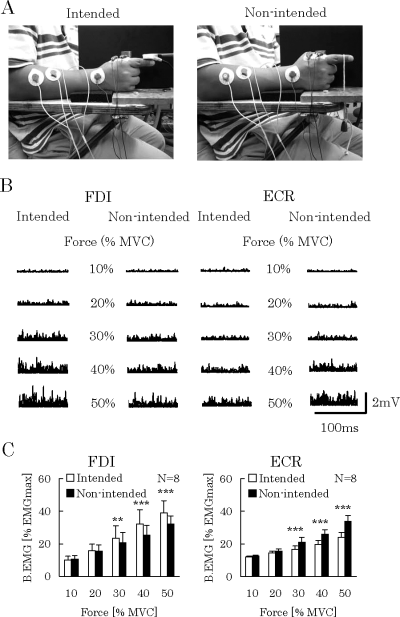
(A) Arm and finger positions during intended (left) and non-intended (right) muscle contractions. (B) Typical electromyogram (EMG) recordings (superimposed five rectified trials) obtained from the first dorsal interosseous (FDI, left panel) and extensor carpi radialis (ECR, right panel) muscles when force was varied from 10% to 50% maximal voluntary contraction (MVC). Left traces in each panel show the recordings for the intended muscle contractions and right traces for the non-intended ones. (C) Means and standard deviations (N = 8) of background EMGs (B.EMGs) in the FDI (left) and ECR (right) muscles. In the FDI muscles, B.EMGs during intended muscle contractions were larger than those during non-intended ones at 30–50% MVC. In the ECR muscles, reverse results were obtained. **P < 0.01, ***P < 0.001, comparing intended and non-intended.
In experiment 1, to investigate whether there were different contributions of agonist and synergist muscles between intended and non-intended muscle contractions, electromyogram (EMG) activities were recorded from the first dorsal interosseous (FDI) and the extensor carpi radialis (ECR) muscles during two muscle contractions. During the intended muscle contraction (left side of Fig. 1A) the right index finger of the subject was attached tightly to an immobile bar. A force sensor, which was fixed to the bar, was connected to a strain gage amplifier (model AS1302, Nihondenkisanei, Tokyo, Japan) for amplifying (50 times) the force signal. After being digitized (same method used for EMG recordings described below), the force signal was stored in a computer. The force value during maximal voluntary contraction (MVC), when the subject abducted the right index finger in the maximum effort, was measured as a standard reference. A beam line, which represented the target force, was displayed on an oscilloscope screen. The target force was adjusted from 10% to 50% MVC in 10% MVC steps. Another beam line, which illustrated real force level generated by the subject, was also displayed on the screen. The subject was instructed to abduct the index finger and to keep the coincidence of two beam lines for several seconds. The EMG activities were recorded from the FDI and ECR muscles. During off-line analysis, EMG activities were transferred to a percentage value of EMGmax (EMG activities during MVC). The value was named background EMG (B.EMG; in the next documents, B.EMG means the percentage value). In order to determine the weight of load for non-intended muscle contraction, force values of each subject were calibrated in advance. For calibration, a load (the weight was varied from 0.2 to 2 kg in 0.2-kg steps) was suspended to the immobile bar. Through the force recordings obtained from the strain gage amplifier, a regression line, which showed the relationship between the force value (percentage of MVC) and the weight of load, could be drawn up. According to the individual regression line of each subject, the optimal weight of load used for non-intended muscle contraction was pre-decided. During the non-intended muscle contraction (right side of Fig. 1A), the pre-decided load was suspended to the first interphalongeal joint of the right index finger. The subject was instructed to maintain the finger horizontally for several seconds for recording the EMG activities. The weight of the load was varied from about 0.2 to 1 kg in 0.2-kg steps, corresponding to the force levels of 10–50% MVC for each subject during the intended muscle contractions. These protocols were repeated in a random order, until 10 trials were recorded for each condition (muscle contractions cross force levels). Adequate break was taken between the trials.
In experiment 2, to examine whether there were different relationships between MEPs and B.EMGs during intended and non-intended muscle contractions, MEPs were recorded from the FDI muscle at several B.EMG levels. Referring to the results of experiment 1 (see the Results), B.EMG was varied from 10% to 40% EMGmax in 10% EMGmax steps. Using the same experimental procedures as in experiment 1 (when the subject maintained the target force or load, TMS was applied), 10 trials for each condition (muscle contractions cross B.EMG levels) were recorded.
In experiment 3, to examine whether there were different practice effects on intended and non-intended muscle contractions, MEPs and EMG activities were recorded from the FDI muscle before and after practice. Because differences in either MEPs or B.EMGs between intended and non-intended muscle contractions were not found at 10% MVC (see the Results), the practice effects were examined at 10% MVC force level. The practice was performed as a repetition of isotonic muscle contractions against a load of about 0.2 kg (10% MVC to each subject). The subject was instructed to lift the load up and down at a frequency of 0.5 Hz, following the rhythm of an acoustic metronome. The practice lasted for 10 rounds successively in one session, and the whole course included 10 sessions. To avoid fatigue, we took a break of 2 min between every two sessions. The total practice time consisted of 10 sessions of 20 s on practice and nine breaks of 2 min. The reason why isotonic muscle contractions were used for practice was that during the repetitive isotonic muscle contractions excitability in M1 could be definitely modulated (Classen et al., 1998; Kaelin-Lang et al., 2005; Yahagi et al., 2005). Before and after practice, the same TMS protocols were performed for the intended and non-intended muscle contractions. When the subject maintained the force or load of 10% MVC, TMS was applied. Twenty successful trials for each muscle contraction (intended and non-intended) were recorded before and after the practice.
TMS
MEPs elicited by TMSs were recorded from the FDI muscle, which was the agonist during the index finger abduction (Kasai & Yahagi, 1999; Hasegawa et al., 2001).
A Magstim 200 stimulator (Magstim, Whitland, Dyfed, UK) and a figure-of-eight-shaped coil (outside diameter of each loop was 9.5 cm) were used for applying TMS. The slightly angulated coil was placed tangentially to the scalp with the handle pointing backward and rotated appropriately 30 ° away from the mid-sagittal line. The current induced in the brain was anterior-medially directed and was perpendicular to the central sulcus, which could activate the pyramidal neurons trans-synaptically and produce early I waves (Kaneko et al., 1996; Di Lazzaro et al., 2001). We determined the optimal position for activation of the right FDI muscle by moving the coil in 0.5-cm steps around the presumed hand motor area in the M1 (approximately 4–6 cm lateral and 2 cm anterior to the vertex). A swimming cap was covered on the scalp and paper tapes were adhered to the cap in 2-cm steps as a reference. The site at which stimulation of slight superthreshold intensity consistently produced the largest MEPs in the FDI muscle was marked with a pen as the motor hot-spot. Special attention was paid to the position and orientation of the coil (the coil was maintained on the scalp by one author). Experiments 2 and 3 were undertaken continuously. The different measurements (kinds of muscle contractions and B.EMG levels) were randomly arranged in each experiment, to minimize the possible influence caused by the inconsistency of motor hot-spot. Additionally, the hot-spot was confirmed before and after practice in experiment 3. It was also checked randomly during the TMS protocols. If an unexpected variation occurred, we would take a break until its recovery.
At the beginning of each experiment, rest motor threshold was determined. The threshold was defined as the minimum output of the stimulator that induced reliable MEPs (above 50 µV in amplitude) in at least five out of 10 consecutive trials when the FDI muscle was completely relaxed. For most subjects, stimulation intensity of 80% threshold was able to elicit MEP of 0.5–1 mV in amplitude when 10% MVC was generated. It is considered that MEP shows great sensitivity in this range of amplitude (Ziemann et al., 1996; Capaday, 1997; Ni et al., 2006).
EMG recordings
The surface EMG activities were recorded from the FDI muscle in experiments 1–3. The active electrode was placed over the muscle belly, and the reference one over the metacarpophalangcal joint of the index finger. Additionally, in experiment 1, to observe the coordinative contribution of the synergist, EMG activities were also recorded from the ECR muscle (Hasegawa et al., 2001). The active electrode was placed over the muscle belly, and the reference electrode was placed approximately 4 cm distal to the active one. The recordings were made with 9 mm diameter Ag–AgCl surface cup electrodes. After being amplified (500 times) and band-pass filtered (5 Hz−1 kHz, model AB-621G, Nihonkohden, Tokyo, Japan), the recordings were then digitized at 5 kHz by an analog-to-digital interface (Wave-master ADX-98E, Canopus, Japan). The final data were stored in a computer for latter off-line analysis.
Data analysis and statistics
The EMG activities just 50 ms prior to the TMS were integrated as the value of B.EMG. Each value was normalized as a percentage of EMGmax. B.EMGs in the ECR muscle were calculated by the same method used in the FDI muscle. MEP, which showed a latency of 19.4 ± 1.2 ms (N = 8), could be recorded in the FDI muscle. The latencies did not change during the intended and non-intended muscle contractions. MEP amplitude was measured as the peak-to-peak value. Maximal muscle potential (Mmax) was measured at the beginning of the experiments. It was elicited by electric stimulation of 1 ms duration pulse with high intensity on ulnar nerve. In experiment 3, Mmax was also measured before and after practice. It remained stable. Mmax is a stationary value under a given experimental condition (property of the muscle, location of the electrodes, etc.). It indicates the sum of the motor pathways that are included in the experiment (all motor nerve fibers can be recruited by the electric stimulation with high intensity). It almost does not vary with the functional changes occurring at the cortical or spinal level. In view of these neurophysiological mechanisms, MEP amplitude was normalized as a percentage value of Mmax (Ni et al., 2006). The variation of the value could indicate the different extent of motor pathways recruited by the TMS, under different measurements (kinds of muscle contractions and B.EMGs).
A two-way anova (experiment 1, force levels cross muscle contractions; experiment 2, B.EMG levels cross muscle contractions; experiment 3, before and after practice cross muscle contractions) and a post hoc test (paired t-test with Bonferroni correction for multiple comparisons) were used for statistical analysis of the B.EMGs and MEPs. All significant levels were set at a criterion of P < 0.05.
Results
Figure 1B showed typical rectified EMG recordings in the FDI and ECR muscles during intended and non-intended muscle contractions. Figure 1C showed the mean values obtained from all subjects (N = 8). During both muscle contractions, B.EMGs increased in the FDI and ECR muscles following the increment of force level (FDI, F4,28 = 27.13, P < 0.001; ECR, F4,28 = 79.61, P < 0.001). But the B.EMGs were different between two muscle contractions at the comparable force levels, especially at higher ones. In the FDI muscles, at 30–50% MVC force levels, B.EMGs were larger during intended muscle contractions than those during non-intended ones (F1,28 = 9.09, P < 0.01; post hoc: 30% MVC, P < 0.01, 40% MVC, P < 0.001, 50% MVC, P < 0.001). In the ECR muscles reverse results were found. B.EMGs were larger during non-intended muscle contractions than those during intended ones (F1,28 = 206.59, P < 0.001; post hoc: 30–50% MVC, P < 0.001). Additionally, there were strong interactions between the force levels and the kinds of muscle contractions (FDI, F4,28 = 18.01, P < 0.001; ECR, F4,28 = 67.99, P < 0.001).
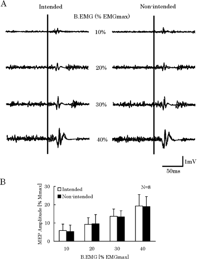
To examine whether there were different relationships between MEPs and B.EMGs during the intended and non-intended muscle contractions, MEPs were recorded from the FDI muscles at several B.EMG levels. Figure 2A showed the typical recordings of MEPs and B.EMGs. It indicated that MEPs increased with the increment of B.EMGs during both intended and non-intended muscle contractions. However, at any comparable levels of B.EMGs in the FDI muscle there was no different MEP facilitation between two muscle contractions. The mean values (N = 8) were summarized in Fig. 2B. The statistical analysis confirmed the data from the typical subject (B.EMG levels, F3,21 = 26.78, P < 0.001; muscle contractions, F1,21 = 0.04, P = 0.84).

(A) Typical motor-evoked potential (MEP) recordings (superimposed five trials) obtained from the FDI muscle when background electromyogram (B.EMG) was varied from 10% to 40% EMGmax, left traces for the intended muscle contractions and right traces for the non-intended ones. (B) Means and standard deviations (N = 8) of MEPs shown in (A). Note that at any B.EMG level there is no different MEP between two muscle contractions.
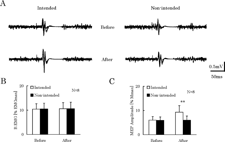
From the results shown in Fig. 2, there were similar relationships between MEPs and B.EMGs during both intended and non-intended muscle contractions. Based on the results shown in Fig. 1B and C, it seemed that there were no different voluntary drives between intended and non-intended muscle contractions at low force levels. We re-examined this point through investigating the practice effects on these two muscle contractions. Figure 3A showed the typical recordings of MEPs and B.EMGs obtained from the FDI muscle before and after practice. B.EMGs obtained from the FDI muscles (N = 8) were summarized in Fig. 3B. They remained stable before and after practice during both muscle contractions at 10% MVC force level. However, MEPs did not show the familiar results. Before practice, the MEP amplitudes (N = 8) did not change during two muscle contractions, which was consistent with the results of experiment 2. After practice, the MEPs became larger during the intended muscle contractions than those during the non-intended ones (anova, F1,7 = 6.43, P < 0.05; post hoc, P < 0.01; Fig. 3C). The practice effects were also statistically significant (F1,7 = 6.56, P < 0.05).

(A) Typical motor-evoked potential (MEP) and electromyogram (EMG) recordings (superimposed five trials) obtained from the FDI muscle before (upper) and after (lower) practice. Left traces were obtained from the intended muscle contractions, and right traces were obtained from the non-intended ones. Note the enlarged MEPs during intended muscle contractions after practice. Means and standard deviations (N = 8) of background EMGs (B.EMGs) (B) and MEP amplitudes (C) obtained from the FDI muscles before and after practice. Open or filled columns indicate the intended or non-intended muscle contractions, respectively. **P < 0.01, comparing intended and non-intended.
Discussion
Internal awareness and external trigger can refer to various aspects of movement. Several reports have suggested that the central neural drives from M1 to a target muscle are different between internally generated and externally triggered movements (Gerloff et al., 1998; Mima et al., 1999; Lotze et al., 2003). However, different electrocortical force-related efforts accompanying with different voluntary drives have never been reported. In the present study, we focused on the binding mechanisms of voluntary drives integrated in M1 during isometric motor actions. Because the intended and non-intended muscle contractions were both isometrically generated in the present study, it was convincing that they could be treated as the reflections of force-related efforts induced by corresponding voluntary drives. The novel findings can be summarized as follows. (i) B.EMGs are different between intended and non-intended muscle contractions in both agonist (FDI) and synergist (ECR), especially at higher force levels (30–50% MVC). Additionally, different B.EMGs during two muscle contractions show reverse relations for the FDI and ECR muscles (when the B.EMG in the FDI muscle is larger, that in the ECR is smaller). (ii) Although MEPs become larger with the increment of B.EMGs during both intended and non-intended muscle contractions, there is no different MEP facilitation between these two different muscle contractions at any comparable B.EMG levels. (iii) After practice, no significant change of B.EMG occurs in the agonist (FDI) muscle. However, MEP can become larger during intended muscle contraction in contrast with the constant value during non-intended one. In the next paragraphs we will try to explain the neurophysiological mechanisms, accordingly.
Motor strategy
Although the definite concepts of voluntary drives concerning internal awareness and external trigger are not quite clear, it is suggested that these voluntary drives are different at the level of voluntary efforts including overt movement and motor image (Lotze et al., 1999). It has a long history in the physiological and psychological field to study the voluntary drives during motor tasks through investigating voluntary performance (Borg, 1982). A potentially useful method for understanding the difference between intended and non-intended voluntary drives can be performed to examine the motor behaviors when subjects keep their voluntary commands constantly. In the present study, it was found that even during the isometric muscle contractions (force or load was kept constant), intended and non-intended voluntary drives could be expressed separately. The force outputs are the same during two muscle contractions; however, the motor pathways of the agonist (FDI) muscle have higher activations when an intended muscle contraction is generated because the B.EMG is larger. It is natural to assume that the FDI muscle can do work more positively during intended muscle contraction than during the non-intended one, for producing force output. Therefore, the problem is which muscle does the remaining work during non-intended muscle contraction (the force outputs are the same between intended and non-intended muscle contractions). The present results showed that when the force level increased, B.EMG in the ECR muscle became larger during non-intended muscle contraction than during intended one. It can be explained that, when internal awareness is required in the central motor system (intended muscle contraction), the agonist muscle will become more active. In contrast, when an external trigger is applied (non-intended muscle contraction), the synergist muscle will become more active for generating the force output. Additionally, when a heavier load is added during the non-intended muscle contraction, the subject is not capable to hold the load and an eccentric movement will be performed. Many previous studies had demonstrated that the neural command controlling eccentric movement was unique and led to different motor strategy (cf. Enoka, 1996). Del Valle & Thomas (2005) reported that the firing rates of motor units during eccentric movement were always lower than those during corresponding concentric one.
Combining the present results and previous studies, the voluntary drives during intended and non-intended muscle contractions may be different and consequently descend different motor strategies to the muscles. Additionally, the results showed that there was a strong interaction between the kinds of muscle contractions and the force levels. It suggests that the different motor strategies induced by different voluntary drives can only be expressed within a suitable range of force level. Indeed, post hoc tests showed that at low (10% and 20% MVC) force levels, there was no different B.EMG between two muscle contractions. It can be explained that for generating low force not all but a limited part of pyramidal neurons and motoneurons can be recruited. The different activation levels between two muscle contractions might be too small to be found out at these force levels.
Relationship between MEP and B.EMG
To reveal why intended and non-intended voluntary drives were different and could lead to different motor strategies during intended and non-intended muscle contractions, relationships between MEPs and B.EMGs were investigated during two muscle contractions. Referring to the principle of recruitment order, when the B.EMG increases, larger pyramidal neurons or motoneurons must be recruited, which induces a larger MEP (Ashe, 1997; Capaday, 1997). It is not surprising, as from the present result, that during both intended and non-intended muscle contractions MEPs become larger as the B.EMGs increase. On the other hand, MEPs during two muscle contractions are the same at comparable B.EMG levels. There is a general issue that EMG activities indicate the amount of motoneurons being recruited and the firing rates of the recruited motoneurons. MEP can estimate the recruited pyramidal neurons and motoneurons as well as the subliminal fringes in the M1 (pyramidal neurons) and motoneuron pools (Rothwell, 1997). Thus, the present results mean that, when comparable motoneurons are recruited during two muscle contractions, the intended and non-intended voluntary drives seem to produce similar subliminal fringes in M1 or motoneuron pools.
Practice effects
From the explanation described above, the question why intended and non-intended voluntary drives are different still remained unproven. The present results showed that only during intended muscle contraction an extra MEP facilitation could be found after practice. Recently, Kaelin-Lang et al. (2005) demonstrated that voluntary drive during active movement could lead to encoding of a motor memory in M1 during motor learning, whereas that during passive movement could not. Additionally, sensorimotor integration is crucial in complex motor tasks, in which excitability of M1 enhances the efficacy of the motor activity (Tamburin et al., 2001). Gating or filtering of sensory inputs is important in removing predictable sources of afferent input associated with feed-forward motor command (Wolpert et al., 2001). The present results of different practice effects on intended and non-intended muscle contractions can be explained in line with the difference of integrative functions of peripheral inputs and voluntary drives within M1.
With regard to effects of integrative functions of voluntary drives on motor learning, it is demonstrated that a voluntary drive concerning internal awareness enhances processing within M1 more than that concerning external trigger (Alary et al., 1998; Carel et al., 2000; Kaelin-Lang et al., 2005). It was also suggested that only voluntary drive of internal awareness can play an effective role in integration of afferent inputs (Classen et al., 1998). Taken together with the previous studies on active and passive movements (Lotze et al., 2003), the present results suggest that intended voluntary drive leads to a more prominent increase of activation, recruitment occurrence and intracortical facilitation in M1 than non-intended one. Indeed, the excitability changes of M1 are dependent not only on the extent of voluntary drive for contracting the target muscle, but also on afferent inputs produced by muscle contractions (Lotze et al., 2003; Kaelin-Lang et al., 2005). The present results showed that practice (repetitive isotonic muscle contractions) was more effective on intended muscle contraction. It suggests that a more prominent increase in the strength of inputs converging onto M1 can be elicited by intended voluntary drive than by non-intended one. That is, the gating or filtering effects of sensory inputs may be stronger during intended muscle contraction than that during non-intended one (Tamburin et al., 2001; Wolpert et al., 2001). Therefore, the integrative function of the intended voluntary drive may be eventually enhanced in M1. After practice, these effects could be strengthened and larger MEP could be elicited during intended muscle contraction.
Other factors
Other possible factors to the present study may not be ignored. It has been reported that the thixotropic properties of muscle can alter the gain of voluntary drive. MEP can be facilitated during muscle shortening and be suppressed during lengthening (Coxon et al., 2005). Somewhat, the altered gain might have occurred after practice although Mmax did not change. Because of the same reason, the extremely small difference in muscle length between intended and non-intended muscle contractions (both muscle contractions used in the present study were generated isometrically) might have also altered the gain of voluntary drives.
The site of motor hot-spot was very important to the present results. Although it was randomly checked during the experiments, the hot-spot might have changed because it was impossible for the subjects to keep their motivation at the same level throughout the experiments. Another point was that the measurement of practice effect (before and after practice) could not be randomly arranged. In compensation, the hot-spot was confirmed before and after practice. In short, the inconsistency of the motor hot-spot might have some influence on the main discussion of the present study (Tyèet al., 2005).
Functional and clinical implications
Once again, the present study focused on the different binding mechanisms of intended and non-intended voluntary drives integrated in M1. First of all, it is found that intended and non-intended voluntary drives cause different excitability changes of the hand motor area in M1 during isometric muscle contractions. When the force outputs increase, the different excitability changes in M1 lead to different motor strategies during intended and non-intended muscle contractions. Therefore, where the effort signals of different voluntary drives generate in the brain becomes an important question. It is already demonstrated that the effort signal of voluntary drive is not simply derived from a copy of the output of M1, but arises somewhere upstream (Carson et al., 2002). The way forward to further research should be created to find the central sites of the origin of the voluntary drive and to find their linkage to the motor output from M1.
Our study suggests that intended voluntary drive plays an important role for contributions of peripheral sensory afferent inputs to excitability changes in the M1 after practice. It can be inferred that only intended voluntary drive can play an effective role in integration of peripheral afferent inputs (the kinematical details of the practiced movements: Classen et al., 1998) for reorganization in M1. This evidence may provide fundamental knowledge of effectiveness of motor adaptation learning processes (Bütefisch et al., 2000, 2004; Ridding et al., 2000; Stefan et al., 2000, 2002; Sawaki et al., 2002) concerning intended voluntary drive. The understandings of these neural mechanisms related to use-dependent plasticity and reorganization in central motor structures could be helpful in optimizing physiotherapeutic approaches for damage of the central motor system.
In conclusion, we prefer that motor strategies and excitability changes of hand motor area in the human primary motor cortex are different between intended and non-intended muscle contractions, and that these differences are due to the different voluntary drives of intended and non-intended.
Acknowledgements
The present study was supported by Research Projects Grant-in-Aid for scientific number T.K.: NO 16500380 from the Ministry of Education, Culture, Sports, Science and Technology of Japan. We thank the anonymous reviewers whose comments improved the manuscript.
Abbreviations
-
- B.EMG
-
- background EMG
-
- ECR
-
- extensor carpi radialis
-
- EMG
-
- electromyogram
-
- EMGmax
-
- EMG activities during MVC
-
- FDI
-
- first dorsal interosseous
-
- M1
-
- primary motor cortex
-
- Mmax
-
- maximal muscle potential
-
- MEP
-
- motor-evoked potential
-
- MVC
-
- maximal voluntary contraction
-
- TMS
-
- transcranial magnetic stimulation


