Strategies for non-responders
SUMMARY
• When improvements in symptoms of hospital-acquired pneumonia are difficult to see, an investigation should be made into whether there truly is no response to antimicrobial treatment.
• Non-infectious disease that results in pneumonia-like shadows needs to be excluded.
• In pneumonia that is unresponsive to antimicrobials, differential diagnoses must be made with consideration of causative microorganism factors, host factors and medication factors, respectively.
• Preferably, the dose, administration frequency and administration time for the administered antimicrobials should be established based on PK/PD.
INTRODUCTION
When no response is seen to antimicrobial treatment, consideration of the possible reasons for this lack of response is better than simply making an abrupt change in the antimicrobial regimen.1 First is the possibility that the response has been judged as poor, despite an actual amelioration in the pneumonia. Improvements in shadows normally lag behind improvements in clinical symptoms, and inflammation parameters, such as CRP, do not necessarily reflect the state of the disease. In hospital-acquired pneumonia in particular, improvements in symptoms can be difficult to see after modification by any underlying disorders. Even fever may represent drug-induced fever from the antimicrobials. Bacteriologically as well, simple colonizing bacteria with antimicrobial resistance that are detected in culture must not be confused with a new infection after the causative microbe has been eliminated.
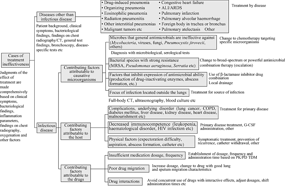
As shown above, when no contributing factors and no improvement in symptoms are seen, causes such as the following may be considered (Fig. VI-1).

Differentiation of causes and measures for non-response to early treatment. ALI, acute lung injury; G-CSF, granulocyte-colony stimulating factor; MRSA, Methicillin-resistant staphylococcus aureus; PK/PD, pharmacokinetics/pharmacodynamics; TDM, therapeutic drug monitoring.
DISEASES OTHER THAN INFECTIOUS DISEASE
The following are respiratory diseases that present pneumonia-like shadows on chest radiography, but are not infectious diseases:
- 1
Drug-induced pneumonia
- 2
Organizing pneumonia (including cryptogenic organizing pneumonia: COP)
- 3
Eosinophilic pneumonia
- 4
Radiation pneumonitis
- 5
Various types of interstitial pneumonia and their exacerbation
- 6
Malignant tumour
- 7
Congestive heart failure
- 8
Acute lung injury (ALI)/ARDS
- 9
Pulmonary infarction
- 10
Pulmonary alveolar haemorrhage
- 11
Foreign body in trachea or bronchus
- 12
Pulmonary atelectasis
In addition to the above, all underlying respiratory disorders that present with shadows in the lung fields may need to be differentiated. When these diseases are suspected, antimicrobial treatment is discontinued and special tests directed at the respective diseases are conducted. CT, including scans of both the chest and other regions, is often useful,2 and bronchoscopy or other invasive tests may also be needed for diagnosis.
NON-RESPONSE TO TREATMENT IN THE CASE OF INFECTIOUS DISEASES
1. Contributing factors attributable to causative microorganisms
The possibility of pneumonia caused by microorganisms against which general antimicrobials are ineffective (mycobacteria, viruses, fungi, Pneumocystis jirovecii, others) should be considered. This is particularly important in immunocompromised patients. Microbiological or serological tests directed at these microorganisms need to be conducted.
With bacterial species that are strongly resistant, including MRSA, Pseudomonas aeruginosa and Serratia, effects can be difficult to see even when antimicrobials are administered within the indication range,3 and a change should be made to the most powerful drug among those to which the microbe is susceptible. The effect is especially poor when Pseudomonas aeruginosa pneumonia is treated using a single drug.4
In cases of abscess or empyema formation, the drug effect may be blocked by mass production of drug-inactivating enzymes, or drug migration may be poor, and local drainage should be considered.
In refractory pneumonia with accompanying bacteremia, location of the infection focus in places other than the lungs (liver abscess, infectious endocarditis, osteomyelitis, catheter infection, etc) should also be considered.
2. Contributing factors attributable to the host
Patients with hospital-acquired pneumonia have underlying disorders or complicating diseases that need to be treated together with antimicrobial treatment for the pneumonia. For example, in cases of lung cancer complicated with obstructive pneumonia, the primary disease must be treated or there is no prospect of improvement in the pneumonia. In infections accompanying diabetes, blood sugar levels need to be controlled.
In immunocompromised patients or patients with leukopenia, the pneumonia will be prolonged or exacerbated until immunocompetence is restored. Conversely, in cases of refractory or recurrent pneumonia, or pneumonia from rare pathogens, the possibility of latent immunodeficiency, including HIV infection, should be investigated.
Pneumonia is often prolonged when the patient is malnourished or immobile. In patients with repeated aspiration, pneumonia will recur unless aspiration is prevented through non-oral nutritional support, such as with high-calorie transfusions, tube feeding or gastrostomy. In patients who have difficulty eliminating sputum, postural drainage or tapping may be necessary.
Biofilms may form on intravascular catheters, gastric tubes, urine balloons and other medical implements and become sources of infection. These implements should therefore be extracted and replaced as necessary.
3. Contributing factors attributable to drugs
Even when appropriate antimicrobials are administered, the anticipated effect may not be achieved for reasons such as insufficient dosage of the antimicrobial or insufficient dosing frequency. In Japan, antimicrobial dosages are generally smaller than in Western countries. In addition, determinations of susceptible (S), intermediate (I), or resistant (R) in drug susceptibility tests use break points of the Clinical and Laboratory Standards Institute (CLSI) in the USA, which assumes dosages used in the USA, and so are unsuited to the situation in Japan (see Chapters III, IV). Time above the MIC (TAM) ≥40–60% is needed if one is to expect maximum bactericidal effect with β-lactams,5,6 and achieving this may require increasing the number of doses and/or prolonging the administration time for each dosage. It is possible that no effect will be obtained with many drugs unless they are used near the maximum dose allowed in Japan. With anti-MRSA antibiotics or aminoglycoside drugs, therapeutic drug monitoring (TDM) is also needed.
Considerations of the tissue migration characteristics of antimicrobials are also important. In general, β-lactams do not migrate well in lung tissue, and raising blood levels with intravenous infusions or increasing number of doses and TAM becomes necessary. In contrast, macrolides, tetracyclines and quinolones migrate well in lung tissue.
Patients with hospital-acquired pneumonia are given a number of other drugs in addition to those administered to treat the pneumonia, and antimicrobial activity can be weakened by interactions between these medications. For example, blood levels of the antifungal itraconazole decline if used in combination with rifampicin or phenytoin, and use of increased doses need to be considered.
CONFLICT OF INTEREST
No conflict of interest has been declared by The Committee for the Japanese Respiratory Society guidelines for the management of respiratory infections.


