Hepatobiliary and Pancreatic: Hemobilia caused by bleeding from hepatic artery aneurysms
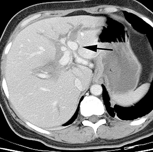
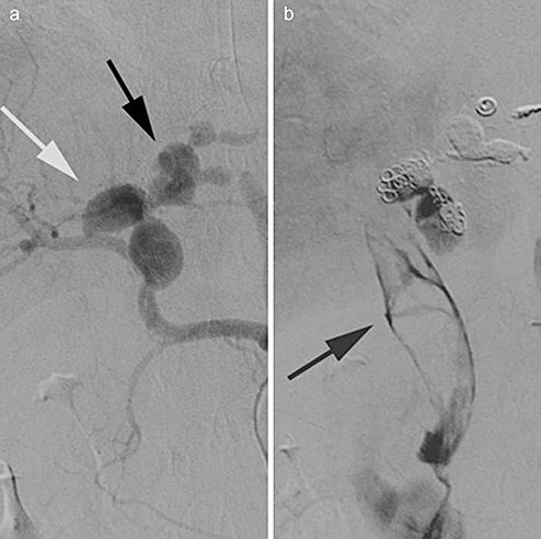
A 50-year-old woman was admitted to hospital with hematemesis and hypotension. She had previously been well. Her hemoglobin was 7.9 g/dL (79 g/l) and she had a mild elevation of alanine aminotransferase (102 u/l) and alkaline phosphatase (187 u/l). At upper gastrointestinal endoscopy, oozing of blood from superficial gastric ulcers was treated with injections of adrenaline. However, bleeding continued and an urgent upper abdominal ultrasound study showed dilatation of the bile duct and a possible gallbladder lesion. A contrast-enhanced computed tomography (CT) scan showed aneurysmal dilatation of hepatic vessels in the hilum of the liver as well as radiological contrast in a mildly dilated bile duct (Figure 1). Because of the possibility of an hepatic artery aneurysm, hepatic angiography was performed and revealed several aneurysms in the liver hilum (Figure 2a). The aneurysms involved the main hepatic artery, middle hepatic artery (white arrow) and left hepatic artery (black arrow). The aneurysms were initially embolized with five thrombogenic coils but there was still passage of some contrast into the bile duct (arrow, Figure 2b). Bleeding subsequently ceased after 1 ml of diluted N-butyl cyanoacrylate was injected into the area. CT-angiography after 3 months showed segmental ischemic changes in the left lobe of the liver but there were no hepatic aneurysms or aneurysms involving the renal arteries.


Common causes for hepatic artery aneurysms include trauma, infections and atherosclerosis. However, in the above patient, we have attributed hepatic artery aneurysms to fibro-muscular dysplasia. This is a rare disease characterized by aneurysmal dilatations in medium-sized arteries, sometimes creating a ‘string of beads’. The renal and the internal carotid arteries are the most frequently affected but other arteries can also be involved including the hepatic artery. As far as we are aware, this is the first report of involvement of the hepatic artery without the renal artery. Fibro-muscular dysplasia is more common in women than in men and presents in a variety of different ways depending on the location of the aneurysms. There are at least three previous cases where the presenting symptom was hemobilia. One of these was successfully treated by transcatheter arterial embolization. An alternative therapy is surgical ligation of the hepatic artery.


