Hepatobiliary and pancreatic: Choledochal cyst
Choledochal cysts are uncommon congenital disorders of the bile duct that manifest as cystic dilatation of extrahepatic or intrahepatic ducts. The incidence varies between countries with the highest reported incidence in Japan (1:1000 individuals). Cysts vary in site, type and number and are usually classified by the nomenclature of Dr Todani and others. The classification includes choledochoceles (type III) and Caroli's disease (type V). At least 40% of choledochal cysts are associated with an anomalous pancreatobiliary ductal junction that permits reflux of pancreatic juice into the biliary system. This reflux may aggravate duct dilatation and seems likely to be responsible for biliary inflammation and an increased risk for cholangiocarcinoma. In relation to symptoms, at least two-thirds of patients seek medical attention before the age of 10 years because of abdominal pain, jaundice or a palpable mass. In adults, presenting symptoms can include cholecystitis and pancreatitis. Choledochal cysts can be diagnosed by a variety of imaging modalities including abdominal ultrasound, computed tomography (CT), magnetic resonance cholangiopancreatography and endoscopic retrograde cholangiopancreatography (ERCP). Without surgery, 10–30% of adults will develop cholangiocarcinoma that arises from either the cyst itself or non-dilated portions of the bile duct. There is also an increased risk for gallbladder cancer. Management usually includes excision of the cyst with an hepaticojejunostomy and cholecystectomy. After surgery, the lifetime risk for cholangiocarcinoma decreases to approximately 1%.
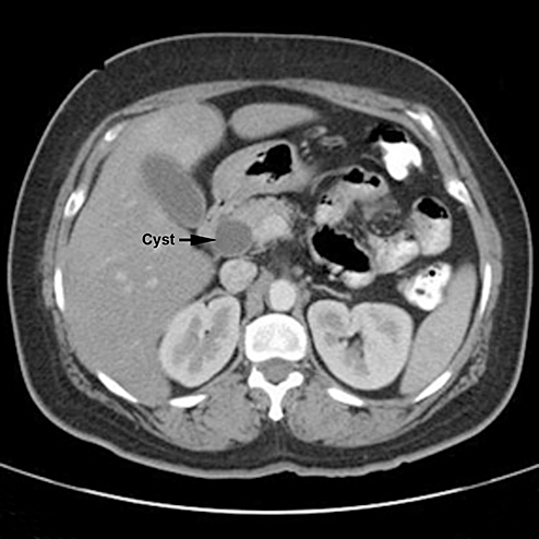
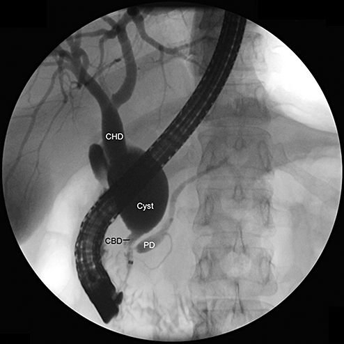
The patient illustrated below was a 45-year-old Hispanic woman who was admitted to hospital with a 2-week history of upper abdominal pain. Blood tests revealed a minor elevation of alanine aminotransferase (89 u/L) and lipase (150 u/L). An abdominal CT scan showed cystic dilatation of the lower bile duct without dilatation of the common hepatic duct or intrahepatic ducts (Fig. 1). ERCP confirmed the presence of a cyst, 2 cm in diameter, in the common bile duct consistent with a choledochal cyst type IB (Fig. 2). She did not have an anomalous pancreatobiliary ductal junction. The patient was treated by cyst excision and hepaticojejunostomy.


