Hepatobiliary and pancreatic: Aberrant left gastric vein
Contributed by H Sugimoto, S Takeda, S Inoue, T Kaneko and A Nakao, Department of Surgery II, Nagoya University School of Medicine, Nagoya 466–8550, Japan.
Contributions to the Images of Interest Section are welcomed and should be submitted to Professor IC Roberts-Thomson, Department of Gastroenterology, The Queen Elizabeth Hospital, Woodville South, South Australia 5011, Australia.
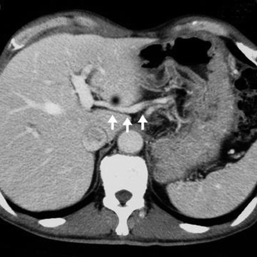
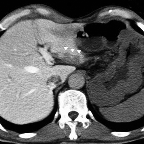
The left and right gastric veins usually enter the portal vein trunk, the splenic vein or the confluence of the two. Entry of gastric veins into other sites in the portal venous system is rare (<1%) and such veins are often called aberrant gastric veins. The images shown in this report were from a 66-year-old man who was admitted to hospital with cancer of the body of the pancreas. An enhanced computed tomography (CT) scan showed that the tumor had invaded the portal vein, splenic vein and celiac artery. The splenic vein was completely occluded and, because of this, a more prominent left gastric vein could be seen in the hepatogastric ligament. This vein entered the left portal vein close to the bifurcation of the portal trunk into left and right portal veins (Fig. 1, arrows). This was diagnosed as an aberrant left gastric vein. A CT scan during arterial portography (CTAP) demonstrated small portal perfusion defects in segment II corresponding to the area of perfusion of the aberrant left gastric vein (Fig. 2, arrowheads).


Various pseudolesions have been reported after CTAP. Some of these have been attributed to venous drainage to the liver that bypasses the portal venous blood. This may arise from veins such as an aberrant right gastric vein, cholecystic vein and epigastric/paraumbilical vein, but has rarely been associated with an aberrant left gastric vein. In the case described above, the aberrant left gastric vein became more prominent as a result of left-sided portal hypertension caused by splenic vein obstruction. Preservation of gastric veins to prevent gastric congestion can be relevant to major pancreatic surgery when distal pancreatectomy is accompanied by resection of the celiac trunk and portal vein. In this setting, preservation of veins can be facilitated by the presence of aberrant gastric veins.


