Liver lipid metabolism
Presented as part of the 10th Congress of the European Society of Veterinary and Comparative Nutrition held in Nantes, France, October 5–7, 2006.
Summary
The liver plays a key role in lipid metabolism. Depending on species it is, more or less, the hub of fatty acid synthesis and lipid circulation through lipoprotein synthesis. Eventually the accumulation of lipid droplets into the hepatocytes results in hepatic steatosis, which may develop as a consequence of multiple dysfunctions such as alterations in β-oxidation, very low density lipoprotein secretion, and pathways involved in the synthesis of fatty acids. In addition an increased circulating pool of non-esterified fatty acid may also to be a major determinant in the pathogenesis fatty liver disease. This review also focuses on transcription factors such as sterol-regulatory-element-binding protein-1c and peroxisome proliferator-activated receptor alpha, which promote either hepatic fatty acid synthesis or oxidation.
Lipid metabolism involves several pathways that are at least in part, inter-dependent and ‘cross-regulated’. There are therefore different possible approaches to review this topic to get an overview. The focus of the present discussion will be fatty acids and triacylglycerols. Fatty acids are the most commonly stored and circulating forms of energy, and triacylglycerols are the most common non-toxic form of fatty acids. Fatty acids/triacylglycerols may originate from four sources (pool input): De novo lipogenesis, cytoplasmic triacylglycerol stores, fatty acids derived from triacylglycerols of lipoprotein remnants directly taken up by the liver, and plasma non-esterified fatty acids (NEFA) released by adipose tissue. The relative importance of these sources depends on species differences (e.g. in ruminants, only modest amounts of hepatic De novo lipogenesis occurs compared with adipose tissue; the inverse is true in birds, with liver being the main site of De novo lipogenesis), and on short- and long-term nutritional status and energy balance. Fatty acids and triacylglycerols may also be used in different ways (pool output). Triacylglycerols may accumulate in hepatocytes (while NEFA or activated forms of NEFA may not) unless NEFA are oxidized (more or less completely) or triacylglycerols are exported as constituents of very low density lipoproteins (VLDL). Two examples of inter-connection may be cited: (i) a low rate of esterification when the oxidation rate is high in response to energy demand, (ii) a strong relationship between VLDL secretion and fatty acid/triacylglycerol availability; this is especially the case in species where De novo lipogenesis is very active, but not in those species where high triacylglycerol concentration may be present and where the liver is not the ‘usual’ site of De novo lipogenesis.
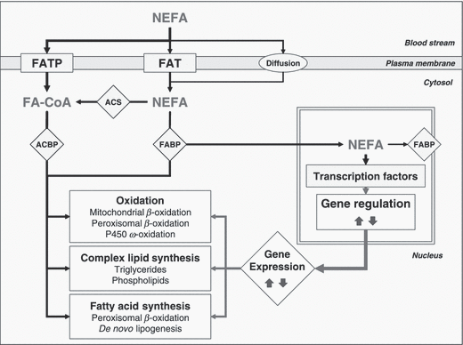
The triacylglycerol content of hepatocytes is regulated by the activity of cellular molecules that facilitates hepatic fatty acid uptake, fatty acid synthesis, and esterification (‘input’) and hepatic fatty acid oxidation and triacylglycerol export (‘output’) (Fig. 1). Moreover, and interestingly, fatty acids regulate overall lipid metabolism by binding nuclear receptors that modulate gene transcription.

Trans-membrane fatty acid transport, fatty acid intracellular activation and main pathways of activated fatty acids that may explain intracellular triacylglycerol accumulation. FA, fatty acid; FATP, FA transport protein; FAT, FA translocase; ACS, acyl-coenzyme A synthase, NEFA, non-esterified FA; FABP, FA binding protein; ACBP, acyl-coenzyme A binding protein; MTP, microsomal triglyceride transfer protein; TAG, triacyglycerol.
Fatty acid uptake and synthesis and glycerolipid synthesis
Fatty acid uptake
Non-esterified fatty acids can arise from the hydrolysis of complex lipids by lipases, or the hydrolysis of fatty acid-CoA by thioesterases. The liver takes up NEFA from the blood in proportion to their concentration. Non-esterified fatty acids enter cells via transporters [fatty acid transport protein (FATP) or fatty acid translocase (FAT), CD36] or diffusion. Within the hepatocytes, long-chain fatty acids of 14 carbons or more are covalently bound and activated by fatty acid binding protein (FABP) or acyl-CoA synthetases (ACS) found in the microsomes and outer mitochondrial membrane (Fig. 1). Several isoforms of ACS have been identified and the further fate of a particular acyl-CoA (especially channeling towards complex lipid synthesis and storage, or toward oxidation) depends on which of the isoforms catalyzes its synthesis (Coleman et al., 2002). Non-esterified fatty acids and fatty acyl-CoA are bound to FABP and acyl CoA binding protein which transport them to intracellular compartments (for metabolism) or the nucleus (to interact with transcription factors). Cells challenged with exogenous fatty acids rapidly assimilate the fatty acids into neutral and polar lipids, and some are oxidized. The result of these metabolic pathways is to keep intracellular NEFA and fatty acyl-CoA very low.
De novo synthesis of fatty acids
De novo lipogenesis (i.e., De novo synthesis of fatty acids) is a key metabolic pathway for energy homeostasis in higher animals. Lipogenic flux is tightly controlled by hormonal and nutritional conditions. Briefly, high carbohydrate diets induce, whereas fasting or fat feeding inhibit, De novo lipogenesis; this is especially dependent on insulin concentration, and tissue insulin sensitivity.
Two major tissues produce fatty acids in the body: the liver and the adipose tissue. Fatty acids synthesized in the liver are exported through lipoprotein production, and thus provide an energy source and structural components for membrane building. In adipose tissue, De novo synthesis of fatty acids directly contributes to in situ fat deposition and long-term energy storage.

The rate-limiting step in this pathway is catalyzed by acetyl-CoA carboxylase that converts acetyl-CoA to malonyl-CoA and is considered to be the chain extender substrate (‘donor’ of acetyl units) in the elongation process (Kim, 1997). Formation of a new C–C bond by condensation of the acetyl and malonyl moieties is coupled with an energetically favourable decarboxylation, so that the carbon originating from CO2 introduced in the reaction catalyzed by acetyl-CoA carboxylase is recycled. The β-ketoacyl formed is reduced and then participates in a second round of condensation with a malonyl moiety.
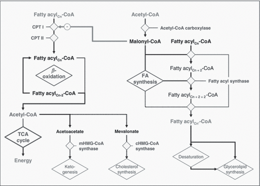
A close relationship exists between the rate of fatty acid synthesis and the activity of fatty acid synthase (FAS) (Fig. 2), a key multifunctional enzyme that catalyzes the entire pathway of palmitate synthesis (Smith et al., 2003). Fatty acid synthase is expressed in the two major sites of fatty acid production in the body, liver and adipose tissue, but the relative contribution of these sites to De novo lipogenesis is highly variable among species. Adipose tissue is the main site of De novo lipogenesis in non-lactating ruminants (Travers et al., 1997), pigs (O’Hea and Leveille, 1969), dogs (Stangassinger et al., 1986) and cats (Richard et al., 1989). In poultry (Leveille et al., 1975), similarly to humans (Patel et al., 1975), the liver is the major site of De novo lipogenesis, while in rodents and rabbits both liver and adipose tissue are important (Pullen et al., 1990). The mammary gland of ruminant animals also actively synthesizes fatty acids (Bergen and Mersmann, 2005).

Main steps of fatty acid oxidation and fatty acid synthesis. These two pathways are mainly regulated (and cross-regulated) by PPARα and LXR/SREBP, respectively, dependent on activation or inactivation by fatty acid binding. The main mitochondrial pathways are β-oxidation, the TCA cycle and ketogenesis; in the cytosol, the main pathways are fatty acid and cholesterol synthesis; while at the cytosolic face of microsomal membranes, glycerolipid synthesis occurs. CPT I, carnitine palmitoyltransferase I; CPT II, carnitine palmitoyltransferase II; TCA cycle, tricarboxylic acid cycle; mHMG-CoA-synthase, mitochondrial hydroxymethylglutaryl-coenzyme A synthase; cHMG-CoA synthase, cytosolic HMG-CoA synthase.
The regulation of FAS by hormones (insulin, glucagon) (Sul et al., 2000) and nutritional state (carbohydrates and polyunsaturated fatty acids) (Volpe and Vagelos, 1974; Clarke and Jump, 1996) has been described in liver and adipocytes. Insulin and substrate (citrate, isocitrate) availability activates the enzyme whereas glucagon and catecholamines inhibit its activity [via 3′,5′-cyclic adenosine monophosphate (cAMP)-dependent phosphorylation]. Increased concentration of fatty acyl-CoA in the cytosol also inhibits the acetyl-CoA carboxylation. Regulation of FAS is also largely determined by intracellular fatty acid concentration, an increase of which lowers FAS activity (Hillgartner et al., 1995; Wiegman et al., 2003). The regulation of lipogenic gene expression by insulin and fatty acids is mainly mediated by transcription factors, such as sterol regulatory element binding proteins SREBPs (Kim et al., 2002), and in part by nuclear receptors such as liver X receptors (LXRs) (Yamamoto et al., 2007). Overexpression of SREBP-1a markedly increases the expression of genes involved in cholesterol synthesis and FAS, and causes a corresponding accumulation of both cholesterol and triacylglycerals. Overexpression of hepatic SREBP-1c causes only a selective induction of lipogenic genes, with no effect on cholesterol synthesis genes (Eberle et al., 2004). The expression of SREBP-1 is increased by insulin, and decreased by glucagon or cAMP. Sterol regulatory element binding proteins-1c would play a direct role in the relative level of FAS protein between tissues and species. Sterol regulatory element binding proteins-1c and FAS genes are both expressed and correlated in tissues that synthesize fatty acids De novo. In pigs, birds and rabbits, there is a close relationship between the tissue (adipose tissue vs. liver) specificity of FAS expression, protein content or activity, and the expression of SREBP-1 mRNA. In chicken liver, SREBP-1c and FAS transcripts and proteins are elevated co-ordinately, whereas in pigs, SREBP-1c and FAS transcripts and De novo lipogenesis are elevated in adipose tissue (Gondret et al., 2001).
De novo lipogenesis also needs the hydrogen donor, reduced form of nicotinamide adenine dinucleotide phosphate (NADPH), which is generated through the metabolism of glucose in the pentose phosphate pathway and in the malic enzyme reaction. In ruminants, cytosolic isocitrate dehydrogenase can generate over 50% of the required NADPH, the remainder being derived from the pentose phosphate pathway.
Glycerolipid synthesis
The enzymes necessary for glycerolipid biosynthesis are found in the microsomal fraction of cells. Acyl chains from acyl-CoA are transferred consecutively to glycerol-3-phosphate produced either via glycolysis (glyceroneogenesis) or through phosphorylation of glycerol released from adipose tissue during lipolysis (Reshef et al., 2003). The enzymes glycerophosphate acyltransferase (GPAT), lysophosphatidate acyltransferase and diacylglycerol acyltransferase transfer acyl moieties respectively to glycerol-3-phosphate then subsequent compounds, leading successively to formation of 1-acylglycerol-3-phosphate (lysophosphatidate), diacylglycerol and triacylglycerols. These enzymes may be regulatory steps for triacylglycerol synthesis, and accumulation of triacylglycerols in the liver (Coleman and Lee, 2004).
Few NEFA are found in the animal body, most are esterified to glycerol which occurs as glycerolipids. Esterification takes place at the cytosolic face of the microsomal membrane. Phospholipids are transferred to membranes whereas triacylglycerols are temporarily transferred to a cytosolic storage pool from which they can be mobilized through a lipolysis/re-esterification process (Gibbons et al., 2000). Results from liver slice studies have suggested that species with limited hepatic lipogenesis have less ability to secrete triacylglycerol from the liver compared with species in which the liver is a major or moderate source of lipogenesis (Pullen et al., 1990). Fatty acids could preferentially be esterified into phospholipids that would be incorporated into membranes, then transferred to pre-high-density lipoprotein particles (Yokoyama, 2006). Nevertheless even in such cases, the liver can also actively synthesize triacylglycerols when high concentrations of NEFA are present and phospholipid transfer to membranes is overloaded.
The first enzyme involved is GPAT, which is regulated (via dephosphorylation and phosphorylation) by 5′-phosphates of adenosine (AMP)-activated kinase (AMPK), a sensor of cellular energy supply. 5′-phosphates of adenosine kinase is activated when cellular 5′(pyro)-triphosphates of adenosine (ATP) concentration is relatively depleted and AMP levels rise. 5′-phosphates of adenosine kinase stimulates fatty acid oxidation and inhibits several synthetic pathways including those of cholesterol, glycogen and fatty acids. The action of AMPK regulates acyl-CoA channeling towards β-oxidation and away from glycerolipid biosynthesis. 5′-phosphates of adenosine kinase phosphorylates and downregulates acetyl-CoA carboxylase, thereby decreasing the production of malonyl-CoA. Without inhibition by malonyl-CoA, the activity of carnitine palmitoyltransferase-1 (CPT-1), the rate-controlling step in β-oxidation increases, as does fatty acid oxidation. Thus, when cellular fuel supplies are low, AMPK increases the flux of acyl-CoA into the pathway of β-oxidation while simultaneously inhibiting GPAT activity and triacylglycerol synthesis. Conversely SREBP-1c responds to high fuel supplies and when it is overexpressed, the expression of genes such as FAS or GPAT increases (Pegorier et al., 2004).
Overall, triacylglycerol synthesis is under the control of transcription factors and nuclear receptors such as SREBP-1c, carbohydrate regulatory element binding protein (ChREBP) (Dentin et al., 2006), peroxisome proliferator-activated receptor γ (PPARγ) and LXRs and their ligands. These play an important role alongside hormonal and nutritional regulators, such as insulin, carbohydrate, and fatty acids (Coleman and Lee, 2004).
Fatty acid oxidation
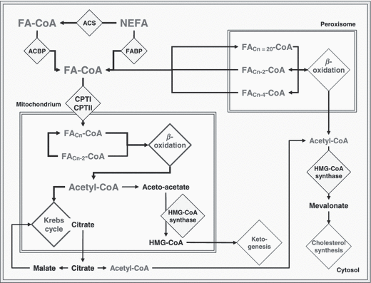
Non-esterified acyl-CoA may be oxidized, either in the mitochondria or peroxisomes. Mitochondrial oxidation may be either complete or incomplete. Incomplete oxidation leads to formation of ketone bodies (Fig. 3).

Fatty acid oxidation: main steps of mitochondrial and peroxisomal β-oxidation, and fate of acetyl-CoenzymeA.
The two main factors regulating the degree to which fatty acids are oxidized by the liver are the supply of fatty acids to the liver (via lipolysis), and the partitioning between oxidation and microsomal esterification.
Intramitochondrial oxidation
Intramitochondrial oxidation of fatty acyl-CoA occurs through the β-oxidation pathways resulting in the formation of acetyl-CoA. During this process, electrons are transferred to flavin-adenine dinucleotide (FAD) and oxidized form of nicotinamide-adenine dinucleotide (NAD+), forming the reduced forms of these coenzymes, which in turn donate electrons to the electron transport chain to drive ATP synthesis. The acetyl-CoA can be oxidized completely to carbon dioxide in the tricarboxylic acid cycle (TCA).
The mitochondrial matrix does not contain any ACS enzyme that could activate fatty acids with 14 carbons or more. Entry of these long-chain fatty acids into the mitochondria is regulated by the activity of the enzyme CPT-I (Kerner and Hoppel, 2000). This enzyme is an integral protein of the outer mitochondrial membrane, and catalyzes the formation of acyl-carnitine molecules which are transported across the mitochondrial membrane by a specific carrier protein and are reconverted to acyl-CoA within the mitochondrial matrix by the action of CPT-II, a peripheral protein of the inner mitochondrial membrane. Short- and medium-chain fatty acids (12 carbons or less) pass through the mitochondrial membrane and are activated by ACS within the mitochondrial matrix. Consequently, oxidation of these fatty acids is not controlled by CPT-I.
The activity of CPT-I is inhibited by interaction with malonyl-CoA, the product of the first step of De novo synthesis of fatty acids catalyzed by acetyl-CoA carboxylase (the activity of which is stimulated by insulin) (Brindle et al., 1985). Negative energy balance results in a decrease in malonyl-CoA, and an increase in fatty acid oxidation. The control of CPT-I by malonyl-CoA would be a way to prevent simultaneous oxidation and synthesis of fatty acids within the liver cell, a potential futile cycle. In ruminants, CPT-I also is highly sensitive to inhibition by methylmalonyl-CoA which is an intermediate in the conversion of propionate to succinyl-CoA in the process of gluconeogenesis.
Peroxisomal and microsomal oxidation
Whereas they function similarly (β-oxidation), there are notable differences between the peroxisomal and mitochondrial pathways for fatty acid oxidation. Peroxisomal β-oxidation is responsible for the metabolism of very long chain fatty acids while mitochondrial β-oxidation is responsible for the oxidation of short, medium and long chain fatty acids. In peroxisomes, the first dehydrogenation of mitochondrial β-oxidation is replaced with an oxidation (acyl-CoA oxidase), resulting in the formation of H2O2 rather than reduced NAD+. Secondly, peroxisomes do not contain an electron transport chain. Peroxisomal β-oxidation therefore results in less ATP-energy than does mitochondrial β-oxidation. Peroxisomal β-oxidation is active with long-chain fatty acids (that are relatively poor substrates for mitochondrial β-oxidation) and provides an ‘overflow’ pathway to help cope with high availability of fatty acids. There are nevertheless wide differences between species. For example, peroxisomal oxidation in liver homogenates from cows may represent 50% and 77% of the total capacity for the initial cycle of β-oxidation of palmitate and octanoate, respectively, but only 26% and 65% for rats (Grum et al., 1994).
Very long chain fatty acids are also metabolized by the cytochrome P450 CYP4A ω-oxidation system to dicarboxylic acids. Indeed, the CYP4A enzymes are especially capable of hydroxylating the terminal ω-carbon and, to a lesser extent the (ω-1) position of fatty acids. Ω-hydroxylation is followed by cytosolic oxidation to produce long chain dicarboxylic acids (Simpson, 1997). These acids cannot be readily metabolized by the mitochondria, whereas they are the preferred substrate for the peroxisomal β-oxidation pathway. They are thus taken up by the peroxisomes and oxidized to fatty acids, which can then be shortened even further by the mitochondria. The induction of this system would be an adaptive response by the hepatocyte to maintain cellular lipid homeostasis. It is important during fatty acid overload of the mitochondrial β-oxidation system with the microsomal CYP4A-mediated ω-oxidation and peroxisomal β-oxidation being co-operatively regulated to achieve fatty acid metabolism in the liver.
Ketogenesis
Under conditions of increased fatty acid uptake, the liver often produces large amounts of the ketone bodies, acetoacetate and β-hydroxybutyrate, in a process known as ketogenesis. Ketogenesis is enhanced in times of increased NEFA uptake by the liver, when low insulin levels cause activation of CPT-I, allowing extensive uptake of fatty acids into mitochondria.
Conversion of acetyl-CoA to ketone bodies, rather than complete oxidation in the TCA cycle, results in the formation of less ATP/mole of fatty acid oxidized (e. g. five times less in the case of palmitate: 129 vs. 27 ATP/mole, TCA and oxidative phosphorylation in the electron transport chain vs. conversion of acetyl-CoA to ketone bodies). Ketogenesis therefore allows the liver to metabolize about five times more fatty acids (for the same ATP yield), and conversion of fatty acids into water-soluble fuels may be an important short-term strategy to redistribute energy.
Ketogenesis (as well as cholesterol biosynthesis) is controlled indirectly by CPT-I (McGarry et al., 1989) and directly by the activity of the mitochondrial key regulatory enzyme 3-hydroxy-3-methylglutaryl-CoA (HMG-CoA) synthase (Hegardt, 1999). The enzyme is regulated by two systems: succinylation in the short term, and transcriptional regulation in the long term (prolonged energy deficit). When the succinyl-CoA pool size increases as a result of an increased flux of glucogenic metabolites, a succinyl group is added to a regulatory sub-unit of HMG-CoA synthase, which inactivates the enzyme. Both control mechanisms are influenced by nutritional and hormonal factors, which explain the incidence of ketogenesis.
Triacylglycerol export (VLDL synthesis and secretion)
The mechanism for synthesis and secretion of VLDL from liver is well known (Adeli et al., 2001). Apoprotein B100 (apoB100; and apoB48 in a few species) is the key component whose rate of synthesis in the rough endoplasmic reticulum controls the overall rate of VLDL production. Lipid components that are synthesized in the smooth endoplasmic reticulum are added by the microsomal triacylglycerol transfer protein to apoprotein B (White et al., 1998) as it moves to the junction of the two compartments. After being carried to the Golgi apparatus in transport vesicles, the apoproteins are glycosylated. Secretory vesicles bud off the Golgi membrane, migrate to the sinusoïdal membrane of the hepatocyte, then fuse with the membrane and release the VLDL into blood.
Animal models have shown that the availability of fatty acids is not the only or the major determinant of the rate of VLDL production (Kendrick et al., 1998; Mason, 1998). Where the limitation in VLDL synthesis or secretion resides is unknown. The rate of synthesis or assembly would be more likely to be limiting than the secretory process per se (White et al., 1998). Inhibition of microsomal triacylglycerol transfer protein (evaluated in the treatment of atherosclerosis) blocks the assembly and secretion of VLDL and chylomicrons but leads to steatosis at least in mice (Liao et al., 2003). Possible limitations also include a high rate of degradation of apoB100, or deficient synthesis of phosphatidylcholine or cholesterol.
There are important species differences in the ability to export triacylglycerols from the liver as VLDL despite similar rates of esterification of fatty acids to triacylglycerols. It has been suggested that among different species, the rate of export of triacylglycerols from the liver is proportional to the capacity of De novo fatty acid synthesis. Cattle (Grummer, 1993), goat (Kleppe et al., 1988) and pigs that do not synthesize fatty acids in the liver also have low rates of triacylglycerol export, whereas poultry that actively synthesize fatty acids in the liver secrete VLDL at very high rates (Pullen et al., 1990). Rates of VLDL export are intermediate for rats and rabbits that undertake lipogenesis in both liver and adipose tissues (Pullen et al., 1990).
The origin of the fatty acids incorporated into triacylglycerols can affect the rate of VLDL export. In obese mice, De novo lipogenesis in the liver does not stimulate VLDL output (Wiegman et al., 2003). In rats, high carbohydrate diets enhance the hepatic output of VLDL triacylglycerols, but this increased secretion of triacylglycerols is accomplished by enhanced formation of VLDL triglyceride from exogenous NEFA rather than from fatty acids synthesised De novo in the liver (Schonfeld and Pfleger, 1971). Plasma NEFA therefore seem to play an important role in enhancing hepatic esterification and stimulating VLDL production (Julius, 2003). The situation would be exacerbated in an insulin-resistant state, which also promotes increased stability of nascent apoprotein B and enhances the expression of microsomal triacylglycerol transfer protein (Taghibiglou et al., 2000; Wiegman et al., 2003).
The mechanism of clearance of accumulated triacylglycerols has not been fully elucidated. In rats, triacylglycerols stored in lipid droplets do not contribute directly to the synthesis of VLDL. Rather it appears that lipolysis of stored triacylglycerol by a microsomal lipase generates NEFA and membrane-bound diacylglycerols, and eventually monoacylglycerols. Usually, as the same lipase hydrolyzes tri- and diacylglycerols, higher triacylglycerol availability and affinity prevents unnecessary formation of monoacylglycerol (Abo-Hashema et al., 1999). Diacylglycerols (and eventually monoacylglycerols) are re-esterified in the microsomal membrane (mainly by a diacylglycerol acyltransferase, and to a lesser extent by monoacylglycerol acyltransferase), translocated into the microsomal lumen, then incorporated into nascent VLDL (Gibbons et al., 2000).
In mice with increased De novo lipogenesis in the liver, VLDL production can be either unaltered or increased, probably depending on the cause of the increase in De novo lipogenesis and the capacity of the liver to increase fatty acid β-oxidation (Wiegman et al., 2003). The inhibition of glucose-6-phosphatase results in an increase in De novo lipogenesis without any stimulation of VLDL production (Bandsma et al., 2001). In contrast, hamsters with increased De novo lipogenesis induced by fructose have increased VLDL production. Therefore, it is likely that different molecular mechanisms are involved explaining the relation between steatosis and the rate of basal VLDL production in different conditions.
Regulation at molecular level
Fatty acids regulate gene expression by controlling the activity or abundance of key transcription factors (Jump et al., 2005), which at the molecular level play a crucial role; this has been particularly illustrated by the link between alterations in their functions and the occurrence of major metabolic diseases. Many transcription factors have been identified as prospective targets for fatty acid regulation, including peroxisome proliferator-activated receptors (PPARα, β and γ) (Schoonjans et al., 1996), SREBP-1c (Xu et al., 1999), retinoid X receptor (RXRα) (Dubuquoy et al., 2002), and LXRα (Zelcer and Tontonoz, 2006). They integrate signals from various pathways and coordinate the activity of the metabolic machinery necessary for fatty acid metabolism with the supply of energy and fatty acids.
Nuclear receptors and transcription factors
Retinoid X receptors (RXRs) play an important regulatory role in metabolic signaling pathways (glucose, fatty acid and cholesterol metabolism) (Ahuja et al., 2003). These receptors activate transcription as homodimers or as obligate heterodimeric partners of numerous other nuclear receptors; especially PPARs and LXRs that belong to the nuclear receptor superfamily of ligand-activated transcription factors and which have been implicated in diverse pathways of lipid metabolism (Barish, 2006; Zelcer and Tontonoz, 2006). Similarly to other nuclear receptors, they interact with nuclear proteins known as co-activators and co-repressors. Activated PPARs or LXRs heterodimerize with RXR (PPAR-RXR or LXR-RXR complex). The heterodimers modulate the transcription of target genes by binding to their promoter region on a specific DNA sequence termed the peroxisome proliferator responsive element (PPRE); this consists of a direct repeat of the nuclear receptor hexameric DNA core recognition (AGGTCA) motif spaced by one nucleotide (Latruffe et al., 2001). Liver X receptors bind to cognate LXR response element (LXRE) sequences that typically consist of a direct repeat of TGACCT spaced by four nucleotides (Willy and Mangelsdorf, 1997). PPARs and LXRs act as key messengers responsible for the translation of nutritional, metabolic and pharmacological stimuli into changes in the expression of genes, especially those genes involved in lipid metabolism.
PPAR-α is highly expressed in the liver and in those tissues that use a lot of lipid-derived energy, where it regulates a set of enzymes crucial for fatty acid oxidation. Indeed, its primary role is to increase the cellular capacity to mobilize and catabolize fatty acids. It increases transcription and expression of proteins and enzymes necessary to transport and catabolize fatty acids (FABP, FAT, CPT-I etc.). It also participates in the regulation of mitochondrial and peroxisomal fatty acid β-oxidation systems, microsomal ω-oxidation system (acyl-CoA oxidase, CYP4A1 and CYP4A6 etc.), and the production of apolipoproteins (Everett et al., 2000). PPAR-α functions as a sensor for fatty acids and ineffective PPAR-α sensing (or PPAR-α null phenotype) can lead to reduced energy burning, resulting in hepatic steatosis. The DNA-binding properties of PPARα and other transcription factors (RXRs) on the PPRE of the mitochondrial HMG-CoA synthase promoter have revealed that ketogenesis can be regulated by fatty acids. Interestingly HMG-CoA synthase can react with PPARα and thus autoregulate its own transcription. PPARγ activation is followed by overexpression of lipogenic enzymes (acetylCoA carboxylase, FAS, GPAT) and FATP. Liver X receptors are master regulators of whole-body cholesterol homeostasis. CYP7a1, which is another member of the cytochrome P450 enzyme family and the rate-limiting enzyme in the pathway of bile acid synthesis is the first direct target of LXRs. Their target genes also include the ATP-binding cassette (ABC) subfamilies (ABCA1 and G1: cholesterol efflux, G5 and G8: bile acid excretion and intestinal cholesterol absorption). In addition to their role in cholesterol metabolism, LXRs are also key regulators of hepatic lipogenesis through the upregulation of the master regulator of hepatic lipogenesis, SREBP-1c, as well as induction of FAS and acyl-CoA carboxylase (Zelcer and Tontonoz, 2006). Sterol regulatory element binding proteins-1 has also been identified as a potent activator of lipogenic gene expression. The regulation of its gene expression by dietary and hormonal factors has already been mentioned. Moreover, polyunsaturated fatty acids suppress SREBP-1c gene expression and inhibit SREBP-1c protein maturation, which results in suppression of its target genes (such as FAS and GPAT) resulting in reduced fatty acid and triglyceride synthesis (Kim et al., 2002).
Nuclear factors and Gene regulation
Nuclear factors play a crucial role in the regulation of lipid metabolism. Indeed, fatty acid metabolism is transcriptionally regulated by two main systems under the control of either LXRs or PPARs. Liver X receptors activate expression of SREBP-1c, an already mentioned dominant lipogenic gene regulator, whereas genes encoding peroxisomal, microsomal and some mitochondrial fatty acid metabolizing enzymes in the liver are transcriptionally regulated by PPARα. An intricate network of nutritional transcription factors with mutual interactions has been proposed, resulting in efficient reciprocal regulation of lipid degradation and lipogenesis. LXR activation, either by overexpression of LXR or its ligand would cause suppression of PPARα signaling, by RXRα competition between PPAR and LXR (Ide et al., 2003). Reciprocally, PPARα activation would suppress the LXR-SREBP-1c pathway through reduction of LXR/RXRα formation (Yoshikawa et al., 2003). As LXRα and PPARα regulate alternate pathways of fatty acid synthesis and catabolism, these nuclear receptors would ‘cross-talk’ to ensure that antagonistic pathways are not simultaneously activated. Other studies have not shown this cross-talk, but some work indicates that PPARα and LXRα activate an overlapping set of genes involved in both fatty acid catabolism and synthesis (Anderson et al., 2004). Hepatic peroxisomal fatty acid β-oxidation would especially be regulated by LXRα, and this process might serve as a counterregulatory mechanism for responding to extreme situations such as hypertriglyceridaemia and liver steatosis (Hu et al., 2005). In such unusual situations, PPARγ expression which is usually low in liver (10–30% of expression in adipose tissue) would nevertheless be capable of selectively upregulating a subset of the lipogenic enzymes in hepatocytes, thus enhancing both lipid synthesis and accumulation leading to steatosis (Coleman and Lee, 2004). PPARγ has been reported to activate LXRα gene expression (Tsukamoto, 2005) which could in turn activate SREBP-1c gene expression and downregulate PPARα, leading to speculation that these four factors could form an auto-loop controlling the activation of adipogenesis and the inactivation of oxidation.
Fatty acids and Gene regulation
Receptors and transcription factors drive liver lipid metabolism. In turn, long chain fatty acids, acyl-CoAs, and other fatty acid-derived compounds (e.g., eicosanoids) are the ligands of nuclear factors and are responsible for their activation, thus acting as metabolic regulators of gene transcription. Fatty acids induce changes in the activity or abundance of at least four transcription factor families: PPARs, LXRs, hepatic nuclear factor 4, and SREBP (Pegorier et al., 2004; Jump et al., 2005). Long chain polyunsaturated fatty acids are strong ligands, while monounsaturated fatty acids are only weak ligands and saturated fatty acids poor ligands. Moreover, downregulation of gene expression by fatty acids would be restricted to polyunsaturated fatty acids, whereas upregulation would be independent of the degree of saturation (Pegorier et al., 2004). Differences might involve differential metabolism (oxidative pathways, kinetics etc.) and selective transport of fatty acids to the nucleus. Table 1 shows some genes involved in lipid metabolism whose expression is regulated by fatty acids. An abundance of polyunsaturated fatty acids regulates numerous PPARα target genes, especially those involved in fatty acid oxidation, while they block the ligand-dependent activation of LXR. Long chain (polyunsaturated) fatty acids suppress SREBP-1c activity, leading to a reduction in liver triacylglycerol content; directly by reducing the nuclear abundance of SREBP-1c, and indirectly because of inactivation of LXR.
| Downregulation | |
| ATP citrate-lyase | Lipogenesis |
| Fatty acid synthase | Lipogenesis |
| Spot 14 | Lipogenesis |
| Stearoyl-CoA-desaturase | Δ9 desaturation |
| Pyruvate kinase | Glycolysis |
| Upregulation | |
| Fatty acid translocase CD36 | Fatty acid transport across cell membrane |
| Fatty acid binding protein | Intracellular fatty acid binding |
| Acyl-CoA-sythetase | Fatty acid activation |
| Acyl-CoA-oxidase | Peroxisomal β-oxidation |
| Carnitine palmitoyltransferase | Transfer into mitochondria |
| Cytochrome P450A2 | Microsomal ω-oxidation |
| Phosphoenolpyruvate carboxykinase | Glyceroneogenesis |
| Uncoupling proteins 2 and 3 | Energy production |
In summary, hepatic lipid metabolism is a highly co-ordinated process, in which many pathways are regulated by nuclear receptors and transcription factors. It is under the tight control of intracellular NEFA levels and composition, but also other metabolic and hormonal factors (e.g. carbohydrate, alcohol, insulin). Despite the complexity and degree of co-ordination of the liver lipid machinery, the overflow of any metabolite pool and/or alteration of nuclear receptors sensing these can lead to organ function impairment and subsequent pathologies.


