Morphology of the Third Eyelid and Superficial Gland of the Third Eyelid on Pig Fetuses
Summary
The morphological and histological examinations of the third eyelid and superficial gland of the third eyelid were conducted in pig fetuses coming from the 20th, 24th, 27th, 30th, 35th, 50th, 63rd, 94th and 112th day of gestation. The morphological examinations were carried out by applying the method of macroscopic preparation with a forehead magnifying glass and binocular (magnification 1.5–5.0×). In order to make anatomical elements more visible, 60–80% absolute alcohol and 0.5–4% acetic acid solution were used for the examinations. On the 20th, 24th, 27th and 30th day of gestation, the whole fetuses were collected for the histological examinations. The whole eyeball with developing accessory organs was collected from the pig fetuses on the 35th day of gestation. On the 50th, 63rd, 94th and 112th day of gestation, only the superficial gland with the third eyelid was collected. Staining with haematoxylin-eosin (H-E) and Azan method was performed. On the 20th, 24th, 27th and 30th day of gestation, the primordia of the glandular epithelium were not found in the examined material. The process of the third eyelid and superficial gland formation starts on the 35th day of gestation. On the 50th and 63rd day of gestation, the gland surrounding the cartilage of the third eyelid is composed of the high amount of loose connective tissue and gland cells which give rise to excretory segments. On the 94th day of gestation, the gland lobes become visible, the efferent ducts form. On the 112th day, the cartilage of the third eyelid assumes the appearance of the mature hyaline cartilage. The excretory segments are composed of simple cuboid epithelium with a large, round nucleus arranged less or more peripherally. Their number increases 2- or even 3-fold at the end of gestation.
Introduction
The superficial gland of the third eyelid belongs to accessory lacrimal glands whose secretion is included in the lacrimal film covering cornea epithelium and palpebral conjunctiva. The superficial gland and the third eyelid are situated in the medial angle of the eye. The third eyelid is more developed in animals. In humans, it constitutes the semilunar fold (Kuryszko and Zarzycki, 2000).
The functions of the third eyelid consist in the mechanical protection of cornea, spreading of lacrimal film whose part (about 30% of the aqueous phase in the dog) is produced by the gland of the third eyelid and the local immunological defence provided by the substances occurring in lymph nodules (Madany, 1999).
The movement of the third eyelid is passive in nature, which is connected with the absence of the muscles actively moving the eyelid. The appropriate location of the free margin of the third eyelid is controlled by the position of the eyeball. The retraction of the eyeball due to the contraction of the retractor bulbi muscle (m.retractor bulbi) results in protrusion towards the temporal angle. The relaxation of the retractor bulbi muscle makes the eyelid move back to the medial canthus (Gelatt, 1981; Lavach, 1990). The purpose of the study is to follow the development of the third eyelid and superficial gland of the pig in the whole gestation period. The study is the continuation of the doctoral thesis (Klećkowska-Nawrot, 2005).
Materials and Methods
The morphological and histological examinations were conducted in pig fetuses coming from the 20th (five fetuses), 24th (five fetuses), 27th (four fetuses), 30th (four fetuses), 35th (four fetuses), 50th (five fetuses), 63rd (five fetuses), 94th (five fetuses) and 112th (five fetuses) day of gestation. The fetuses came from one large scale farm with similar zootechnic and veterinary conditions. The study material was genetically homogenous; it came from the polska biała zwisłoucha (pbz) × the wielka biała polska (wbp) sows and the wielka biała polska (wbp) × the polska biała zwisłoucha (pbz) sows covered by the Hampshire and Duroc boars or their cross breeds with the Pietrain breed. The age of fetuses was established on the basis of cattle paper and confirmed by the application of the method worked out by Marrable (1971)– the measurement of the elongated occiput-caudal section measured from the external occiput eminence to the base of the tail.
The morphological examinations were conducted applying the macroscopic preparation with the use of a forehead magnifying glass and binocular (magnification 1.5–5.0×). In order to make anatomical elements more visible, 60–80% absolute alcohol and 0.5–4% acetic acid solution were used for the examinations. The methods applied in the topographical anatomy: holotopy, syntopy were employed in the study. On the 20th, 24th, 27th and 30th day of gestation, the whole fetuses were collected for the histological examinations. The whole eyeball with developing accessory organs was collected from the pig fetuses on the 35th day of gestation. On the 50th, 63rd, 94th and 112th day of gestation, only the superficial gland with the third eyelid was collected. The collected material was subjected to fixing in 4% buffered formaldehyde solution, washing in running water for 24 h, dehydration in a graded series of alcohols (75% for 20 h, 96% for 8 h, 99.9% for 12 h), embedding in the paraffin and then – cutting into longitudinal cross sections at 7 micron thickness with a Leica RM 2045 microtome. The sections were next stained with haematoxylin-eosin (H-E) and Azan methods. The sections were evaluated in the light microscope 5×, 10×, 40× and 100×. The histological pictures were taken with an Olympus BX 41 microscope (OLYMPUS DP-12 digital camera; OLYMPUS, Tokyo, Japan) applying analySIS 5.0 programme. The following magnifications of pictures were made: 40×, 100× and 200×.
Results
Morphological analysis
The third eyelid and the superficial gland of the third eyelid are macroscopically invisible on the 20th, 24th, 27th, 30th and 35th day of gestation but well visible on the 50th day of gestation. The superficial gland is situated around the cartilage of the third eyelid. The superficial gland lies between the medial rectus muscle (m.rectus medialis) and the ventral rectus muscle (m.rectus ventralis). It contacts ventrally the anterior margin of the deep gland of the third eyelid and is partially covered by the ventral oblique muscle (m.obliquus ventralis). The third eyelid is composed of cartilage which consists of an upper and lower branch and a crossbar. The third eyelid resembles an anchor in shape (1-3).
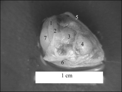
The superficial gland of the third eyelid from the 50th. The right eyeball. The superficial gland of the third eyelid from the 50th. The right eyeball. 1 – upper branch of the cartilage, 2 – crossbar of the cartilage, 3 – superficial gland, 4 – deep gland, 5 – dorsal oblique muscle, 6 – ventral oblique muscle, 7 – cornea.
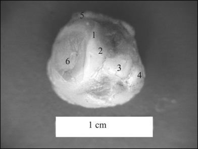
The superficial gland of the third eyelid from the 63rd. The right eyeball. The superficial gland of the third eyelid from the 63rd. The right eyeball. 1 – upper branch of the cartilage, 2 – crossbar of the cartilage, 3 – superficial gland, 4 – deep gland, 5 – dorsal oblique muscle, 6 – cornea.
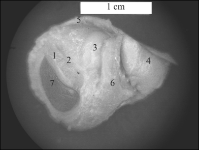
The superficial gland of the third eyelid from the 112th. The right eyeball. The superficial gland of the third eyelid from the 112th. The right eyeball. 1 – third eyelid (upper and lower branch), 2 – third eyelid (crossbar), 3 – superficial gland, 4 – deep gland of the third eyelid, 5 – dorsal oblique muscle, 6 – ventral oblique muscle, 7 – cornea.
The upper and lower branch of the third eyelid cartilage stiffen the fold of the third eyelid, while the crosswise part extends towards the periorbit cone. The crosswise part of the third eyelid is surrounded by the superficial gland of the third eyelid.
The research into the morphology of the third eyelid and the superficial gland showed that the location of the third eyelid and the superficial gland during the whole fetal development of the pig remains unchanged both in the relation to the orbit, eyeball and the neighbouring accessory organs. Similarly, the cartilage of the third eyelid remained anchor-shaped during the whole fetal period.
Histological analysis
On the 20th, 24th, 27th and 30th day of gestation, the primordia of the glandular epithelium were not found in the mesenchymal tissue. The glandular epithelium was observed only on the 35th day of gestation in the form of the strips which will give rise to the superficial gland of the third eyelid in the later part of gestation (Fig. 4).

The superficial gland of the third eyelid from the 35th. 200×. The superficial gland of the third eyelid from the 35th. 200×. 1 – primordia of the gland epithelium, 2 – mesenchymal tissue.
On the 50th and 63rd day of gestation, the superficial gland surrounds the cartilage of the third eyelid in the shape of a ring. The cartilage of the third eyelid is composed of embryonic hyaline cartilage. The hyaline cartilage is made up of cartilaginous cells and the intercellular substance of homogenous appearance. Numerous, regularly arranged, single, round chondrocytes surrounded by a scarce amount of cartilage matrix have been observed (Fig. 6). The superficial gland of the third eyelid is surrounded by the loose connective tissue which penetrates the gland but it does not form lobes yet (Fig. 5). In the loose connective tissue, there occur centres of 2–4 forming excretory segments composed of gland cells. The forming efferent ducts have also been observed (Fig. 7). Another observation has been a high amount of interlobular loose connective tissue composed of collagenous fibres with numerous fibroblasts.

The superficial gland of the third eyelid from the 50th. 200×. The superficial gland of the third eyelid from the 50th. 200×. 1 – hyaline cartilage, 2 – excretory segments, 3 – connective tissue.
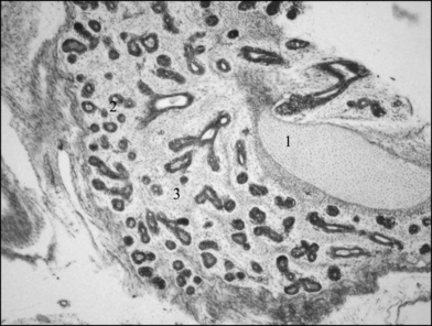
The superficial gland of the third eyelid from the 50th. 40×. The superficial gland of the third eyelid from the 50th. 40×. 1 – hyaline cartilage, 2 - excretory segments, 3 – connective tissue.
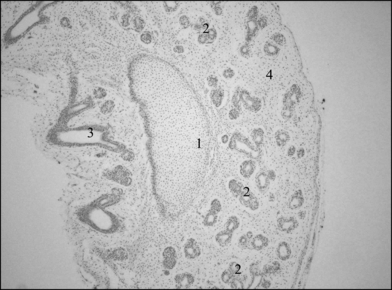
The superficial gland of the third eyelid from the 63rd. 40×. The superficial gland of the third eyelid from the 63rd. 40×. 1 – hyaline cartilage, 2 – excretory segments, 3 – efferent duct, 4 – connective tissue.
On the 94th day of gestation, the chondrocytes of the third eyelid hyaline cartilage are round, regularly and densely arranged in the cartilage matrix. The amount of the loose connective tissue decreases; gland lobes-hardly visible at this stage of development – start to form (Fig. 8) The superficial gland is composed of numerous excretory segments (from 2 to 8–10), made up of the glandular epithelium with a large, round nucleus arranged less or more peripherally.
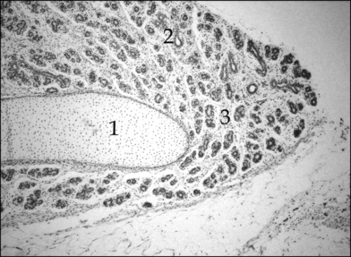
The superficial gland of the third eyelid from the 94th. 40×. The superficial gland of the third eyelid from the 94th. 40×. 1 – hyaline cartilage, 2 – excretory segments, 3 – connective tissue.
On the 112th day of gestation, the chondrocytes of the third eyelid hyaline cartilage are small, flattened and densely situated. In deeper layers, they are larger and spherical, and the distance between each other increases. The superficial gland of the third eyelid is composed of distinctly formed lobes which contain 7–16 excretory segments (Fig. 9). Those excretory are made up of simple cuboid epithelium with a large, round nucleus situated less or more peripherally (Fig. 10). In this stage of gestation, there is a small amount of interlobar and interlobular loose connective tissue forming the connective tissue stroma with numerous fibroblasts.

The superficial gland of the third eyelid from the 112th. 40×. The superficial gland of the third eyelid from the 112th. 40×. 1 – hyaline cartilage, 2 – lobes, 3 – connective tissue.

The superficial gland of the third eyelid from the 112th. 200×. The superficial gland of the third eyelid from the 112th. 200×. 1 – hyaline cartilage, 2 – excretory segments, 3 – connective tissue.
Discussion
The conducted morphological and histological examinations showed that the process of the third eyelid and superficial gland formation in the pig starts on the 35th day of gestation.
According to Aguirre et al. (1972) the superficial gland with the third eyelid in the dog develops from the 32nd–33rd day of gestation.
The third eyelid cartilage in the examined pig fetuses consisted of the dorsal and ventral branches and a crossbar resembling an anchor. According to Getty (1975) in the pig it is T- or anchor-shaped. According to Nickel et al. (1999) and Schlegel et al. (2001), it takes on the shape of an anchor.
In the dog it is T-shaped (Constantinescu and McClure, 1990; Evans, 1993), but Schlegel et al. (2001) think that the upper and lower branch are cone- or crescent-shaped.
According to Acheson (1938) and Nuyttens and Simoens (1995), the third eyelid in the cat is crescent-like in shape. Thompson (1958) describes it as T-shaped. According to Schlegel et al. (2001) it is shaped like a reverse S-form.
The third eyelid cartilage in small ruminants looks like a thin rod with a slightly curved form ending in an oval plate; the crossbar is crescent-like.
The third eyelid cartilage in the horse has a unique structure. The base of the cartilage is surrounded by a thick layer of fatty tissue which gives it a shape of a wide rod. But the crossbar takes on a characteristic hook-form. Its ventral branch supports the free margin of the third eyelid; the dorsal branch has a large incision which gives it a triangular shape (Schlegel et al., 2001) the cartilage – the main component of the third eyelid – is composed of embryonic hyaline cartilage. On the 50th and 63rd day of gestation numerous, regularly arranged cartilaginous cells were found in the cartilage matrix.
On the 112th day of gestation, chondrocytes are arranged in the manner typical of mature cartilage. Near perichondrium they are small, flattened and densely arranged, while in deeper layers they are larger and spherical, and the distance between each other increases (Klećkowska-Nawrot, 2005).
According to Liebich (1999), Nickel et al. (1999), Kuryszko and Zarzycki (2000) and Schlegel et al. (2001), the cartilage of the third eyelid in the pig, dog, cattle and small ruminants is composed of hyaline cartilage. Kuryszko and Zarzycki (2000) and Liebich (1999) characterize the cartilage in the horse as hyaline, but Schlegel et al. (2001) note that it is composed of elastic cartilage. Bisaria and Bisaria (1978) indicate that the third eyelid cartilage in the Albino rat is made up of hyaline cartilage.
The spherical gland of the third eyelid is composed of lobes which become microscopically visible only on the 112th day of gestation. The lobes are made up of excretory segments composed of simple cuboid epithelium with a round nucleus situated less or more peripherally. The number of excretory segments increases 2- or even 3-fold at the end of gestation (Klećkowska-Nawrot, 2005).


