Papular dermatitis due to Leishmania spp. infection in dogs with parasite-specific cellular immune responses
Information in this article has been partially presented as a poster at the 17th European Society of Veterinary Dermatology–European College of Veterinary Dermatology Congress of Veterinary Dermatology, Copenhagen 2001.
Abstract
Abstract Papular dermatitis due to Leishmania spp. infection was diagnosed in three boxers and two Rottweilers with Leishmania-specific cellular immunity. Diagnosis was based on histological and immunohistochemical examination of papules in four dogs and on cytological examination in one dog. Serum protein electrophoresis was within reference ranges and low antibody levels to Leishmania infantum were detected. Delayed-type hypersensitivity (DTH) reaction to leishmanin was evaluated before treatment in three dogs with positive results. After meglumine antimoniate therapy for 3 to 4 weeks and allopurinol treatment for 6 to 10 months, all dogs were clinically normal, had positive DTH reactions to leishmanin and reduced antibody titres. In conclusion, we suggest that this previously unreported cutaneous presentation of canine leishmaniosis appears to be associated with specific immunocompetence and, consequently, with a favourable prognosis.
INTRODUCTION
Canine leishmaniosis is a disease caused by a protozoan parasite of the genus Leishmania, which is transmitted by sand flies.1 The disease is endemic in the Mediterranean basin where the prevalence of infection is much greater than the prevalence of overt Leishmania-related disease.2 Canine Leishmania-infection in the Mediterranean basin is mostly attributed to Leishmania infantum and rarely, to Leishmania tropica.3
Skin lesions are the most common manifestation of canine leishmaniosis although the clinical spectrum of the disease includes local to generalized lymphadenopathy, chronic wasting, anaemia, anorexia, diarrhoea, lameness, epistaxis, splenomegaly and renal failure.1
Typical cutaneous problems in canine leishmaniosis are localized to generalized scaling and nonpruritic alopecia, and nonpainful ulcers most commonly located on the head and limbs.4 Less common dermatological problems are mucocutaneous nodular dermatitis, sterile pustular dermatitis and onychogryphosis.4–6 This wide variability in clinical manifestations is the consequence of the numerous disease pathomechanisms and of the diversity of immune responses of individual hosts.1
The immune response to Leishmania infection in the dog is both humoral and cellular and a whole spectrum of immune responses exists, ranging from predominantly cellular to predominantly humoral. Several studies performed in the dog have demonstrated that, as occurs in the murine model and in humans, a predominantly T-cell (Th1) mediated immune response is associated with a protective immunity, whereas a predominant production of antibodies and absence of cell-mediated immunity is associated with disease susceptibility.7,8 Leishmanin skin test (LST), which detects the delayed-type hypersensitivity (DTH) to Leishmania antigen, has proven to be a useful tool in identifying dogs with a specific cellular immune response.9
The purpose of this article is to describe a previously unreported distinctive papular dermatitis due to Leishmania spp. infection in five dogs with Leishmania-specific cellular immunity.
CASE REPORTS
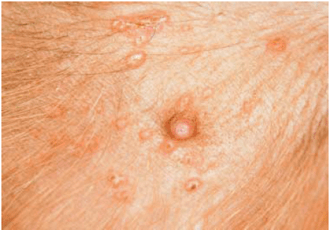
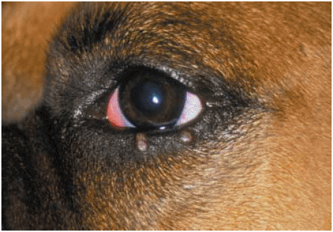
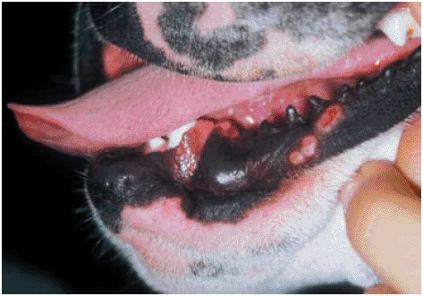
Five dogs (Table 1) were examined at the Veterinary Teaching Hospital of the Veterinary School of Barcelona (Universitat Autonoma de Barcelona) for nonpruritic cutaneous lesions of 2 weeks to a few months duration, appearing from the beginning of May to the end of October. The time of lesion onset for case 4 was unknown. On dermatological examination, multiple coalescent, erythematous and firm umbilicate papules were observed at various sites including the skin of the abdomen, the bridge of the nose, the inner surface of pinnae, the inner aspect of anterior and posterior extremities, the prepuce, the eyelids and the lower lips (1-3; Table 1). In addition, case 1 had multifocal alopecia on the face, including the peri-ocular region, and case 5 had a nodular lesion on the chin. No other abnormalities were found on physical examination, except for case 4 where a mild generalized lymphadenopathy was noted. Fine needle aspiration (cases 1, 2 and 3) and skin biopsies (cases 1, 2, 4 and 5) from several papules were performed. In addition, serum protein electrophoresis and an enzyme-linked immunosorbent assay (ELISA) to detect Leishmania-specific IgG antibodies were carried out in all cases. The ELISA technique was performed, as previously described,10 in the Department of Pharmacology, Veterinary School of Barcelona. The serum from a sick dog with confirmed disease was used as a positive control and the serum from a healthy dog as a negative control. Serological results were considered positive, negative or inconclusive based on the cut-off absorbance values established from 28 noninfected dogs that were living in an endemic region.10 In addition, bone marrow aspiration was performed in case 2 and skin scrapings in case 1.
| Case | Breed | Sex | Age | Time of onset of Leishmania-related cutaneous lesions | Distribution of Leishmania- related cutaneous lesions |
|---|---|---|---|---|---|
| 1 | Rottweiler | F | 16 months | August | Abdomen (Fig. 1), bridge of the nose |
| 2 | Boxer | M | 18 months | End of October | Inner surface of one pinna and of the thigh, prepuce |
| 3 | Boxer | F | 24 months | October | Inner surface of the anterior and posterior extremities, eyelids (Fig. 2), lower lips (Fig. 3), inner surface of one pinna |
| 4 | Boxer | F | 17 months | Unknown | Inner surface of one thigh |
| 5 | Rottweiler | M | 22 months | Beginning of May | Abdomen |
- F, female; M, male.

Abdomen; canine cutaneous leishmaniosis: case 1. Multiple, firm and umbilicate papules.

Peri-ocular region; canine cutaneous leishmaniosis: case 3. Multiple papules due to Leishmania infection are visible on the lower eyelid.

Peri-oral region; canine cutaneous leishmaniosis: case 3. Multiple erythematous and crusted papules are visible on the lower lip.
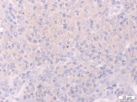
Pathological features are summarized in Table 2. Cytological examination of the papules showed a pyogranulomatous inflammation in which infectious agents were not visualized, except for case 3 in which intramacrophagic bodies compatible with Leishmania amastigotes were observed. Histopathological examination of skin biopsies revealed similar findings in all four cases (cases 1, 2, 4 and 5). Nodular to diffuse pyogranulomatous dermatitis (Fig. 4) with low numbers of intramacrophagic amastigotes was observed by using an immunohistochemical technique (Fig. 5). This was performed, as previously described,11 in the Diagnostic Pathology Service, Veterinary School of Barcelona using rabbit polyclonal anti-Leishmania antibody provided by Dr Dominguez (CSIC, Madrid, Spain).11 Skin sections were also evaluated by periodic acid shift (PAS), Zhiel–Neelsen and Gram stains, with negative results. In case 5, histopathological examination of the nodular lesion on the chin revealed an eosinophilic perivascular dermatitis. Serum protein electrophoresis was within reference ranges in all dogs. Serological testing results were positive, with results slightly above the cut-off in cases 2, 3 and 5 and inconclusive in cases 1 and 4. The bone marrow aspirate performed in case 2 revealed hyperplasia of the myeloid line, but Leishmania organisms were not observed. Parasites of the genus Demodex were identified from skin scrapings performed from the alopecic areas in case 1.
| Case | Cytology | Biopsy | IHC | Special stains* | BM | SPE | ELISA | LST | |||
|---|---|---|---|---|---|---|---|---|---|---|---|
| bt | pt | bt | pt | bt | pt | ||||||
| 1 | PGD | N to D PGD | + | – | NP | U | U | I | – | + | + |
| 2 | PGD | N to D PGD | + | – | Hyperplasia ofmyeloid line | U | U | + | – | + | + |
| 3 | PGD with Leishmania organisms | NP | NP | NP | NP | U | U | + | I | + | + |
| 4 | NP | N to D PGD | + | – | NP | U | U | I | – | NP | + |
| 5 | NP | N to D PGD | + | – | NP | U | U | + | I | NP | + |
- IHC, immunohistochemistry; *PAS, Zhiel–Neelsen, Gram; BM, bone marrow; SPE, serum protein electrophoresis; ELISA, enzyme-linked immunosorbent assay; LST, leishmanin skin test; bt, before therapy; pt, post therapy; PGD, pyogranulomatous dermatitis; N to D, nodular to diffuse; NP, not performed; U, unaltered; I, inconclusive.

Skin; canine cutaneous leishmaniosis: case 1. Diffuse to nodular pyogranulomatous dermatitis. Haematoxylin and eosin, bar = 100 µm.

Skin; canine cutaneous leishmaniosis: case 1. Pyogranulomatous infiltrate with Leishmania amastigotes in the cytoplasm of few macrophages. Indirect immunoperoxidase stain, bar = 25 µm.
Based on the clinicopathological data, a diagnosis of papular dermatitis due to Leishmania spp. infection was made in all dogs. The disease was associated with localized demodicosis in case 1 and with a probable arthropod bite reaction (chin nodule) in case 5.
LST was performed before the beginning of anti-leishmanial therapy in cases 1, 2 and 3, as previously described.12 Briefly, 100-µL solution of inactivated suspension of 3 × 10⁸L. infantum promastigotes/mL in 0.4% phenol-saline (kindly provided by Dr Alvar, Instituto de Salud Carlos III, Madrid, Spain) was intradermally injected in the skin of the groin. Any skin reaction was recorded after 72 h, and an induration or erythematous area > 5 mm in diameter was considered positive. Leishmanin diluent served as control.12 All three dogs had a positive reaction to leishmanin (Fig. 6).

Inguinal region, canine cutaneous leishmaniosis: case 1. Note erythema of the positive reaction to intradermal inoculation to leishmanin after therapy.
All cases were treated with meglumine antimoniate (Glucantim®, Aventis Pharma, Leganes, Madrid, Spain) at 75 mg kg−1 body weight, subcutaneously every 24 h for 1 month, except for case 5 in which injections were discontinued after 19 days because of inflammatory reactions at the inoculation sites. Allopurinol (Zyloric®, Glaxosmithkline SPA, Madrid, Spain) at 10 mg kg−1 body weight, orally every 12 h, was used for a period ranging from 4 to 6 months and concurrently to meglumine antimoniate during the first month in all cases. The exception for this was case 1 in which allopurinol treatment was introduced 1 month after the diagnosis was made. At the time of allopurinol discontinuation all dogs were clinically normal, and had unaltered serum protein electrophoresis. In all dogs a decrease of anti-Leishmania IgG antibody levels was detected, being negative in cases 1, 2 and 4 and inconclusive in cases 3 and 5. In addition, a positive reaction to LST was obtained in all the cases from 0 to 5 months after allopurinol therapy withdrawal.
DISCUSSION
This study describes a series of five cases of cutaneous canine leishmaniosis presenting with a distinctive papular dermatitis. Dogs described here had a history of persistent multiple papular lesions on sparsely haired skin such as the inner aspect of the pinnae, the bridge of the nose, peri-ocular and peri-oral areas, and the ventral abdomen. Clinically, papules were firm, slightly erythematous with a scaling, pale brown surface and umbilicate shape. Lesions were neither painful nor pruritic. This clinical form was characterized by a very low number of intralesional parasites detected by an immunohistochemical technique. This technique is a highly sensitive and specific tool for the diagnosis of canine lesihmaniosis.11 Although immunohistochemistry is considered by some authors as less sensitive than polymerase chain reaction (PCR),13 the technique appeared to be sensitive enough to detect a small number of organisms in the skin sections of cases described herein. The presence of Leishmania amastigotes in skin lesions was considered to play a central role in infection, rather than coincidental exposure, because in clinically normal skin from healthy dogs living in an endemic area, microscopic lesions are absent and immunohistochemistry is negative, even with positive PCR.14
To the authors’ knowledge, this clinical form of canine cutaneous leishmaniosis has not been described to date. However, some considerations need to be made. Nodular forms of canine cutaneous leishmaniosis have been described with variable aspects, from simple papules to tumour-like nodules.4 We believe that these two dermatological forms, namely papular and nodular dermatitis, should be considered as two distinct cutaneous presentations of canine leishmaniosis. This is because of the distinctive clinical appearance of the papular lesions described herein. Moreover, histologically, nodular lesions have been described as a granulomatous dermatitis with giant cells and large numbers of amastigotes.4 These histopathological features differ from the papular dermatitis described in this study, which is characterized by the absence of giant cells and a low number of parasites.
In this report, the papular dermatitis due to Leishmania spp. infection was diagnosed in short-haired dogs, and lesions were localized to scarcely haired skin, more susceptible to sand fly biting. This observation, together with the fact that in 4/5 dogs papules appeared during the active season of the sand fly vector (from early spring to late autumn), leads us to hypothesize that these papules might represent inoculation and multiplication sites of the parasite as it occurs in Old World human localized cutaneous leishmaniosis. This is a self-healing benign form of leishmaniosis limited to the skin.15
The cutaneous manifestation of canine leishmaniosis reported here is suggestive of a benign form of the disease, supported by the lack of systemic signs, the good clinical response to therapy and the absence of relapses. These clinical features might be a consequence of Leishmania-specific immunocompetence, taking into account both the low anti-Leishmania IgG antibody levels and the positive response to LST detected in these dogs. In addition, the low number of intralesional parasites observed in this cutaneous manifestation of leishmaniosis might have been the consequence of parasite-specific cellular immunity, indicating an efficient control of parasite multiplication.
In conclusion, we suggest that this cutaneous presentation of Leishmania-related disease is associated with specific immunocompetence and, consequently, with a favourable prognosis.
REFERENCES
Résumé Une dermatite papuleuse due àLeishmania spp.a été diagnostiquée chez trois Boxers et deux Rottweilers. Le diagnostic a été fait par l’examen histopathologique et immunohistochimique de papules chez 4 chiens et par la cytologie dans un cas. L’électrophorèse des protéines sériques était dans les normes et des taux faibles d’anticorps anti-leishmania ont été détectés. Une hypersensibilité retardée (DTH) à la leishmanine a étéévaluée avant le traitement chez trois chiens, avec un résultat positif. Un traitement avec l’antimoniate de méglumine pendant 3 à 4 semaines et avec l’allopurinol pendant 6 à 10 mois a permis une guérison dans tous les cas. Tous les chiens avaient alors une réaction positive à la leishmanine et une diminution des titres en anticorps. En conclusion, nous suggérons que cette présentation nouvelle de leishmaniose semble être associée à une immunocompétence spécifique et par conséquence un pronostic favorable.
Resumen La dermatitis papular debido a la infección por Leishmania spp. Fue diagnosticada en tres Boxers y dos Rottweilers con inmunidad celular Leishmania-específica. El diagnóstico se basó en el examen histológico e inmunohistoquímico de las pápulas de cuatro perros y en el examen citológico en un perro. La electroforesis de las proteínas séricas se encontraba dentro de los márgenes de referencia y se detectaron niveles bajos de anticuerpos contra L. infantum. Se evaluó una reacción de hipersensibilidad retrasada (DTH) a leishmanina antes del tratamiento en tres perros con resultados positivos. Después del tratamiento con antimoniato de meglumina durante tres a cuatro semanas y alopurinol durante seis a 10 meses, todos los perros eran clínicamente normales, mostraban reacciones positivas DTH a leishmanina y redujeron los títulos de anticuerpos. En conclusión, sugerimos que esta presentación cutánea no descrita anteriormente de la leishmaniosis canina parece estar asociada a inmunocompetencia específica y, consecuentemente, con un pronóstico más favorable.
Zusammenfassung Bei drei Boxern und zwei Rottweilern wurde eine papuläre Dermatitis aufgrund einer Infektion mit Leishmania spp. mit Leishmania-spezifischer zellulären Immunität diagnostiziert. Die Diagnose basierte auf histologischer und immunhistochemischer Untersuchung von Papeln bei vier Hunden und auf zytologischer Untersuchung bei einem Hund. Serumprotein-Elektrophorese war innerhalb des Referenzbereiches und es wurden niedrige Antikörpertiter gegen L. infantum nachgewiesen. DTH-Reaktion (delayed-type hypersensitivity) gegen Leishmanin wurde vor der Behandlung bei drei Hunden mit positivem Ergebnis überprüft. Nach Meglumin Antimonat-Behandlung für drei bis vier Wochen und Allupurinol-Behandlung für sechs bis zehn Monate waren alle Tiere klinisch normal, hatten positive DTH-Reaktion gegen Leishmanin und verminderte Antikörpertiter. Folglich behaupten wir, dass diese bislang nicht berichtete cutane Präsentation caniner Leishmaniose mit spezifischer Immunkompetenz und folglich mit einer günstigen Prognose verbunden zu sein scheint.


