Targeting proteins to the cell wall of sporulating Bacillus anthracis
Summary
Dormant spores of Bacillus anthracis germinate during host infection and their vegetative growth and dissemination precipitate anthrax disease. Upon host death, bacilli engage a developmental programme to generate infectious spores within carcasses. Hallmark of sporulation in Bacillus spp. is the formation of an asymmetric division septum between mother cell and forespore compartments. We show here that sortase C (SrtC) cleaves the LPNTA sorting signal of BasH and BasI, thereby targeting both polypeptides to the cell wall of sporulating bacilli. Sortase substrates are initially produced in different cell compartments and at different developmental stages but penultimately decorate the envelope of the maturing spore. srtC mutants appear to display no defect during the initial stages of infection and precipitate lethal anthrax disease in guinea pigs at a similar rate as wild-type B. anthracis strain Ames. Unlike wild-type bacilli, srtC mutants do not readily form spores in guinea pig tissue or sheep blood unless their vegetative forms are exposed to air.
Introduction
Bacillus anthracis, the causative agent of anthrax, is a spore-forming, Gram-positive microbe that requires oxygen for vegetative growth and spore formation (Roth et al., 1955; Koch, 1876). Upon entry into host tissues, spores, the infectious form of B. anthracis, germinate within phagocytes and the resulting vegetative forms then replicate in the cytosol of immune cells (Koch, 1876; Ruthel et al., 2004; Hu et al., 2006; Ribot et al., 2006). B. anthracis dissemination and replication in all organ tissues of the infected host generates up to 1010 vegetative bacilli per gram of blood or tissue (Koch, 1876; Ivins et al., 1994). Vegetative bacilli secrete lethal factor (LF), protective antigen (PA) and oedema factor (OF) (Smith et al., 1955; Leppla, 1982; 2000; Vodkin and Leppla, 1983; Robertson and Leppla, 1986; Klimpel et al., 1994; Duesbery et al., 1998). Assembly of LF and PA into lethal toxin and of OF and PA into oedema toxin (OT) are critical events in the pathogenesis of anthrax disease, enabling toxin-mediated killing of infected hosts (Milne et al., 1994; 1995; Collier and Young, 2003).
At the time of death, anthrax-infected animals do not harbour spores (Koch, 1876). B. anthracis spore formation in carcasses of anthrax-infected small animals, such as rabbits, guinea pigs or mice, occurs within 24–48 h (Toschkoff and Veljanov, 1970). Spore formation in large animals, cows or sheep, however, requires cadaver opening and exposure of infected tissues to air (Koch, 1876). These observations are consistent with the general hypothesis, initially derived from bacterial growth studies in laboratory media, whereby B. anthracis sporulation occurs only in the presence of oxygen (Roth et al., 1955).
The cell wall envelope of Gram-positive bacteria represents a unique cellular structure, which contains proteins that require specific sorting signals for their anchoring (Marraffini et al., 2006). Sortases cleave motif sequences of sorting signals and form amide bonds between the C-terminal carboxyl groups of proteins and amino groups of peptide cross-bridges within cell wall peptidoglycan (Schneewind et al., 1995; Ton-That et al., 1999). The genome of B. anthracis encodes three sortases (Read et al., 2003; Gaspar et al., 2005). Sortase A cleaves LPXTG motif sorting signals of seven surface proteins destined for the cell wall of vegetative bacilli: BasA, BasB, BasC, BasD, BasE, BasF and BasG (B. anthracissurface protein). Physiological functions are thus far known for BasC, a collagen adhesin that promotes bacterial binding to connective tissues (Xu et al., 2004), and BasD, the surface protein receptor of γ-bacteriophage (Davison et al., 2005). B. anthracis sortase B recognizes the NPKTG motif of BasK (IsdC, iron-regulated surface determinant C), a haem-binding protein involved in uptake of iron during infection (Gaspar et al., 2005; Maresso et al., 2006). The third sortase-like gene, sortase C (srtC) (BA5069), is located in the basI-srtC-sctR-sctS operon, where basI (BA5070) encodes for a surface protein with an LPNTA-motif sorting signal and sctR-sctS encodes for a two-component regulatory system (Fig. 1A). A second gene specifying a polypeptide with LPNTA-motif sorting signal, basH (BA0397), is positioned elsewhere in the B. anthracis genome (Gaspar et al., 2005) (Fig. 1A). Although bioinformatic analysis provides a guide for the discovery of physiological gene function, the assignment of sortase/substrate relationships does require experimental proof. Further, the contribution of sortases and surface proteins to the pathogenesis of anthrax and the replication cycle of B. anthracis cannot be gleaned from bioinformatics analysis alone. This report examines the molecular and physiological properties of sortase C and its substrates.
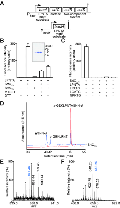
Biochemical characterization of B. anthracis sortase C.A. The basI-srtC-sctR-sctS operon contains basI, a sortase substrate with a LPNTA-motif sorting signal (B. anthracissurface protein I), sortase C (srtC), sctR response regulator (sortase Ctwo-component system regulator) and sctS (sensory kinase). basH encodes a second sortase substrate with LPNTA sorting signal.B. SrtCΔN was purified from E. coli (Coomassie-stained SDS-PAGE, inset) and incubated with peptide substrate (a-GEKLPNTASNN-d) in the presence or absence of thiol reagent MTSET or reducing agent DTT. Peptides were conjugated to the fluorophore 2-aminobenzoyl (a) and the quencher 2,4-dinitrophenyl (d) groups at the N- and C-terminus respectively. 2-aminobenzoic acid fluorescence increases as a result of peptide cleavage due to loss of intramolecular quenching. Peptide cleavage was measured as arbitrary fluorescence units.C. SrtCΔN was incubated with different peptides harbouring pentapeptide motifs of C-terminal cell wall sorting signals of B. anthracis surface proteins (Gaspar et al., 2005). SrtCΔN cleaved only LPNTA, but not LPATG (SrtA substrate), LGATG nor NPKTG (SrtB substrate) peptides.D. rpHPLC separation and mass spectrometry of SrtCΔN substrate and reaction products reveals that sortase C cleaves the LPNTA motif between the threonine and the alanine residues.E. MALDI-TOF mass spectrometry of the compound in the 42 min fraction (D). A species with m/z 877.46 was detected that could be assigned the compound structure a-GEKLPNT-OH (calculated m/z 877.98). Asterisks indicate signals present in matrix control spectra.F. MALDI-TOF mass spectrometry of the compound in the 40 min fraction (D). The main cleavage product with m/z 656.25 was detected and assigned the compound structure NH2-ASNN-d with a calculated m/z of 656.59.
Results
Sortase C (SrtC) cleaves LPNTA motif peptides
Due to their genetic linkage (Fig. 1A), we hypothesized that sortase C may specifically recognize and cleave the BasI LPNTA motif. Recombinant SrtCΔN, with a replacement of the N-terminal signal peptide of sortase C for six histidyl, was purified from Escherichia coli lysate by affinity chromatography on Ni-NTA (Fig. 1B, inset) and its activity was tested towards substrate peptides encompassing sorting signal motif sequences of different types of B. anthracis surface proteins – LPNTA, LPATG, LGATG and NPKTG. A fluorescence-based cleavage assay was used to measure sortase activity. Fluorescence of 2-aminobenzyl-GEKLPNTASNN-dinitrophenyl (a-GEKLPNTASNN-d) is quenched due to the close proximity of the fluorophore (amino-benzyl) and the quencher (dinitrophenyl). Incubation of a-GEKLPNTASNN-d with SrtCΔN increased sample fluorescence more than 30-fold above background, indicating that in vitro peptide cleavage did occur (Fig. 1B). SrtCΔN cleaved only LPNTA, but not LPATG, LGATG and NPKTG peptides (Fig. 1C). In contrast, SrtAΔN and SrtBΔN, which recognize LPKTG and NPKTG, respectively, did not cleave LPNTA motif peptide (Fig. 1B) (Gaspar et al., 2005; Maresso et al., 2006). Incubation of SrtCΔN with methylmethane-thiosulphonate (MTSET) abolished substrate cleavage (Fig. 1B), suggesting MTSET may form a disulphide with the active site cysteine (Cys181) of sortase C, a property observed for all other sortases examined thus far (Ton-That and Schneewind, 1999; Ton-That et al., 1999; Mazmanian et al., 2002; Gaspar et al., 2005; Maresso et al., 2006).
Reaction products of SrtCΔN or mock-treated peptide substrate were separated by reversed phase HPLC and elution monitored by absorbance (Fig. 1D). Mock-treated peptide eluted at 49 min (39% acetonitrile). MALDI-TOF mass spectrometry of the eluted compound confirmed substrate integrity. Incubation of substrate with SrtCΔN generated two product peaks eluting at 40 (36% acetonitrile) and 42 min (32% acetonitrile). MALDI-TOF mass spectrometry of the compound in the 40 min fraction (Fig. 1F) revealed the main cleavage product with m/z 656.25 and assigned the compound structure NH2-ASNN-d (calculated m/z of 656.59). MALDI-TOF mass spectrometry of the compound in the 42 min fraction (Fig. 1E) revealed a species with m/z 877.46 and assigned compound structure a-GEKLPNT-OH (calculated m/z 877.98). Together these data indicate that SrtCΔN cleaves peptides encompassing the LPNTA motif between threonine (T) and alanine (A). This reaction is inhibited by MTSET, consistent with the hypothesis that the thiol of Cys181 functions as the active site SrtCΔN (Fig. 1B). These results suggest further that basH and basI, whose precursor products harbour LPNTA-motif sorting signals, likely encode in vivo substrates of sortase C.
srtC is expressed during sporulation
A hallmark of sporulation in the soil bacterium Bacillus subtilis is the formation of an asymmetrically positioned (polar) septum that divides the developing cell into dissimilarly sized progeny called the forespore (the smaller cell) and the mother cell (Losick and Stragier, 1992). Both compartments further differentiate by activating different sigma (σ) factors (Losick and Pero, 1981), each of which directs transcription of a specific set of genes (Losick and Stragier, 1992). Activation of σF in the forespore leads to the expression of forespore-specific genes as well as activation of σE in the mother cell (Karow et al., 1995; Londono-Vallejo and Stragier, 1995). Differential gene expression in the two cell types brings about engulfment of the forespore as the mother cell membrane migrates around the forespore (Rubio and Pogliano, 2004). Following engulfment, the forespore matures into a fully formed spore (Chada et al., 2003), which is released after mother cell lysis (Nicholson et al., 2000).
To examine sporulation of B. anthracis, an overnight culture of B. anthracis strain Sterne in brain–heart infusion (BHI) broth was diluted 1:10 into modG sporulation medium (Kim and Goepfert, 1974) and incubated at 30°C. Optical density (OD600) of culture aliquots was used as a measure for vegetative growth values in hourly intervals. T0 marks the time when exponential growth reaches plateau and sporulation commences (Fig. 2A). FM4-64 membrane dye was used to monitor sporulation by fluorescence microscopy (Fig. 2B). Polar septation of B. anthracis strain Sterne begins at T+1 and forespore engulfment neared completion at T+3. Bright endospores were observed at T+5. Henceforth, optical density measurements of B. anthracis cultures and fluorescence microscopy of FM4-64-stained samples were used to examine gene expression during spore development.
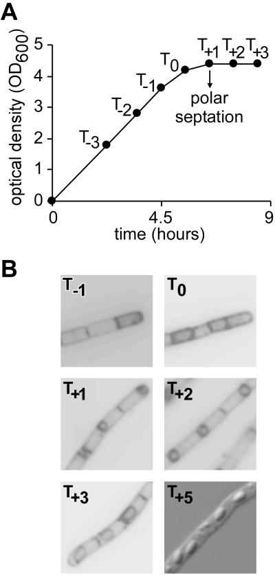
Bacillus anthracis sporulation analysed by fluorescence microscopy.A. Typical sporulation curve of B. anthracis Sterne. An overnight culture of B. anthracis in brain–heart infusion broth was diluted 1:10 into modG sporulation medium (Kim and Goepfert, 1974) and incubated at 30°C. OD600 values were taken every hour. T0 marks the time when exponential growth reaches plateau.B. Forespore development. FM4-64 membrane dye was used to monitor sporulation by fluorescence microscopy. Polar septation begins at T+1 and forespore engulfment neared completion at T+3. Bright endospores were observed at T+5.
Specific antibodies [rabbit antibodies raised against purified SrtC (αSrtC) and BasI (αBasI)] and gfp transcriptional fusions were used to probe expression of srtC::gfp and its substrates genes, basH::gfp and basI::gfp, in B. anthracis Sterne under sporulating conditions. SrtC was detected by immunoblotting in bacillus extracts at T−1, T0 and T+1 (Fig. 3A), but only when basI-srtC-sctR-sctS expression was increased in sporulating cells harbouring a multicopy plasmid with the sctR response regulator (pLM218, Fig. 3B). SrtC was not detected in B. anthracis with vector control plasmid (data not shown). Immunoblot analysis indicated that SrtC abundance peaked at T0 and declined at T+1 (Fig. 3A). Disappearance of SDS-soluble SrtC appears to be at least in part due to SrtC sequestration within developing spores (vide infra). Fluorescent signal generated from a chromosomal srtC::gfp transcriptional fusion was first detected in sporulating cells at T−1, 2 h prior to asymmetric septum formation (T+1) (Fig. 3B). As a test for the regulatory function of SctR, the sctR gene was placed under control of the IPTG-inducible Pspac promoter and expression induced by the addition of IPTG during exponential growth (Fig. 3C). GFP fluorescence was observed in vegetative srtC::gfp bacilli during exponential growth, indicating that the SctR response regulator is sufficient to induce expression of basI-srtC-sctR-sctS in the absence of other sporulation-specific factors (Fig. 3C).
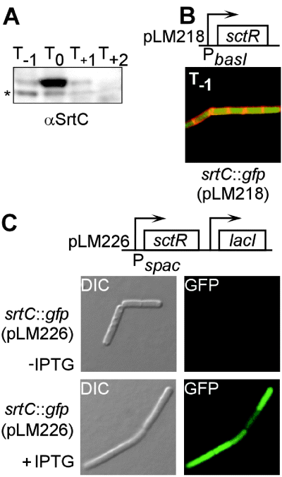
Expression of srtC in sporulating B. anthracis.A. srtC expression begins at T−1 (2 h before polar septation) as measured by immunoblotting of sporulating B. anthracis extracts with SrtC-specific antiserum (αSrtC). A faster-migrating species (*) constitutes a SrtC degradation product.B. srtC expression begins at T−1 as measured by fluorescence microscopy of B. anthracis carrying a srtC::gfp transcriptional fusion and pLM218, which contains sctR under the control of the basI-srtC promoter (PbasI). Red fluorescence of the membrane dye FM4-64 delineates cell boundaries.C. IPTG-inducible expression of the two-component response regulator SctR promotes srtC::gfp expression in bacilli during vegetative growth, as judged by microscopic detection of GFP fluorescence.
Sortase C substrate BasH is expressed in the forespore
Nucleotide sequence upstream of basH matches the σF consensus (Wang et al., 2006), i.e. the promoter recognition sites of σF RNA polymerase for genes that are only transcribed in the forespore (Schmidt et al., 1990; Fig. 4A). To visualize basH expression, bacteria were transformed with a plasmid containing gfp under control of the basH promoter (Fig. 4B). Fluorescence microscopy of sporulating bacilli revealed forespore-restricted expression of PbasH–gfp (4, 5). Previous work using DNA microarray analysis also revealed basH transcription during sporulation (Liu et al., 2004). Fluorescence of a translational hybrid, BasH–GFP (Fig. 5B), was detected at forespore boundaries, consistent with signal peptide-mediated targeting of BasH–GFP to the forespore plasma membrane (Fig. 5C and D). To determine whether σF RNA polymerase is sufficient to drive basH expression, σF (spoIIAC) expression was induced via the IPTG-inducible Pspac promoter (pLM223, Fig. 4B) during vegetative growth of bacilli. GFP fluorescence was indeed observed during exponential growth in the presence but not in the absence of IPTG, indicating that σF RNA polymerase directs basH expression (Fig. 4D) and that its product, BasH, must be targeted to the cell wall compartment of B. anthracis forespores (Fig. 5).
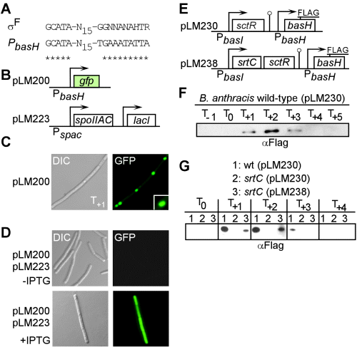
SrtC-dependent targeting of BasH to the cell wall of forespores.A. Alignment of basH promoter with σF consensus sequence (Wang et al., 2006). Asterisks indicate conserved nucleotides.B. Plasmid pLM200 encodes for a transcriptional fusion between basH promoter (PbasH) and gfp coding sequence. pLM223 harbours B. anthracis spoIIAC (σF) under control of the IPTG-inducible Pspac promoter.C. DIC and fluorescence microscopy images of B. anthracis harbouring pLM200 under sporulation conditions (T+1).D. Images of B. anthracis harbouring pLM200 and pLM223 during exponential growth in the presence or absence of IPTG.E. pLM230 harbours basH, carrying the FLAG epitope three codons upstream of coding sequence for the LPNTA motif (BasHFLAG), as well as the response regulator sctR. pLM238 encodes BasHFLAG, SrtC and SctR.F. FLAG immunoblot of cell wall extracts generated from sporulating cultures (T−1 to T+5) of wild-type B. anthracis carrying pLM230 grown under sporulation conditions.G. Anti-FLAG immunoblot of cell wall extracts generated from sporulating cultures of wild-type B. anthracis carrying pLM230 (lane 1) or srtC mutants harbouring pLM230 (lane 2) or pLM238 (lane 3).
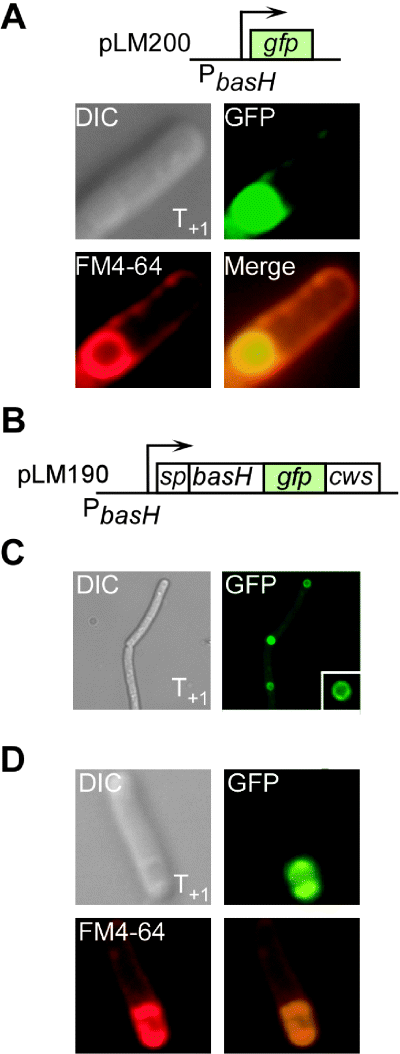
basH is expressed within B. anthracis forespores.A. Plasmid pLM200 encodes for a transcriptional fusion between basH promoter (PbasH) and gfp coding sequence. B. anthracis Sterne bearing pLM200 was analysed by fluorescence microscopy to reveal the basH expression pattern. GFP fluorescence was only detected during sporulation, starting at T+1. GFP expression was restricted to the forespore compartment, which was delineated with membrane dye FM4-64.B. Plasmid pLM190 encodes for a basH–gfp translational fusion. gfp coding sequence was inserted three codons upstream of nucleic acid sequence specifying the BasH cell wall sorting signal (cws). sp indicates basH sequences encoding for an N-terminal signal peptide that directs secretion of the protein product.C. Fluorescence of BasH–GFP was localized to forespore boundaries. B. anthracis Sterne harbouring pLM190 was analysed by fluorescence microscopy at T+1. Green fluorescence was confined to forespore boundaries (inset).D. BasH–GFP is initiated into the secretion pathway. Bacilli expressing BasH–GFP during sporulation (T+1) were stained with FM4-64, revealing colocalization of BasH–GFP (green) and membrane (FM4-64, red) fluorescence at forespore boundaries. The fusion protein is presumably initiated into the Sec pathway, a prerequisite for entering the sorting pathway (Marraffini et al., 2006); however, tight folding of GFP precludes secretion of the hybrid.
BasH is anchored to the forespore cell wall envelope by sortase C
To examine whether BasH is indeed targeted to forespore cell wall, codons for the FLAG epitope were inserted into the basH open reading frame, upstream of nucleotide sequences that specify the LPNTA sorting signal. basHFLAG was cloned on a plasmid and expressed from its σF-dependent promoter in sporulating bacilli (pLM230, Fig. 4E). Culture aliquots were treated with PlyL N-acetylmuramoyl-l-alanine amidase (Low et al., 2005) and lysates of the cell wall compartment analysed by immunoblotting with anti-FLAG monoclonal antibody. BasHFLAG was detected at T+1, when the first polar septa appeared (Fig. 4F). Abundance of BasHFLAG peaked at T+2, declined at T+3 and, upon further incubation (T+4 and T+5), no immunoreactive species could be detected (Fig. 4F). Similar to SrtC, BasH polypeptide appears to be sequestered upon spore maturation, presumably preventing PlyL-mediated solubilization of spore peptidoglycan with anchored BasH. In B. subtilis, cortex peptidoglycan biosynthesis has not yet commenced during the T+1–T+3 interval (Meador-Parton and Popham, 2000). Assuming that this may also be true for B. anthracis, BasH may be anchored to primordial peptidoglycan surrounding the newly formed forespore. BasH targeting to forespore cell wall occurred in wild-type B. anthracis, but not in an isogenic srtC mutant (Fig. 4G). This defect was complemented in sporulating srtC mutant bacilli harbouring plasmid-encoded wild-type srtC (pLM238, Fig. 4G).
BasI is anchored to the cell wall envelope by sortase C
Expression and anchoring of BasI were also analysed in sporulating bacteria using optical density measurements and fluorescence microscopy of FM4-64-stained B. anthracis cultures. When measured with GFP fluorescence generated from a chromosomal basI::gfp transcriptional fusion in sporulating bacilli, basI expression commenced at T−1 (Fig. 6B). PlyL digestion of the cell wall followed by immunoblotting revealed that BasI is anchored to peptidoglycan of sporulating bacilli in a srtC-dependent manner (Fig. 6C). The defect in anchoring of BasI was restored by plasmid encoded srtC, whose expression was activated by the sctR response regulator also encoded on the same plasmid (pLM233, Fig. 6A and C). As a fractionation control, ribosomal subunit protein L6 was not found in the cell wall compartment (Fig. 6C).
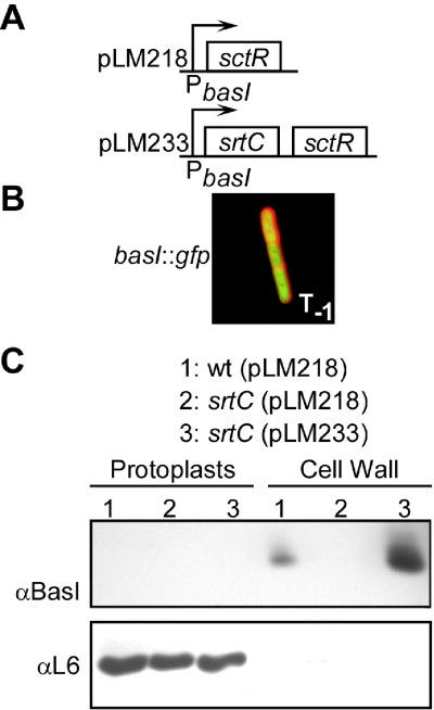
SrtC-dependent targeting of BasI to the cell wall of sporulating B. anthracis.A. Plasmid pLM218 contains sctR under control of the basI promoter (PbasI), thereby promoting expression of chromosomal basI-srtC via sctR response regulator. pLM233 provides for expression of srtC and sctR under PbasI control.B. basI expression commences 2 h prior to polar septation (T−1) in a fluorescence microscopy experiment with B. anthracis harbouring a transcriptional basI::gfp fusion on the chromosome as well as plasmid pLM218. Red fluorescence of the membrane dye FM4-64 delineates mother cell boundaries.C. BasI is anchored to the cell wall of sporulating bacilli in a srtC-dependent manner. Wild-type cells containing pLM218 (lane 1) and srtC mutants containing either pLM218 or pLM233 (lanes 2 and 3 respectively) were grown under sporulation conditions (T−1). BasI was detected by immunoblotting in cell wall fractions only in the presence (lanes 1 and 3), but not in the absence of srtC (lane 2). Overexpression of plasmid-encoded srtC appeared to increase the amount of cell wall-anchored BasI. As a fractionation control, cell wall and protoplast fractions were analysed by anti-L6 immunoblot (ribosomal protein control).
SrtC–mCherry is inherited by B. anthracis forespores
srtC is expressed by sporulating cells prior to septum formation between mother cell and forespore, i.e. before the cellular compartment that represents the final destination of BasH has been formed. Forespore inheritance of SpoIIIJ, a regulatory factor, has been described in B. subtilis (Serrano et al., 2003). To test whether SrtC is also inherited by the forespore compartment, plasmid-encoded translational srtC–mCherry fusion was expressed via the basI-srtC promoter. As expected, SrtC–mCherry was detected in sporulating bacilli that had not yet formed an asymmetric septum (T−1, Fig. 7). Upon septum formation (T+1) SrtC–mCherry was detected by fluorescence microscopy in both forespore and mother cell compartments. Upon further incubation, SrtC–mCherry fluorescence was detected in mature spores (T+20, Fig. 7). Thus, even though basH is expressed in a cell that does not synthesize SrtC, mother cell membranes ensure inheritance of sortase C into the forespore compartment, thereby enabling BasH anchoring to the forespore cell wall envelope.
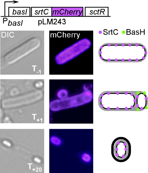
Sortase C is inherited by the B. anthracis forespore. Plasmid pLM243 provides for the expression of SrtC–mCherry in wild-type bacilli grown under sporulation conditions and analysed by fluorescence microscopy. The drawing depicts a model for SrtC and BasH localization during spore development, integrating data displayed in 1-5.
srtC contributions during the pathogenesis of anthrax in guinea pigs
basI-srtC-sctR-sctS operon and basH homologues were found only in genomes of members of the Bacillus cereus group (Jensen et al., 2003) (B. cereus, B. anthracis and Bacillus thuringiensis), but not in the genome of other Bacillus species. Isogenic variants of the non-virulent vaccine strain, B. anthracis Sterne (Sterne, 1937), carrying a deletion of the srtC gene, displayed no defect in germination, growth, spore formation or virulence (data not shown). This prompted us to derive srtC mutants from the fully virulent isolate B. anthracis Ames (Welkos et al., 1989). Similar to Sterne variants, srtC mutants of B. anthracis Ames displayed no defect in spore formation and germination in laboratory media (data not shown) and no defect in the ability to cause lethal anthrax disease in a guinea pig model of subcutaneous infection (Ivins et al., 1994; Fellows et al., 2001; Fig. 8A). At the time of death, organ tissues of animals infected with B. anthracis Ames or its isogenic srtC mutant were teeming with vegetative bacilli in spleen and blood (Table 1). Within 3 weeks, tissues of animals that had been infected with wild-type bacilli generated infectious spores (∼20% of vegetative bacilli), whereas tissues infected with the srtC mutant strain did not (Table 1). To investigate whether deletion of srtC renders spores heat-sensitive or even abolishes spore formation, tissue samples were stained for spores and viewed by microscopy. Abundant spores were detected in heart blood and in spleens from animal carcasses that had been infected with B. anthracis Ames, but not in animals that succumbed to infection by srtC mutants (Fig. 8B).
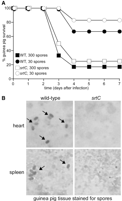
Virulence and sporulation of srtC mutants derived from B. anthracis Ames in a guinea pig infection model.A. Kaplan–Meier analysis for guinea pigs infected with B. anthracis Ames wild type or srtC spores. Six animals each were injected subcutaneously with wild-type or srtC mutant spore preparations containing 30 or 300 spores (Ivins et al., 1994) and monitored for lethal anthrax disease over 14 days. LD50 values were calculated for wild type (66 spores) and srtC mutants (47 spores).B. Heart and spleen tissues harvested from carcasses of guinea pigs that had been infected with B. anthracis Ames (wild type) or isogenic srtC mutant spores (see Table 1) were Schaeffer–Fulton stained to detect spores (Schaeffer and Fulton, 1933). Spores (arrows) were detected only in tissues harbouring B. anthracis Ames.
| Strain | Organa | Vegetative bacillibc | Colony-forming units (day 21)c | Heat-resistant spores (day 21)c | Sporulation efficiencyd |
|---|---|---|---|---|---|
| Wild type | Heart blood | 1.05 × 10⁸ | 2.60 × 10⁸ | 5.10 × 10⁷ | 19.6 |
| Wild type | Heart blood | 3.25 × 10⁷ | 1.90 × 10⁷ | 3.80 × 10⁶ | 20.0 |
| Wild type | Heart blood | 7.75 × 10⁸ | 1.40 × 10⁷ | 2.50 × 10⁶ | 17.9 |
| Wild type | Spleen | 1.05 × 10⁹ | 2.00 × 10⁷ | 7.10 × 10⁶ | 35.5 |
| Wild type | Spleen | 1.92 × 10⁹ | 1.80 × 10⁷ | 4.10 × 10⁶ | 22.8 |
| Wild type | Spleen | 6.00 × 10⁹ | 3.50 × 10⁷ | 9.00 × 10⁶ | 25.7 |
| srtC | Heart blood | 2.57 × 10⁷ | 3.60 × 10⁶ | < 100 | < 0.001 |
| srtC | Heart blood | 4.00 × 10⁷ | 7.60 × 10⁵ | < 100 | < 0.001 |
| srtC | Heart blood | 4.00 × 10⁶ | 3.10 × 10⁵ | < 100 | < 0.001 |
| srtC | Spleen | 7.75 × 10⁸ | 2.70 × 10⁶ | < 100 | < 0.001 |
| srtC | Spleen | 1.00 × 10⁸ | 3.00 × 10⁷ | < 100 | < 0.001 |
| srtC | Spleen | 1.20 × 10⁹ | 3.50 × 10⁶ | < 100 | < 0.001 |
- a. Organs were removed from guinea pigs (infected with B. anthracis Ames or isogenic srtC mutant spores) at time of death (4 days following infection), homogenized and incubated in sterile tubes at room temperature.
- b. Enumerated as colony-forming units per millilitre of tissue harvested at time of death. No spores were detected at this time and no statistically significant difference in the number of vegetative bacilli between wild type and srtC mutant were detected.
- c. Numbers represent the average of two measurements of tissues samples from the same animals. < 100 indicates below the limit of detection.
- d. Per cent amount of colony-forming units resistant to heat treatment (65°C for 30 min).
Delayed spore formation of srtC mutants in sheep blood
To monitor spore formation in animal blood, B. anthracis cultures were inoculated into sheep blood and grown to a density of 107 vegetative bacilli per millilitre of tissue. Within 24 h in sheep blood, B. anthracis Ames generated heat-resistant, viable spores, a process that was essentially completed within 2–3 days (Fig. 9A). srtC mutants did not generate heat-resistant spores during the first 12 days; however, spore formation in sheep blood rose eventually to the same level as that observed for B. anthracis Ames (Fig. 9A). Microscopic analysis of bacilli stained with FM4-64 revealed that srtC mutants failed to form polar septa during the first 10 days of incubation in sheep blood at room temperature (Fig. 9B). Nevertheless, although spore formation was significantly delayed in sheep blood, srtC mutants eventually completed the developmental programme.
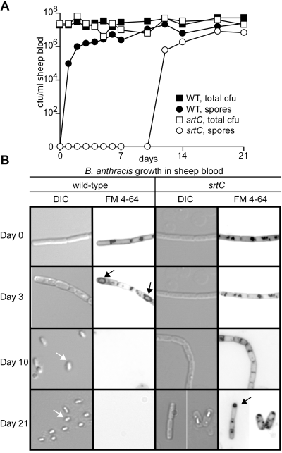
Sporulation of B. anthracis in sheep blood.A. Sporulation of B. anthracis in sheep blood without rotation (air exposure). B. anthracis Ames (WT, wild type) and srtC mutants were grown to OD600 0.7 (107 cfu ml−1), mixed with defibrinated sheep blood and 600 μl of aliquots incubated in 1.5 ml tubes at room temperature. Total colony-forming units (cfu), i.e. vegetative bacilli and spores, and heat-resistant spores were enumerated.B. Microscopic analysis of wild-type and srtC bacilli grown in defibrinated sheep blood without rotation. At timed intervals (see A), culture aliquots were fixed with formalin, stained with FM4-64 and viewed by fluorescence microscopy. Wild-type bacilli developed polar septa (black arrows) and endospores (white arrows) within 1–3 days, whereas srtC mutants failed to form polar septa and endospores for 12–21 days. Similar results were obtained with other srtC mutant alleles that were independently derived from B. anthracis Ames.
Earlier work revealed B. anthracis spore formation in carcasses of anthrax-infected small animals, such as rabbits, guinea pigs and mice (Toschkoff and Veljanov, 1970). Spore formation in large animals, cows or sheep, however, requires opening of cadavers and exposure of infected tissues to air (Koch, 1876). These and other observations support a general hypothesis, whereby B. anthracis sporulation occurs only in the presence of oxygen (Roth et al., 1955). We wondered whether the sporulation defect of srtC mutants can be rescued by exposure of bacilli to air and measured vegetative growth and spore formation in sheep blood cultures with rotation. Exposure to air indeed restored spore formation of srtC mutants to a level similar to that observed for wild-type B. anthracis Ames (data not shown). Moreover, microscopic analysis revealed synchronous formation of polar septa and endospores in wild type and srtC mutants (Fig. 10).
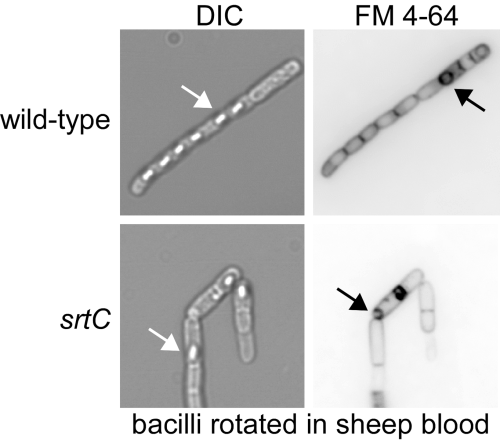
Sporulation of B. anthracis in sheep blood with rotation. Spore formation in sheep blood with rotation (air exposure). Experiments were carried out as described in Fig. 9, but with rapid agitation. After 18 h of rotation, culture aliquots were fixed with formalin, stained with FM4-64, and viewed by fluorescence and phase contrast microscopy. B. anthracis Ames (wild type) and srtC mutant bacilli developed polar septa (black arrow) and endospores (white arrow) at similar rates (data not shown).
Discussion
Dramsi et al. (2005) used bioinformatic tools and assigned 14 sortases as class D with three subclusters as a reflection of host phylogeny (bacilli, clostridia and actinomycetales). Members of the Bacillus (B. anthracis, B. cereus and B. thurigensis), Streptomyces (S. coelicolor, S. avermitilis) and Clostridium spp. (C. difficile, C. perfringens, C. tetani) all engage in a developmental programme that leads to the formation of spores (Dramsi et al., 2005). Only Corynebacterium diphtheriae, a member of the actinomycetales and the causative agent of diphtheria, encodes a class D sortase yet this species does not form spores.
Previous work examined the role of class D sortases in S. coelicolor, a Gram-positive, filamentous soil bacterium that is capable of producing many natural antibiotics (Hodgson, 2000). The life cycle of S. coelicolor commences with the formation of a feeding (submerged) mycelium, which differentiates into aerial hyphae that septate and develop into spore chains. The hyphal surface becomes hydrophobic, a property that promotes outgrowth into the air and facilitates the dispersion of spores, eventually giving rise to new mycelia. Six surface proteins of S. coelicolor, collectively called chaplins, are involved in aerial hyphae formation (Claessen et al., 2003; Elliot et al., 2003). All chaplins are synthesized during mycelium formation as precursors with an N-terminal signal sequence and a short hydrophobic domain (chaplin domain) of about 40 residues. Chaplins D–H are small polypeptides, whereas chaplins A–C are larger and carry a C-terminal sorting signal (LAXTG motif). Deletion of all six chaplin genes (chpA–H) hinders the formation of aerial hyphae, a defect that can be rescued by addition of purified chaplin proteins (Elliot et al., 2003). As mixtures of ChpD–H isolated from cell walls of aerial hyphae are highly surface active (Claessen et al., 2003), it seems plausible that these proteins lower aqueous surface tension for the emergence of aerial hyphae at the air–water interface. Although this has not yet been demonstrated, large chaplins ChpA–C are thought to be anchored by sortase to the bacterial peptidoglycan, serving as scaffold for assembly of small chaplins. Furthermore, chaplins may provide a hydrophobic surface for deposition of RdlA and RdlB, a highly insoluble, outer protein layer that facilitates spore dispersion (Claessen et al., 2004).
In this report, we have used purified sortase C to analyse its substrate requirements. SrtCΔN, lacking the N-terminal signal peptide and membrane anchor, cleaved peptides bearing the LPNTA motif, but not peptides with LPXTG, NPKTG or LGATG sequences. LPXTG and NPKTG motif sorting signals are the substrates of sortase A and sortase B respectively (Gaspar et al., 2005; Maresso et al., 2006). A sorting signal-like sequence with LGATG motif has been identified at the predicted C-terminal end of BasJ; however, a sortase enzyme responsible for cell wall anchoring of this internalin A homologue has thus far not been revealed (Gaspar et al., 2005). Two substrates, BasH and BasI, are anchored by sortase C to the cell wall of sporulating B. anthracis. basI, along with sortase C, is expressed by the basI-srtC-sctR-sctS operon. Both srtC::gfp and basI::gfp translational products were first detected at T−1, 2 h prior to the formation of the asymmetric septum that separates the mother cell from the developing forespore. The abundance of immunoreactive BasI and SrtC in B. anthracis extracts peaked at T0 and rapidly declined at T+1 (Fig. 3A, data not shown). BasI and SrtC species were no longer detectable at T+2. A similar, although delayed, expression pattern was observed for BasHFLAG; here the peak occurred at T+2, declined at T+3 and BasHFLAG was no longer detectable at T+4 (Fig. 4F). Nevertheless, fluorescence microscopy allowed detection of SrtC–mCherry in forespores and endospores even at T+20, suggesting that SrtC, and perhaps even BasH and BasI, may be sequestered within developing spores in a manner that resists treatment with PlyL enzyme and hot SDS.
sctR and sctS, which encode a two-component regulatory system, appear to autoregulate the basI-srtC-sctR-sctS operon, as IPTG-induced synthesis of SctR from another promoter is sufficient to induce expression of the sortase C operon. Expression of basH, which encodes another sortase C substrate, is driven by σF RNA polymerase and the translation product of basH::gfp is located only in the forespore. As cell wall anchoring of BasHFLAG absolutely requires srtC, it follows that sortase C must act in the intermembranous space that encloses forespore peptidoglycan.
What could be the role of protein targeting to the cell wall of developing B. anthracis spores? Initial pathogenesis experiments with srtC mutants derived from either B. anthracis strain Sterne in A/J mice (data not shown) or B. anthracis strain Ames in guinea pigs revealed no defect in spore-mediated virulence and development of lethal anthrax disease. Surprisingly, vegetative bacilli derived from srtC mutants failed to form spores in guinea pig carcass tissues (spleen, liver or heart's blood) that had been excised post-mortem and incubated for 21 days, whereas wild-type bacilli sporulated with remarkable efficiency. Microscopic analysis of guinea pig carcass tissue revealed the absence of spores with the Schaeffer–Fulton stain and suggested that spore formation must be hindered in mutants lacking srtC. This phenotype was also observed when srtC mutants were incubated in defibrinated sheep blood, although that spore formation eventually commenced after 12 days of incubation. It should be noted that spore formation of wild-type B. anthracis in sheep blood is essentially complete within 2–3 days. Fluorescence microscopy of FM4-64-stained bacilli revealed that srtC mutants do not form asymmetric division septa and hence cannot develop spores. This phenotype of arrested sporulation was completely rescued when B. anthracis srtC mutants were rotated in sheep blood to increase their exposure to air and oxygen.
We wonder how srtC and its substrate genes (basH and basI), whose products are targeted to the cell wall envelope of sporulating bacilli, can enable formation of infectious B. anthracis spores under oxygen-limiting conditions as has been observed in carcasses of small animals. BasI and BasH are small polypeptides, seemingly unstructured when analysed with protein fold predictions and one of them harbours GXGS sequence repeat. In future experiments, we will need to consider how sortase C deposition of BasH and BasI may increase exposure to molecular oxygen, for example, by altering the physical properties of developing forespores, as has been reported for sortase-dependent deposition of chaplins by S. coelicolor. Other work will need to consider the two-component regulator sctR/sctS, whose physiological function may be to sense of environmental conditions in the host that permit spore formation.
Experimental procedures
Bacterial strains and plasmids
Bacillus anthracis strains Sterne 34F2, Ames or its variants were grown in BHI or LB broth at 30°C. Sporulation was induced by diluting overnight BHI cultures 1:10 into modG medium (Kim and Goepfert, 1974) and further incubation at 30°C. FM4-64 membrane dye (Invitrogen) (Pogliano et al., 1999) was added to monitor sporulation (1:1000 dilution). Growth media were supplemented with kanamycin (20 μg ml−1) or chloramphenicol (10 μg ml−1) to provide for plasmid selection. Isopropyl-β-d-thiogalactopyranoside (IPTG) was added at 1 mM where indicated. B. anthracis chromosomal DNA was extracted with the Wizard Genomic DNA Purification Kit (Promega).
Plasmids
Table 2 lists primers used for plasmid construction. PCR with primers P23/P24 was used to append BamHI and XmaI restriction sites to srtC and the product was inserted into pQE-30 (Qiagen) to generate pLM116 (encoding SrtCΔN). Insertion of P18/P19 PCR product flanked by NdeI/BamHI sites into pET15b (Novagene) generated pLM114, which provided for expression His-tagged BasI and affinity chromatography as well as rabbit antibody production. Plasmid pLM4 was used for allelic replacement and constructed as follows. pTS1 (McDevitt et al., 1994) sequence containing the cat gene was excised using HpaI and StuI and replaced by the SmaI fragment of pUT618 containing the aphA3 gene (gift of Dr T. Koehler) to generate pLM3. The bla gene was excised from pLM3 using AdhI and EcoNI, protruding ends blunted with Klenow polymerase and ligated, thereby generating pLM4. For IPTG-inducible expression, spac promoter and lacI repressor sequences were amplified from pMutin-HA (Kaltwasser et al., 2002) using primers P146/P149 and inserted into pLM4 using EcoRI and XmaI restriction sites to generate pLM5. Insertion of P85/P86 (gfp mut2 coding sequence) (Cormack et al., 1996) and P107/P120 (basH promoter) PCR products into pOS1 via EcoRI, BamHI and PstI restriction sites produced pLM200. Insertion of P141/P142 (spoIIAC coding sequence) PCR product into pLM5 via KpnI sites generated pLM223. Insertion of P4/P88 (basI promoter sequence) and P95/P80 (sctR coding sequence) PCR products into pLM4 (EcoRI and BamHI sites) generated pLM218. Insertion of P132/P126 (sctR coding sequence) PCR product into pLM5 (KpnI sites) generated pLM226. Insertion of P12/P131 (srtC 1 kb 5′ flanking sequence) and P132/P13 (srtC 1 kb 3′ flanking sequence) and P112/P111 (gfp coding sequence) PCR products into pLM4 (EcoRI, KpnI and XmaI sites) generated pLM212; this plasmid was used for replacement of srtC coding sequence with gfp in strain LAM012 (srtC::gfp). Insertion of P12/P58 (srtC 1 kb 3′ coding sequence) and P14/P13 (srtC 1 kb 3′ flanking sequence) PCR products into pLM4 (EcoRI, KpnI and XmaI sites) generated pLM201 for in-frame deletion of srtC (strain LAM005). Insertion of the EcoRI fragment of pLM218 (basI promoter–sctR fusion), P107/P108 PCR product (basH promoter and 5′ coding sequence) and PCR product P109/P110 (basH sorting signal and terminator) into pOS1 (EcoRI, KpnI and XmaI sites) generated pLM230. Replacement of the pLM230 EcoRI fragment with the P12/P13 PCR product (on pLM233 template) generated pLM238. Insertion of P73/P52 PCR product (basI 1 kb 5′ flanking sequence) and P9/P13 product (basI 1.5 kb 3′ flanking sequence containing srtC and sctR) into pLM4 (EcoRI, KpnI and XmaI sites) generated pLM233. Insertion of P112/P111 product (gfp) into pLM233 (KpnI sites) generated pLM234, a plasmid that was used for replacement of basI with gfp in strain LAM010 (basI::gfp). Insertion of P12/P154 product (basI promoter and coding sequences as well as srtC coding sequences), P159/P158 product (mCherry orf from pRSET-B-mCherry; Shaner et al., 2004) and P132/P13 product (sctR) into pLM4 (EcoRI, XbaI, KpnI and XmaI sites) generated pLM243 for SrtC–mCherry expression. PCR reactions were performed with Pfu DNA polymerase (Stratagene). Ligation products were transformed into E. coli K1077(dam–dcm–) and purified (non-methylated) plasmid DNA was transformed into B. anthracis following a previously developed protocol (Kim et al., 2004).
| Primer | Restriction site added | Sequencea |
|---|---|---|
| P4 | EcoRI | aaGAATTCcaatgaaatggagtaagtggag |
| P9 | KpnI | aaGGTACCcaatgatggcactaagcgcg |
| P12 | EcoRI | aaGAATTCaggtttgaagaagagacccc |
| P13 | XmaI | aaCCCGGGgatctaatgtcgtaatccacg |
| P14 | KpnI | aaGGTACCacactgacgacttgttaccc |
| P18 | BamHI | aaGGATCCttaagttgcagcattttttggttg |
| P19 | NdeI | aaCATATGattgaagaagagaaaaagag |
| P23 | BamHI | aaaGGATCCgcgcaagaattaacgaatgagg |
| P24 | XmaI | aaaCCCGGGtcattttgaataactaccagttaac |
| P52 | KpnI | aaGGTACCcattttatttctctccttttatac |
| P58 | KpnI | aaGGTACCctttatttcctcattcgttaattc |
| P73 | EcoRI | aaaGAATTCaaataaagtcgattgttacatcg |
| P80 | PstI | aaaCTGCAGttcaagagcatcaatgcttatc |
| P85 | BamHI | aaaGGATCCagtaaaggagaagaacttttcac |
| P86 | PstI | aaaCTGCAGttatttgtatagttcatccatgcc |
| P88 | BamHI | aaaGGATCCcattttatttctctccttttatac |
| P95 | BamHI | aaaGGATCCaattgtagtttcgaaaagcggg |
| P107 | EcoRI | aaaGAATTCccatatttaatttgtgtaccacc |
| P108 | KpnI | aaaGGTACCcttatcgtcgtcatccttgtaatcttgttttttcgcattatcttttacb |
| P109 | KpnI | aaaGGTACCatgcatcaccatcaccatcacggcgataagttaccgaatactgc |
| P110 | BamHI | aaaGGATCCttttgaacaagaacatttagcag |
| P111 | KpnI | aaaGGTACCatgagtaaaggagaagaacttttcac |
| P112 | KpnI | aaaGGTACCtttgtatagttcatccatgccatg |
| P120 | BamHI | aaaGGATCCtttcgtcattgatttcctcctg |
| P131 | KpnI | aaaGGTACCcatatttcatccctccatgcc |
| P132 | KpnI | aaaGGTACCtgaaattgtagtttcgaaaagcg |
| P141 | KpnI | aaaGGTACCaaggaggatagcctatggacatag |
| P142 | KpnI | aaaGGTACCttgcgtttgatctttatagtagcg |
| P146 | XmaI | aaaCCCGGGaggttttcaccgtcatcaccg |
| P149 | EcoRI | ttatggGAATTCtacacagccc |
| P154 | XbaI | aaaTCTAGAttttgaataactaccagttaacttcg |
| P158 | KpnI | aaaGGTACCagcaagggcgaggaggataac |
| P159 | XbaI | cgTCTAGAgtgagcaagggcgaggaggataacatg |
- a. Capital letters indicate the sequence of the restriction site inserted.
- b. Underlined nucleotides encode for the FLAG epitope (Hopp et al., 1988).
Bacillus anthracis mutants
-
Bacillus anthracis variants were generated with pLM4, containing a thermosensitive origin of replication. Plasmids with 1 kb DNA sequence flanking each side of the mutation were transformed into B. anthracis and transformants grown at 30°C (permissive temperature) in LB broth (20 μg ml−1 kanamycin). Cultures were diluted 1:100 and incubated at 43°C overnight (restrictive temperature). After four growth cycles at 43°C, cultures were diluted 1:100 into LB broth without antibiotics and grown overnight at 30°C. To ensure loss of pLM4-based plasmid, cultures were diluted four times into fresh media and propagated at 30°C. Bacteria were plated on LB agar and colonies examined for kanamycin resistance. DNA from kanamycin-sensitive colonies was analysed by PCR for the presence or absence of mutant alleles. Nucleic acid sequences of wild-type and mutant alleles were verified by DNA sequencing.
SrtCΔN cleavage of LPNTA peptide
Expression of SrtCΔN in E. coli XL-1 Blue (pLM116) was induced with IPTG. Six-histidyl-tagged enzyme was purified from cleared lysate by affinity chromatography on Ni-NTA agarose. Activity was assayed in buffer A (50 mM Tris-HCl pH 7.5, 150 mM NaCl) after 16 h incubation at 37°C (50 μM SrtCΔN and 10 μM peptide substrate). Modified peptide substrates (BiopeptideCo., LLC) were conjugated to 2-aminobenzoyl fluorophore (a) and 2,4-dinitrophenyl quencher (d) at the N- and C-terminus respectively. Reactions were quenched by boiling samples for 5 min and peptide cleavage monitored by quantification of 2-aminobenzoyl fluorescence at 320 nm excitation and 420 nm emission. Mean and standard deviation of three independent measurements are reported. SrtCΔN was treated with 5 mM MTSET ([2-(trimethylammonium)ethyl]methanethiosulphonate) (Smith et al., 1975) and 10 mM dithiothreitol (DTT). For chromatography, sortase reactions were quenched by centrifugation at 7500 g through Centricon-10 membrane (Millipore) to separate enzyme from substrate and product. Filtrate was separated by rpHPLC (2 × 250 mm, C18 Hypersil, Keystone Scientific) and elution (1–60% linear gradient in acetonitrile) monitored at 215 nm absorbance. Vacuum-dried fractions were analysed by MALDI-TOF and tandem mass spectrometry (ABI 4700, operated in reflection mode) to determine peptide structures.
Immunoblotting
Bacillus anthracis cultures were grown in modG sporulation media and aliquots removed at 1 h intervals until spores were detectable. Bacilli were sedimented by centrifugation at 10 000 g, washed and suspended in 300 μl of PlyL buffer (0.5 M sucrose, 1 mM DTT, 1 mM PMSF, 1 mM EDTA, 5 mM sodium phosphate, pH 6.0) with 250 μg ml−1 PlyL amidase (Low et al., 2005). After digestion of cell wall for 16 h at 37°C, protoplasts were sedimented by centrifugation at 10 000 g. Aliquots of supernatant (cell wall lysate) were mixed with sample buffer, separated on SDS-PAGE and electrotransferred to PVDF membrane. Proteins were detected with specific serum (αBasI, αSrtC or αL6 – raised against purified proteins in rabbits) or anti-FLAG mouse monoclonal antibody (Stratagene), followed by species-specific secondary antibody conjugate to horseradish peroxidase and chemiluminescent detection.
Microscopy
Bacillus anthracis culture aliquots were viewed by DIC or fluorescence microscopy under Olympus Provis axial microscope (1000-fold magnification) equipped with CCD camera and digital images of FM4-64, GFP or mCherry fluorescence captured.
Sporulation
Overnight cultures of bacilli in BHI were diluted 1:10 into modG sporulation medium and incubated at 30°C for 48 h. Culture aliquots were heated for 30 min at 65°C to kill vegetative bacilli and 10-fold serial dilutions of heat-and non-heat-treated cultures were spread on LB agar followed by incubation and enumeration of total colony-forming units (cfu) (no heat treatment) and viable spores (heat treatment). Sporulation efficiency was calculated as the ratio (viable spores)/(total cfu).
Animal experiments
Bacillus anthracis spores (wild type and srtC) were washed three times with sterile distilled water and examined for the presence of vegetative bacilli by microscopy. Contaminating bacilli, typically < 1% of spore preparations, were heat-killed by incubating the spore preparation at 65°C for 30 min prior to additional washes. Colony-forming units were enumerated after serial dilution and plating on LB agar. To investigate the virulence of B. anthracis Ames wild-type and srtC spores, six guinea pigs each were infected subcutaneously with 30 or 300 spores for each of the two strains (wild-type and srtC). Animals were monitored for anthrax disease over 7 days following infection and LD50 values were calculated (Reed and Muench, 1938).
Sporulation in host tissues
Shortly following anthrax death, guinea pig organs were removed from three animals infected with either wild-type or srtC mutant bacilli. Tissue aliquots were homogenized (under aseptic conditions) and total cfu as well as spore concentrations enumerated. Following incubation of homogenized tissues for 3 weeks, total cfu as well as spores were enumerated [homogenized tissues of all animals tested generated B. anthracis colonies (ground glass appearance) and no contaminations on LB agar]. For sporulation in sheep blood, 300 µl of culture (wild type or srtC at OD600 0.7, ∼107 cfu ml−1) was mixed with an equal volume of defibrinated sheep blood in a 1.5 ml tube and samples were incubated at room temperature for 21 days. At timed intervals, total cfu and viable spores were enumerated. To determine spore formation in rotating blood, the same experiment was carried out in 15 ml tubes with rapid agitation. Homogenized tissues and blood samples were fixed with 10% buffered formalin prior to microscopic analysis.
Acknowledgements
We thank Dominique Missiakas, Bill Blaylock, Justin Kern and Anthony Maresso for experimental help or critical reading of this manuscript. We are indebted to the Great Lakes RCE Small Animal Core for help with B. anthracis guinea pig infections and to Jonathan Budzik for PlyL purification. This work was supported by Grants AI38897 and AI52474 from the National Institute of Allergy and Infectious Diseases (Infectious Diseases Branch). O.S. acknowledges membership within and support from the Region V ‘Great Lakes’ Regional Center of Excellence in Biodefense and Emerging Infectious Diseases Consortium (GLRCE, National Institute of Allergy and Infectious Diseases Award 1-U54-AI-057153).


