Recurrent thymoma: evidence for histological progression
Abstract
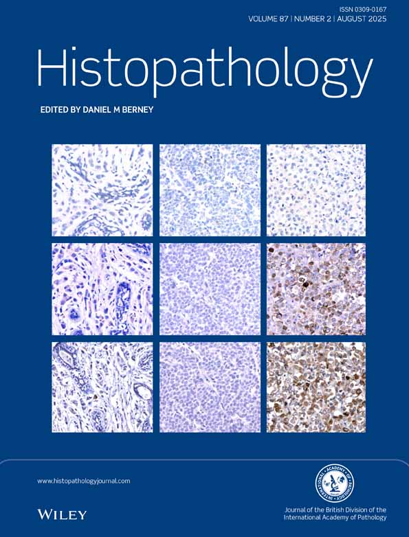
The clinicopathological features of nine cases of recurrent thymomas have been studied. At presentation, all cases were histologically classified as thymomas with cortical differentiation, including predominantly cortical thymoma, cortical thymoma and well-differentiated thymic carcinoma. In five cases the morphological features of the recurrence(s) were suggestive of a histological progression of the tumour from predominantly cortical thymoma to cortical thymoma and/or well-differentiated thymic carcinoma, usually associated with a more advanced clinical stage, the latter indicating a clinical progression. These findings suggest that all types of thymoma with cortical differentiation are histologically and histogenetically related neoplasms, associated with a more aggressive clinical behaviour and a significant risk of recurrence. The overall outcome of patients with recurrent thymoma in this series was poor, since six patients (66.6%) died due to the disease, 2–14 years after the first diagnosis. The clinical implication of our findings is that thymomas with cortical differentiation always need careful follow-up, even in those cases which are not obviously invasive at onset.