A reassessment of the morphology and taxonomic status of ‘Crocodylus’depressifrons Blainville, 1855 (Crocodylia, Crocodyloidea) based on the Early Eocene remains from Belgium
Abstract
The name Crocodilus depressifrons appears in the literature as the caption of a table, published by Blainville in 1855, depicting crocodylian remains from France. Although a proper diagnosis and description have never been published, this species has been frequently used to identify some European Eocene crocodyloids with a generalized, not elongated, rostrum. In the last 50 years, C. depressifrons has been often referred to the genus Asiatosuchus. This genus, erected by Mook in 1940 on the basis of fossil remains from the Middle Eocene of Mongolia, actually contains a rich and apparently nonmonophyletic assemblage of Palaeogene crocodyloids. In order to help clarify the morphology and the relationships of the Asiatosuchus-like taxa, it is here described a rich collection of crocodyloid remains, including skulls and a nearly complete skeleton, from four different Early Eocene localities of the Belgian Tienen Formation: Dormaal, Erquelinnes, Leval, and Orp-le-Grand. All these remains belong to one single taxon which clearly represents the long known but never properly described ‘C.’depressifrons. They allow, for the first time, the diagnosis this species on the basis of an unequivocal set of characters, contributing to the long awaited revision of the Asiatosuchus-like taxa. © 2009 The Linnean Society of London, Zoological Journal of the Linnean Society, 2009, 156, 140–167.
INTRODUCTION
According to the present knowledge of the Cenozoic crocodylians, crocodyloids with a generalized rostrum (i.e. non-elongated; see Brochu, 2001a, for a definition of the term ‘generalized crocodylian’) show a significant chronological and phylogenetic gap in Europe. Uncritical analysis of the literature could give the false impression that these crocodyloids inhabited Europe for nearly all of the Cenozoic because of the fact that the genus name Crocodylus frequently appears in the old literature. In fact, such a name (or better, the name Crocodilus in older literature) was erroneously applied to nearly any generalized crocodylian not showing close alligatoroid affinities (Brochu, 2000). Actually, generalized crocodyloids are unquestionably present in Europe only during the late Neogene (latest Miocene – earliest Pliocene) and during the Eocene, but few data come from Late Palaeocene and perhaps Early Oligocene localities. Late Neogene generalized crocodyloids have been recently referred on a phylogenetic basis to the extant genus Crocodylus and are known only from a few southern European (continental but close to the sea) localities that they probably reached from north Africa (Kotsakis, Delfino & Piras, 2004; Delfino, Böhme & Rook, 2007; Delfino, 2008, in press; Delfino & Rook, 2008). However, the Palaeogene forms are rather common in the fossil record and although they have been known since the first decades of the 19th century, their taxonomy is still uncertain. The oldest generalized crocodyloid remains from Europe seem to be those from the Late Palaeocene of Mont de Berru /Cernay-les-Reims, France [material already mentioned as ‘Asiatosuchus’ by Vasse (1993), and currently under study in collaboration with France de Lapparent]. The youngest could be those from the Early Oligocene of Monteviale (north-east Italy) if the referral of ‘Crocodilus’monsvialensisFabiani, 1914 to crocodyloids by Franco & Piccoli (1993) is upheld against the view expressed by Rauhe & Rossmann (1995), who considered these remains as belonging to the alligatoroid genus Diplocynodon (see also Kotsakis et al., 2004).
The Western European Eocene remains were originally ascribed to several different species but mostly to the genus Crocodylus (as C. depressifronsBlainville, 1855) based on superficial cranial similarities with this living genus. In the second half of the 20th century, following the indications of Berg (1966), these remains were referred to AsiatosuchusMook, 1940, as Asiatosuchus depressifrons (Blainville, 1855), whereas new, apparently related, fossils from Germany (Messel) and France (Issel) were referred to a new species, Asiatosuchus germanicusBerg, 1966 (Vasse, 1992).
As will be shown in the following sections, the reassessment of the relationships of the European basal crocodyloids reveals a confused nomenclature and a doubtful taxonomic status. These are direct consequences of a mixture of problems affecting the preservation of part of the type material and the diagnoses of some of the taxa involved, as well as the unavailability of descriptions adequately supporting a correct analysis of the topic.
The purpose of this paper is to begin resolving these problems. After some introductory notes concerning Crocodylus depressifronsBlainville, 1855 and the complex taxonomic landscape of Asiatosuchus, abundant well-preserved crocodyloid remains from the Early Eocene Belgian localities Dormaal, Erquelinnes, Leval, and Orp-le-Grand will be described (or partly re-described; see Vasse, 1993; Lambrecht, 2003) in order to offer an updated morphological basis for a general revision of the Asiatosuchus-like taxa and, if possible, for the reassessment of their phylogenetic relationships.
Crocodylus depressifronsBlainville, 1855
The ‘Atlas du genre Crocodilus’ published by Blainville does not only contain illustrations and information about extant Crocodylus. In the volume dedicated to the ‘explications de planches’[1855; for the proper publication date of this section see Swinton (1938), and quoted reference] new fossil taxa were also introduced in the literature: among others, Crocodilus depressifrons, C. temporalis, and C. macrorhynchus.
The name C. depressifrons appears in the work by Blainville only as the caption ‘C. depressifrons, du Soissonnais’ of some figures in table 6, figures portraying an incomplete skull and a lower jaw from the Early Eocene, ‘Sparnacian’, of France (Fig. 1). Despite the fact that no description is associated to such a name, for a period of about a century, various remains from the Eocene of north-west Europe were referred, either in publications or simply in museum labels, to C. depressifronsBlainville, 1855. The reasons for such identifications were usually based on the number of teeth/alveoli involved in the dentary symphysis (which in the figure by Blainville are clearly six in number), or based on the flattened frontal area, a character that although not obvious in the figure, is clearly implicit in the specific epithet. No attempts to provide a complete diagnosis have ever been made and the material formerly described by Blainville is nowadays so strongly pyritized (Fig. 2; MNHN G 160; whose label reports the locality as ‘lignites de Muirancourt’– Oise) that the original morphology is no longer visible. The casts of the skull and lower jaw (MNHN G 156), apparently produced before the pyritization started to alter them, are of little help in understanding the finer morphology of the taxon.
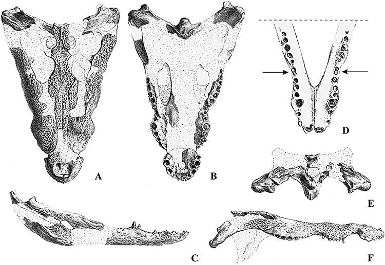
‘Crocodylus’depressifronsBlainville, 1855, type specimens from ‘Soissonnais’, original drawings by Blainville (1855: plate 6), A, B, skull in dorsal and ventral view; C, D, right lower jaw in lateral view and enlarged detail of the symphysis in dorsal view, the arrows in (D) indicate the small and nearly confluent eight and ninth alveoli; E, F, skull in occipital and right lateral view; note that the reconstructed squamosal ‘horns’ are moderately developed in (E) but nearly flat in (F). The scale was not reported in the original drawing by Blainville but, on the basis of the remains still preserved in Paris (MNHN G 156, MNHN G 160), the total skull length can be estimated to be about 500 mm.
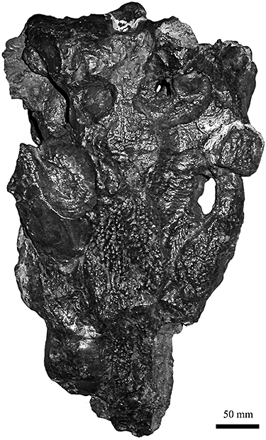
‘Crocodylus’depressifronsBlainville, 1855, type specimen (MNHN G 160) in dorsal view showing the current state of preservation; the original label reports ‘Lignites de Muirancourt (Oise)’.
As for the validity of C. depressifrons, its possible status as a junior synonym was noted by Gervais (1859), who listed the name C. coelorhinusPomel, 1847, in the section dedicated to the former species, and added Laonnais to Soissonnais as if they were the ‘typical’ localities for it. If C. coelorhinus was a valid name it should have priority over C. depressifronsBlainville, 1855, but as already discussed by Swinton (1938) and Vasse (1993), Pomel (1847) neither described nor figured C. coelorhinus, and therefore this name is considered a nomen nudum.
In conclusion, the validity of the species C. depressifrons is firmly granted by the figures published by Blainville, by the name and the locality associated to them, and therefore by the information they provide.
Berg (1966), in his comprehensive work concerning the crocodylians of Messel, the renowned German Eocene locality particularly rich in well-preserved crocodylian remains belonging to seven different taxa (Morlo et al., 2004), marginally discussed the taxon erected by Blainville and suggested that it should be included in the genus Asiatosuchus (see also Berg, 1969).
A siatosuchusMook, 1940
The genus Asiatosuchus was erected by Mook (1940) on the basis of an incomplete lower jaw and a few skull fragments coming from the Middle Eocene of Mongolia (Irdin Manha Formation). According to Mook, Asiatosuchus should be characterized by being similar to Crocodylus, but with at least 17 teeth in each ramus of the lower jaw and with splenial bones not reaching the symphysis. The diagnosis of the type species, Asiatosuchus grangeriMook (1940), is as follows: ‘symphysis extending back to the level of the sixth mandibular teeth, the two rami of the mandible diverging at a moderately wide angle, dental row shorter than postdental portion of jaw, teeth stout and faintly striated, interfenestral plate flat, sutures of nasals with lacrimals considerably shorter than sutures with prefrontals’(Mook, 1940: 1). Steel (1973: 59) strengthened the genus definition as follows: ‘dentition comprising five premaxillary teeth, fourteen maxillary (of which the fifth is the largest), and at least seventeen (possibly up to twenty) mandibular elements. Palatine bones extending forward only as far as the anterior margin of the palatal vacuities. Splenial excluded from mandibular symphysis, which reaches back to the 6th or 7th dentary tooth’. Vasse (1993) suggested that the specific characters should be applied to the genus.
Such a list of characters includes some features which are shared, although not exclusively, by all the species ascribed to this genus in the following decades (such as the elongation of the dentary symphysis) and also features that are present only in some of the species (such as the exclusion of splenial from dentary symphysis) or that have a wide distribution among crocodylians (such as the number of premaxillary teeth).
Brochu (1997) added significant information to the morphology of A. grangeri, explicitly stating that the frontoparietal suture was completely excluded from the supratemporal fossae, and that the medial jugal foramen is small, as well as implicitly offering morphological information thanks to the character coding of this species [characters evidently assessed on remains not described by Mook (1940), as Mook does not mention, for example, any jugal].
The referral of fossil remains to the genus Asiatosuchus has usually been based mostly on the elongated dentary symphysis despite the presence of some discrepancies in the skull morphology. This is the case for two other species that have been described from the Palaeocene of Asia, Asiatosuchus volgensis (Efimov & Yarkov, 1993) and Asiatosuchus zajsanicusEfimov, 1982 (later on referred by its author to the tomistomine genus Dollosuchus; see Efimov, 1988; but see Brochu, 2007a, for the synonymy of Dollosuchus). According to Angielczyk & Gingerich (1998: 185), the naming of these species is premature because of ‘the lack of any truly diagnostic material’.
In the absence of a thorough revision, the complex relationships among these taxa are mostly based on speculations so that Asiatosuchus has been defined as a ‘wastebasket taxon’ (Angielczyk & Gingerich, 1998: 185). Vasse (1992, 1993) in his ‘bilan’ on the knowledge concerning this genus (he did not take into consideration A. zajsanicus and, of course, A. volgensis which was described later), accepted as valid only three species: the European A. depressifrons (Blainville, 1855), the Asian A. grangeriMook, 1940 (the type of the genus) and A. nanlingensisYoung, 1964. He listed the following taxa as synonyms of A. depressifrons: ‘Crocodile des gravières de Castelnaudary’ (partim; Cuvier, 1824); Crocodylus coelorhinusPomel, 1847; Crocodylus dodunniGray, 1831; Pyrenodon sp. Dujardin, 1843; C. doduniGiebel, 1847; C. vicetinusLioy, 1865 (sometimes misspelled in the literature as C. vicentinus); Isselo-saurus dodunniFilhol, 1877; Diplocynodon haeckeliSeidlitz, 1917; Atacisaurus crassiproratusAstre, 1931; Asiatosuchus germanicusBerg, 1966. According to Ortega, Buscalioni & Gasparini (1996), Atacisaurus crassiproratus is not a crocodylian and therefore it is not a synonym of Asiatosuchus depressifrons.
The affinities of Asiatosuchus are not limited to Asian and European material: Berg (1966) and Vasse (1993) underlined the apparent close relationships between the Asian–European Asiatosuchus and some taxa from the middle Eocene Bridger Formation in Wyoming: ‘Crocodilus’affinisMarsh, 1871 (see Norell & Storrs, 1986, for a discussion of the synonyms of this taxon) and ‘Crocodilus’clavisCope, 1872, but both Berg and Vasse discussed the diagnosis of the genus Asiatosuchus on the basis of characters that in most cases can be now considered as devoid of any phylogenetic relevance.
If some of the proposed synonymies can be accepted simply on the basis of educated guesswork (i.e. most of the 19th century species), others are still debatable: issues still being discussed include the synonymy between A. depressifrons and A. germanicus, and the possible inclusion of the North American taxa into the genus Asiatosuchus.
Brochu (1997), emphasizing the presence of frontals participating in supratemporal fenestrae and of splenials slightly participating in the symphysis in A. germanicus as an opposite condition to that shown by A. depressifrons, maintained as separate these two taxa. As for the relationships of the North American taxa mentioned above, he reported that the ‘C.’affinis morphology of splenials and frontals, as well as the size of the medial jugal foramen, is congruent with that of the type species of Asiatosuchus, A. grangeri, and that, as suggested by Berg (1966), the inclusion of ‘C.’affinis in Asiatosuchus could be reasonable but better material of A. grangeri should be found to support such a referral.
At present, character codings for the following species are currently available: ‘C.’affinis, A. depressifrons, A. grangeri, and A. germanicus (based on the remains from Messel) (see, among others, Brochu, 2007b). Because the type of A. depressifrons is heavily altered and therefore of little help, the character coding now available for this taxon has been realised on the basis of some practically unpublished putative A. depressifrons remains coming from Orp-le-Grand in Belgium (IRSNB IG 9875 and 9912); such coding has been variously named in the literature as Dormaal crocodyloid, cf. Crocodylus depressifrons, European basal crocodyloid (?= ‘Crocodylus’depressifrons), or Belgian crocodyloid (Brochu, 1997, 1999, 2000, 2003, 2007a, b; Jouve, 2004; Delfino, Piras & Smith, 2005; Piras et al., 2007; Vélez-Juarbe, Brochu & Santos, 2007).
The cladistic analysis based on these character codings fails to recognize Asiatosuchus as a monophyletic group (Brochu, 1997, 1999, 2000, 2001b, 2003, 2007a, b; Brochu & Gingerich, 2000; Jouve, 2004; Delfino et al., 2005; Piras et al., 2007; Vélez-Juarbe et al., 2007; for different matrixes including only ‘Crocodylus’affinis and A. germanicus see Salisbury & Willis, 1996; Salisbury et al., 2006) and the cladogram topology can be simplified as follows: A. germanicus is a very basal crocodyloid, and ‘C.’affinis, A. depressifrons, and A. grangeri form a polytomy with more derived crocodyloids. As a result of such nonmonophyletic grouping, in the following sections each species, except the type one, will be referred to its description genus encircled by inverted comas to indicate a provisional status.
Abbreviations
Anatomical abbreviations
a, alveolus; a.c, axis centrum; a.h, axial hypapophysis; a.i, atlas intercentrum; an, angular; a.n.a, atlas neural arch; a.n.s, axis neural spine; ar, articular; bo, basioccipital; c.r, choanal rim; d, dentary; di, diastema; e.n, external naris; eo, exoccpital; e.s.a, ectopterygoid sutural area; f, frontal; f.a, foramen aerum; f.i, foramen incisivum; f.m, foramen magum; f.v, foramen vagi; f.V, foramen Vth cranial nerve; f.XII, foramen XIIth cranial nerve; it.f, infratemporal fenestra; j, jugal; l, lacrimal; l.c.f, lateral carotid foramen; m, maxilla; n, nasal; o, orbit; o.c: occipital condyle; o.p, occlusal pit; od.p, odontoid process; p, parietal; pf, prefrontal; pm, premaxilla; po, postorbital; q, quadrate; qj, quadratojugal; qj.s, quadratojugal spine; sa, surangular; sb.f.r, suborbital fenestra rim; so, supraoccipital; sp, splenial; sq, squamosal; st.f, supratemporal fenestra; sy, symphysis; t.c, temporal canal.
Institutional abbreviations
DIIA R-RS, Richard Smith's collection, Bruxelles; IRSNB, Institut royal des Sciences naturelles de Belgique, Bruxelles; MNHN, Muséum National d'Histoire Naturelle, Paris.
GEOLOGICAL SETTING
In Belgium, Eocene generalized crocodyloids are known from two relatively limited regions (Fig. 3). One is in eastern Belgium (localities of Orp-le-Grand and Dormaal) and the second is in the Mons Basin (localities of Erquelinnes and Leval). These two restricted regions are the only ones having preserved continental deposits of the Tienen Formation in Belgium. Figure 4 indicates the stratigraphical position of the Tienen Formation, which corresponds to fluvio-lacustrine deposits belonging to the basal Ypresian (earliest Eocene, NP9b). The Tienen Formation is locally overlaid by the marine Kortrijk Formation belonging to the classical Ieper group (NP10-13) and it is underlaid by the late Palaeocene marine Hannut Formation (NP6-8) or locally by the latest Palaeocene Bois Gilles Formation in the Erquelinnes area (NP9a). Among the four localities that yield generalized crocodyloid remains, the important Dormaal fauna (see Smith & Smith, 1996; Steurbaut et al., 1999; Smith, 2000) was specified for reference-level MP7 of the mammalian biochronological scale for the European Palaeogene (Schmidt-Kittler, 1987). The MP7 fauna is characterized by the first occurrence of modern mammal groups, among which the most typical species are: Teilhardina belgica (euprimate), Diacodexis gigasei (artiodactyl), Microparamys nanus (rodent), Miacis latouri (miacid carnivoran), Prototomus minimus, and Arfia gingerichi (hyaenodontid creodont). These co-occur with species of persisting primitive groups, such as Paschatherium dolloi (condylarth), Landenodon woutersi (arctocyonid), Platychoerops georgei (plesiadapiformes), Palaeosinopa russelli (pantolestid), and Apatemys teilhardi (apatotherid). Similar mammal species have been identified at Erquelinnes. Dormaal and Erquelinnes belong to the lower part of the Tienen Formation that is generally fluviatile and erosive at its base. This lower part has recorded at least partly the carbon isotope excursion (CIE) marking the Palaeocene–Eocene thermal maximum (Steurbaut et al., 2003; Grimes et al., 2006) and it is the onset of the CIE that defines the Palaeocene/Eocene boundary (Gradstein et al., 2004). The best records of the CIE in north-west Europe are the Cap d'Ailly outcrop near Dieppe in the Paris Basin and the Doel and Kallo boreholes near Antwerp in north Belgium.
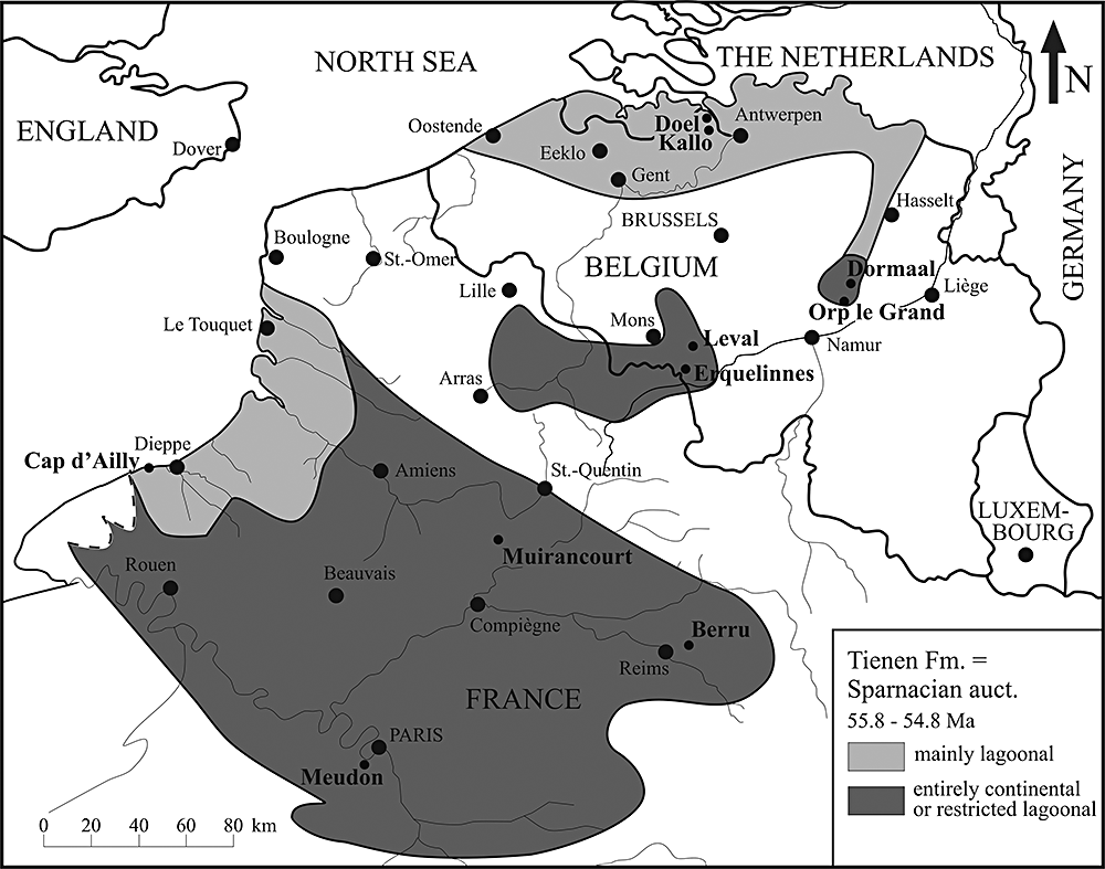
Distribution of the continental lagoonal deposits of the Tienen Formation in Belgium and the Sparnacian facies of the Mont Bernon Group in northern France, with the location of Early Eocene crocodyloid-bearing localities referred to in this study. The remains of ‘Crocodylus’depressifrons come from Muirancourt (holotype), Meudon, Dormaal, Orp-le-Grand, Leval, and Erquelinnes. Undescribed Asiatosuchus-like remains have also been found in the Late Palaeocene locality of Berru.
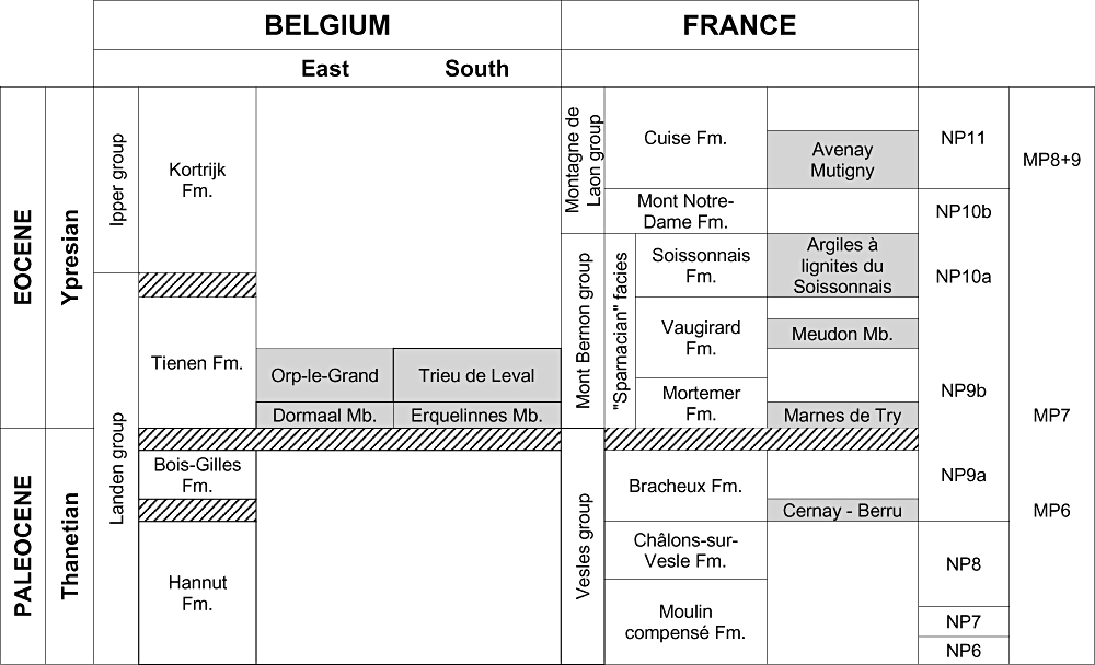
Relative position of the Late Palaeocene–Early Eocene stratigraphical units in the French–Belgian Basin with the position of the main continental vertebrate localities. NP, Nanoplankton zones; MP, mammal Palaeogene reference-levels.
The stratigraphical positions of Orp-le-Grand and Leval are more complex and, following the lithostratigraphy, seem to correspond to higher levels within the Tienen Formation. Deposits at Orp-le-Grand and Leval (Trieu) are rich in organic matter and correspond to swampy palaeoenvironments.
The Tienen Formation is mostly coeval with the ‘Sparnacian’ facies of the Mont Bernon group in the Paris Basin and the lower part of the Tienen Formation is the equivalent of the Mortemer Formation (Aubry et al., 2005). The vertebrate locality of Try (France) is thus probably coeval with Dormaal and Erquelinnes. Several mammal species are shared with these two Belgian localities.
The stratigraphical position of the holotype of ‘Crocodylus’depressifronsBlainville, 1855 (skull MNHN G 160; casts of skull and lower jaw MNHN G 156) from the ‘Lignites de Muirancourt’ is unclear. It seems to be the equivalent of the ‘Argiles à lignites du Soissonnais’ from the upper part of the Sparnacian facies called the Soissonnais Formation (NP10a). The holotype of ‘C.’depressifrons could be therefore at least 1 or 2 million years younger than the Dormaal and Erquelinnes specimens.
SYSTEMATIC PALAEONTOLOGY
CrocodyliaGmelin, 1789 CrocodyloideaFitzinger, 1826‘Crocodylus’depressifronsBlainville, 1855
Referred material and localities: IRSNB R251 (IG 1567), fragmentary skull, Erquelinnes (5-7); IRSNB R252 (IG 9912A), fragmentary skull and lower jaw, Orp-le-Grand (6-9); IRSNB R253 (IG 9912B), fragmentary skull and lower jaw, Orp-le-Grand (10-14); IRSNB IG 9875, 21 vertebrae or vertebral fragments, ilium, ischium, pubis, several metapodials, nine ribs, four gastralia, 40 osteoderms or osteoderm fragments, Orp-le-Grand; IRSNB R254 (IG 8368), incomplete skeleton, Leval (7, 14-16); IRSNB IG 8558, dorsal vertebra, Leval; IRSNB IG 8699, cervical vertebra, Leval; IRSNB IG 8792, four caudal vertebrae and one large phalanx, Leval; IRSNB IG 8979, femur and seven fragmentary vertebrae (and 10 fragments), Leval; IRSNB IG ‘uncatalogued’, highly fragmentary humerus, Leval; IRSNB R255 right maxilla, Dormaal (Fig. 17A); IRSNB R256 right maxilla, Dormaal (Fig. 17B); IRSNB R257 parietal, Dormaal (Fig. 17C); DIIA R50RS parietal and supraoccipital, Dormaal; IRSNB R258 ectopterygoid, Dormaal; IRSNB R259 caudal vertebra, Dormaal (Fig. 17D); IRSNB R260 osteoderm, Dormaal (Fig. 17E). (See online supplementary information for further figures.)
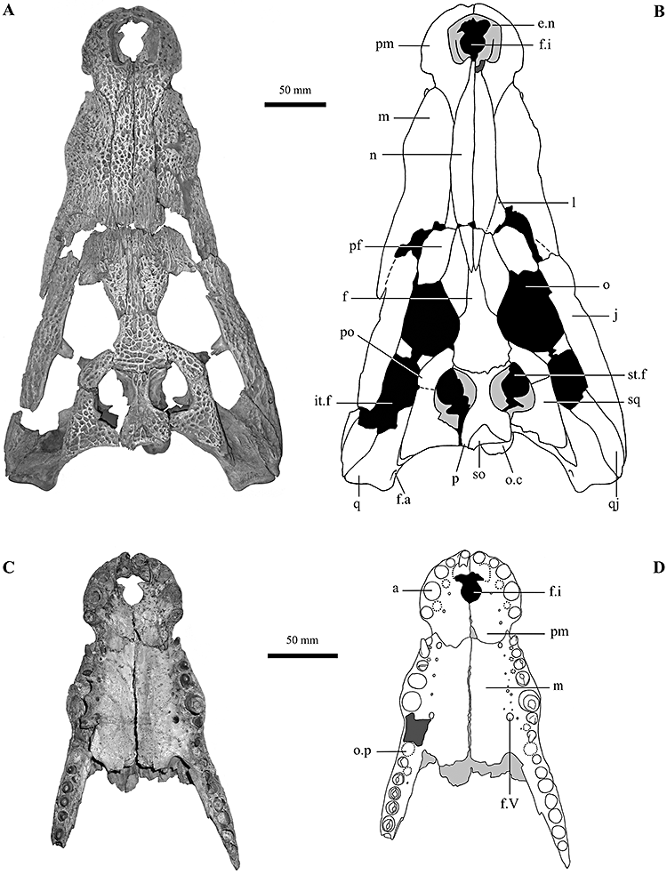
‘Crocodylus’depressifronsBlainville, 1855, specimen IRSNB R251 from Erquelinnes. A, B, photograph and interpretative drawing of the skull in dorsal view; C, D, anterior palatal region of the same specimen in ventral view. See main text for abbreviations.
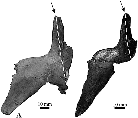
‘Crocodylus’depressifronsBlainville, 1855. Left ectopterygoids in ventral view. (A) IRSNB R251 from Erquelinnes; (B) IRSNB R252 from Orp-le-Grand. Note the pointed anterior process (indicated by the arrow). The dashed lines indicate the limit between the area exposed on the ventral surface of the skull and the sutural area with the maxilla.
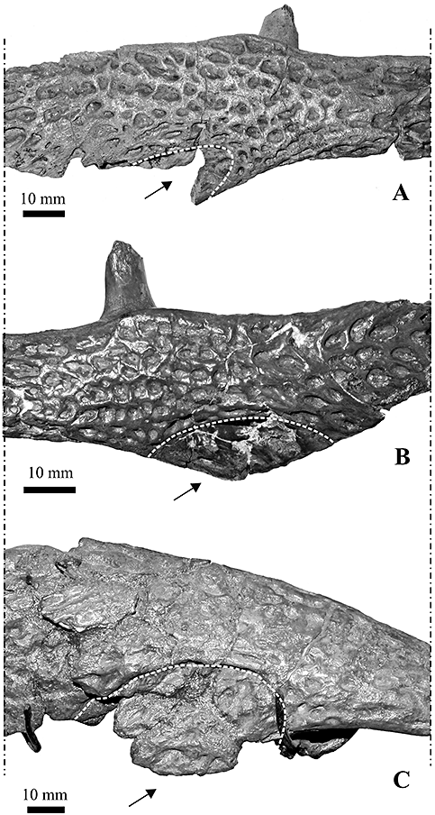
‘Crocodylus’depressifronsBlainville, 1855. A, left jugal in lateral view, specimen IRSNB R251 from Erquelinnes; B, right jugal in lateral view, specimen IRSNB R252 from Orp-le-Grand; left jugal in ventrolateral view, C, specimen IRSNB R254 from Leval. The arrows and the arched lines indicate the depressed and less ornamented area marked by a continuous ridge and located on the lateral surface of the jugal, at the base of the descending process that reaches the ectopterygoids.
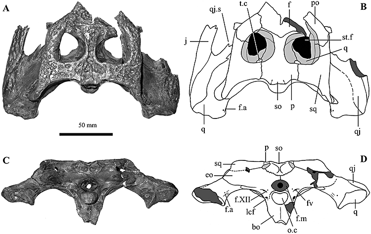
‘Crocodylus’depressifronsBlainville, 1855, specimen IRSNB R252 from Orp-le-Grand. A, B, photograph and interpretative drawing of the posterior sector of the skull in dorsal view and (C, D) in occipital view. See main text for abbreviations.
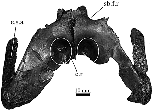
‘Crocodylus’depressifronsBlainville, 1855, IRSNB R252 from Orp-le-Grand. Pterygoids in ventral view. The ellipses schematically indicate the marked concavities placed anteriorly to the choana. A small section of the choanal rim is preserved where indicated. See main text for abbreviations.
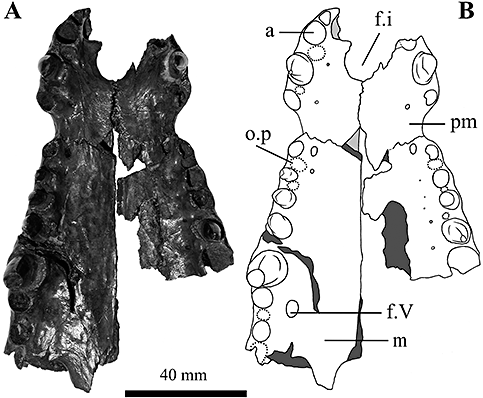
‘Crocodylus’depressifronsBlainville, 1855, specimen IRSNB R253 from Orp-le-Grand. A, B, photograph and interpretative drawing of the anterior palatal region in ventral view. See main text for abbreviations.
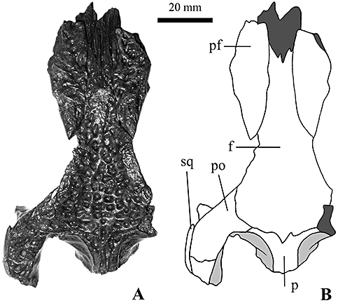
‘Crocodylus’depressifronsBlainville, 1855, specimen IRSNB R253 from Orp-le-Grand. A, B, photograph and interpretative drawing of a fragment of the skull (mostly the interorbital region) in dorsal view. See main text for abbreviations.
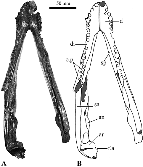
‘Crocodylus’depressifronsBlainville, 1855, specimen IRSNB R253 from Orp-le-Grand. A, B, photograph and interpretative drawing of the lower jaw in dorsal view. See main text for abbreviations.
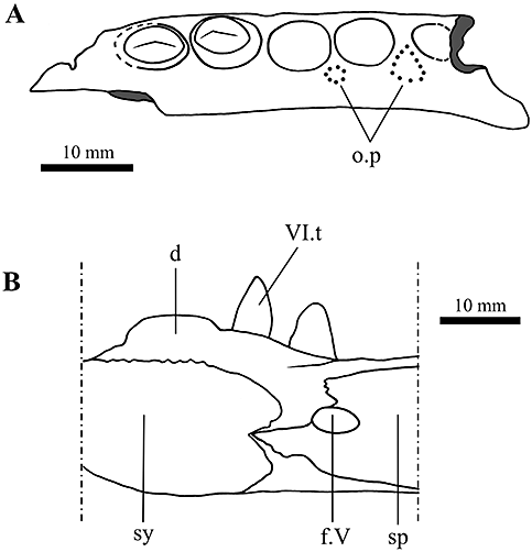
‘Crocodylus’depressifronsBlainville, 1855, specimen IRSNB R253 from Orp-le-Grand. A, fragment (corresponding to the suborbital fenestra) of the right maxilla showing the position of the occlusal pits; B, detail of the symphysary area of the right lower jaw showing the splenial reaching but not entering the symphysis. See main text for abbreviations.
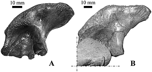
‘Crocodylus’depressifronsBlainville, 1855. A, right ilium of specimen IRSNB R253 from Orp-le-Grand mirrored in order to aid comparison; B, left ilium (digitally isolated from the background) of specimen IRSNB R254 from Leval.
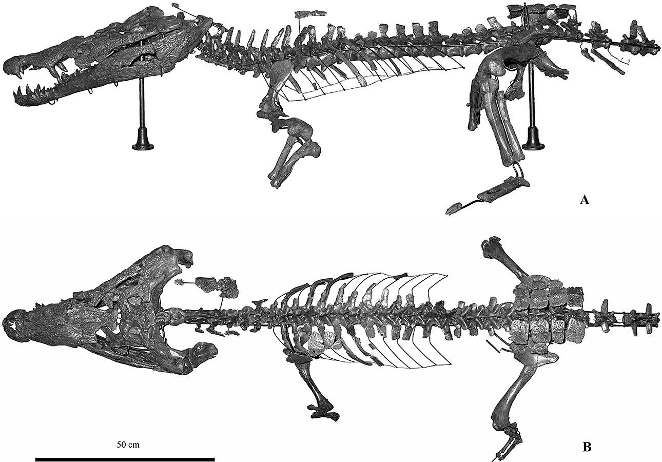
‘Crocodylus’depressifronsBlainville, 1855, specimen IRSNB R254 from Leval. A, skeleton in left lateral view; B, in dorsal view. This adult specimen was a medium sized crocodylian reaching at least a total length of about 3 m.
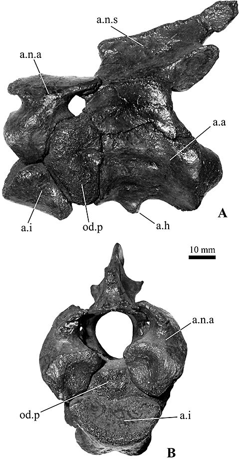
‘Crocodylus’depressifronsBlainville, 1855, IRSNB R254 from Leval. Axis–atlas complex, in left lateral (A) and anterior (B) views. See main text for abbreviations.
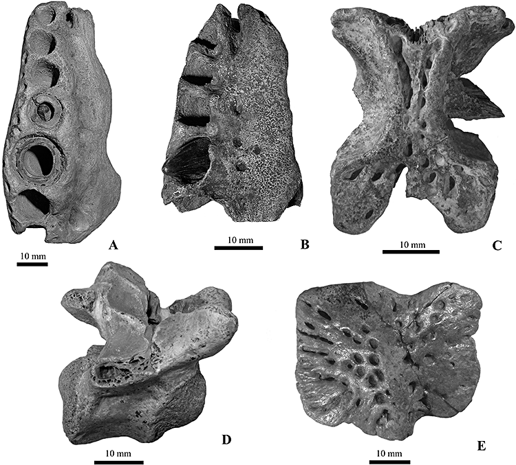
‘Crocodylus’depressifronsBlainville, 1855 from Dormaal. A, right maxilla in ventral view, specimen IRSNB R255; B. right maxilla in ventral view, specimen IRSNB R256; C. parietal in dorsal view, specimen IRSNB R257; D, first caudal vertebra in right lateral view, specimen IRSNB R259; E, dorsal osteoderm in dorsal view, specimen IRSNB R260.
Age: Tienen Formation, Early Ypresian, earliest Eocene.
Emended diagnosis: A basal generalized crocodyloid characterized, as in the other Asiatosuchus-like taxa, by an elongated dentary symphysis (reaching the sixth alveolus), but differing from Asiatosuchus grangeriMook, 1940 (the type species of Asiatosuchus), as well as from ‘Asiatosuchus’germanicusBerg, 1966 and ‘Crocodylus’affinisMarsh, 1871 because of the large medial jugal foramen; from ‘A.’germanicus by the fronto-parietal suture not entering the supratemporal fenestrae, the intermediate (not overbiting) occlusal pattern, and the splenial not participating to the symphysis; and from ‘C.’affinis by the exposure of the postorbital on the lateral surface of the skull table.
Moreover, this basal crocodyloid is also characterized by the presence of an evident depression on the lateral surface of the jugal, a shallow depression located anteriorly to the choana on the ventral surface of each pterygoid, absence of caecal recesses in the narial canal of the maxillae, anterior ectopterygoid process not deeply forked, and a particular trapezoidal shape of the external naris. There are five teeth on the premaxillae, 13 teeth on the maxillae and 16–17 teeth on the dentaries (but maybe up to 18 on the left dentary of the Leval specimen). Such additional characters, at least in part shared by other Asiatosuchus-like taxa, could be useful for the identification of Palaeogene fragmentary remains mixed with those of other taxa.
Description
The following descriptions are focused on the incomplete skull from Erquelinnes (IRSNB R251), which is by far the most informative specimen. The specimens from Orp-le-Grand, Leval, and Dormaal will be briefly commented on in order to prove their conspecificity with the one from Erquelinnes; their descriptions will also focus on the morphological characters that are not available on the Erquelinnes skull (or the minor incongruences).
Specimen irsnb R251 from Erquelinnes
Skull (5, 6)
Preservation, form, and general features: The skull is largely preserved with only the posterior sector of infratemporal fenestrae, the occipital and posterior-palatal areas severely damaged or incomplete. The only major breakage crosses the snout transversally at the level of anterior tips of the prefrontals: a few millimetres of bone are lacking along this fracture. The preserved elements of the skull do not show any sign of deformation but several bones are separated along the sutures (that are therefore generally well visible), allowing the appreciation of their internal morphology (such as the narial canal of the maxillae). Small restoration integrations with a gypsum-like substance were carried out in the past in order to join some fragments. The estimated length of the skull (from the tip of the premaxillae to the quadrates) is about 400 mm.
The skull outline is markedly festooned: in dorsal view, concavities correspond to the premaxilla–maxilla suture and to the eighth alveolus, whereas convexities correspond to the last premaxillary teeth and the fifth dentary tooth; in lateral view, the convexities correspond to the fourth premaxillary and fifth maxillary teeth (but also the last maxillary teeth) and concavities correspond to the premaxilla–maxilla suture and the seventh interalveolar space on the maxilla.
With the exception of the moderate convexity on the maxillae corresponding to the fifth tooth, the dorsal skull surface is particularly smooth, not showing any median boss, canthi rostralii, or preorbital ridges. The skull table and the sloping interorbital area are rather flat (although not coplanar).
In dorsal view, the skull table has rather long squamosal prongs and nearly straight lateral edges, not parallel but diverging posteriorly about 30°, whereas in posterior view, its dorsal edge is approximately planar.
All the dorsal and lateral surfaces, with the exceptions of the quadrates, are markedly ornate with irregular pits that are particularly well defined, wide and deep, on the frontal and the skull table in general.
Cranial fenestrae and openings: The naris is rather large, about as wide as long, with nearly straight and slightly divergent lateral sides and an anteriorly convex rim. It can be defined as dorsally orientated although the anterior rim is located slightly ventrally to the posterior one. The posterior rim of the naris hosts the tip of the nasals, whose precise development inside the naris cannot be evaluated but seems to be modest. The lateral walls of the naris are not vertical but markedly sloping. The foramen incisivum is anteriorly incomplete but the preserved portion is nearly circular in shape, rather small and placed relatively near to the tooth row. The orbits have a vaguely semicircular shape (with a deeply concave medial margin and a nearly straight lateral one) and are characterized by having margins flush with the skull surface. Although the anterior portion of both orbits is partly damaged, the anterior edge seems to be not vertical but slanting. Palpebral bones are not preserved and, although there are no traces of ligamentary attachment on the prefrontals (such as those visible in extant Osteolaemus spp.), their presence has not been excluded a priori. The infratemporal fenestrae were much smaller than the orbits; their posterior and medial rims are now broken off on both sides. The supratemporal fenestrae are not completely preserved because the anterior edges and the posterior (postero-medial) walls are partly missing. Their preserved rims do not overhang the fossae and their medial and anteromedial walls are markedly sloping and not pierced by any foramen. The anteromedial corner of each supratemporal fenestra is smooth. The otic aperture is preserved only on the right side of the skull. Although the dorsal sector of its posterior margin is broken, it is clear from the remaining (ventral) part, made by the quadrate, that the posterior margin was not straight. The ventral surface of the otic aperture shows a median convexity. Information on the shape of suborbital fenestrae and the relationships among the surrounding elements is offered by the fragments representing the margins of the fenestrae. The lateral margins are nearly straight or slightly concave. The preserved posterior corner of the right fenestra is not large enough to assess the presence or absence of a notch. However, a modest convexity of the anterior edge of the preserved ectopterygoid could suggest the former presence of such a notch (as testified by the material from Orp-le-Grand). The anterior corners of suborbital fenestrae reach the seventh interalveolar space. An inner section of the trigeminal foramen (= foramen ovale) is visible on the right side of the skull. Because the lateral surface of the braincase is not preserved, it is not possible to assess the configuration of the bones around the foramen. Post-temporal fenestrae and fossae are not entirely preserved: their medial sector is inclined at about 45° and they are likely to have been relatively small. The median Eustachian foramen is only partly preserved but, because of its position, it is clear that it opened ventrally to the small, slit like, right lateral Eustachian foramen (the only one preserved). The foramen magnum is represented by its lower rim, made by the occipital condyle of the basioccipital. With the exception of a small exoccipital fragment of its left lateral edge, all of the bones around the foramen are missing. The cranio-quadrate passage opens under a blade-like development (slightly eroded) of the exoccipital and is visible in occipital view; the other end of the passage reaches the postero-ventral corner of the otic recess and is not visible in lateral view.
Skeletal elements: The premaxillae are nearly complete because only a small area anterior to the foramen incisivum (and not reaching the tooth row) is damaged. The premaxillary surface at the postero-lateral corner of the external naris is not raised in any particular ridge. A modest, very low, convexity is located on both sides of the skull along the premaxilla–maxilla suture. A deep lateral notch is developed where the premaxilla–maxilla suture reaches the lateral edge of the bone. The premaxillae preserve all the five alveoli and some of them host the remnant of a tooth (there are no crowns). The fourth premaxillary alveolus is by far the biggest. The dorsal premaxillary processes are moderately long and reach the level of the third maxillary alveolus; the ventral ones are not elongated being simple convexities reaching the level of the first maxillary alveolus.
The maxillae are rather well preserved. The right one shows a partial restoration on the dorsal surface. The left maxilla is complete and hosts 14 alveoli, the left one is posteriorly broken and hosts 13 alveoli. Seven teeth are preserved on each maxilla and nearly all have crowns. The largest alveolus is the fifth and it corresponds to a fairly marked convexity (although not as developed as in some extant crocodylid taxa) on the dorsal surface of the maxillae. A shallow concavity is present on each maxilla between this boss and the mild convexity developed along the premaxillary–maxillary suture. Thanks to the fact that maxillae become disarticulated from each other ventrally and from the nasals dorsally, it is possible to see clearly the smooth lateral wall of the narial canal in both maxillae; no caecal recesses are present, but a large foramen (actually two foramina confluent in one single opening) is visible approximately at the level of the fourth tooth. On the palatal surface, the foramen for the palatine ramus of the fifth cranial nerve opens posteriorly to the fifth maxillary alveolus and is significantly larger than the other foramina aligned lingually to the maxillary tooth row. The left foramen (the right one is partly occluded because of the restoration) is nearly as big as the smallest alveolus (the first one; therefore character 111 has been tentatively scored as 1).
Nasals extend from the frontal process (they laterally extend a little posteriorly to the anterior median tip of the frontal process) to the external naris, which they enter slightly. Although their anterior tips are broken off, it is highly probable they did not bisect the naris (as confirmed by the remains from Orp-le-Grand, IRSNB R252, and Leval, IRSNB R254). For most of their length they are rather wide compared to the width of the snout, and roughly symmetrically reduce in size in the proximity of both the anterior and posterior tips.
The lacrimals constitute the anterior edge of the orbits and, although they are not perfectly preserved, an elliptic, dorsoventrally depressed lacrimal duct is present medially to the anterior tip of the orbit. The lateral suture with the jugal could correspond to the edge of the preserved lacrimal, whereas the suture with the maxilla is not perceivable with confidence except close to the lacrimals' anterior tip; however, it is clear that they extend anteriorly more than the prefrontals.
The prefrontals are widely separated by nasals anteriorly and by the frontal process posteriorly. The prefrontal pillars are broken off at mid height but their preserved portions show that they were dorsally expanded in an anteroposterior direction; their medial processes are not preserved.
The dorsal surface of the frontal is heavily pitted and nearly flat (almost imperceptibly concave). Its lateral edges, forming the medial margin of the orbits, are not raised. The frontal process (the region anterior to the suture with prefrontals) is approximately as long as half of the total length of the frontal. Its anterior tip is tripartite on the dorsal surface. Although the anterior edges of both the supratemporal fenestrae are not complete, it is possible to state with confidence that the fronto-parietal suture develops entirely on the skull table because the frontal, which is not firmly joined with the parietal, clearly shows a posterior edge entirely represented by sutures (therefore the frontal does not constitute the anterior rim of the supratemporal fossae). The fronto-parietal suture is slightly concave anteriorly.
The postorbital bar is slender and presents a small and low spine in an anterolateral position in its dorsal sector. The postorbital is exposed above the anterior process of the squamosal on the lateral surface of the skull table.
The squamosals are particularly flat without dorsolateral convexities (the so-called ‘horns’); they markedly overhang posteriorly on the occipital surface. In lateral view, the dorsal and ventral rims of the squamosal are approximately parallel. The anterior process of the squamosal, along the lateral margin of the skull table, does not reach the middle of the postorbital bar and is not dorsally developed.
The parietal is nearly complete (its posterior left lateral area is broken off). The area exposed on the skull table is X-shaped, with small anterior branches and wider and longer posterior branches. The lateral margins of the parietal, constituting the medial vertical wall of the supratemporal fossae, widen in a ventrolateral direction forming a gently sloping surface. The anterolateral edges merge rather gradually with the sloping anteromedial surface of the supratemporal fossae. On both lateral sides of the large supraoccipital, the parietal constitutes a small section (about 5 mm long) of the posterior edge of the skull table. The posterior region of the dorsal surface, along with the dorsal surface of the supraoccipital, is slightly concave. Very small ridges are developed along the medial rim of the supratemporal fenestrae (not significantly altering the nearly flat aspect of the skull table).
The right quadrate is better preserved than the left one, although its condyle is medially eroded. Its dorsal surface is nearly entirely devoid of any ornamentation but shows some rugosities in its medial sector. Anteriorly to the otic aperture, it hosts a rather large foramen (foramen aerum; after Iordansky, 1973). The left condyle has an expanded medial hemicondyle. The foramen aerum on the dorsal surface of the element is rather small and placed close to the medial edge.
Only the posterior part of both the quadratojugals is preserved. Although the right element is better preserved than the left one, we are not able to assess its relationships with the jugal at the posterior corner of the infratemporal fenestra, the position and development of the quadratojugal spine, and the development of the quadratojugal anterior process along the lower temporal.
The right jugal is better preserved than the left, whose caudal region is missing. The medial jugal foramen is extremely large (it is about 14 mm long) on the left jugal and is represented by two large openings on the right one. The jugal extends on the lateral surface of the postorbital bar up to the spine placed on its anterolateral surface. The postorbital bar is inset from the lateral surface of the jugal; it originates from the medial surface of the jugal and is separated from the dorsal edge of the latter by a gutter. In lateral view, the dorsal edge of the jugal is approximately straight anteriorly to the postorbital bar and is modestly concave at the level of and posterior to the bar itself, without developing a distinct notch. A very well-marked depression (Fig. 7A) is located on the lateral surface of the jugal at the base of the descending process that reaches the ectopterygoid. This depression is at least dorsally and posteriorly limited by a continuous evident ridge (like the ridges separating the pits on jugal outside the depression but larger). The ornamentation of the jugal surface inside the depression is represented by large and shallow pits contrasting with the smaller and deeper ones on the rest of the jugal surface.
The supraoccipital is perfectly preserved. Its dorsal exposure is considerable because although it does not exclude the parietal from the posterior edge of the skull table, it is a triangle with a base of 28 mm and a height of 15 mm. Its posterior edge markedly overhangs on the posterior (occipital) surface (note that, despite not being delimited by sutures, the area corresponding to the supraoccipital exits from the posterior profile of the skull in the original drawing by Blainville, 1855, here reproduced in Fig. 1A). The occipital exposure of the supraoccipital is rather wide and narrow (about 45 mm wide and approximately 15 mm tall); it hosts two small lateral concavities symmetrically placed, but not a marked longitudinal ridge.
The vomer is not exposed on the palate.
Only the right palatine is preserved. It is rather narrow and its lateral edge is approximately straight and slightly flared anteriorly (not producing any lateral process) but not posteriorly. The palatine–pterygoid suture is developed anteriorly to the posterior end of the suborbital fenestra (although laterally not far from the posterior corner of the fenestra, character 85 has been tentatively scored as 1). The anterior tip is missing and therefore the shape of the maxillary–palatine suture cannot be evaluated, but because of the presence of part of such a suture on the left maxilla it seems that it was broad and rounded. The palatine appears not to have extended anteriorly to the suborbital fenestrae because the maxillae extend (medially) beyond the anterior corner of the fenestrae.
The left ectopterygoid is nearly completely preserved (Fig. 6A), but as its posteroventral tip is broken off and the corresponding pterygoid is not preserved, it is not possible to evaluate the relationships between these two elements. The anterior process of the ectopterygoid nearly touches the last three alveoli of the maxillary: its tip, pointed and not deeply forked, reaches the anterior edge of the third last alveolus and it is quite close to the last two alveoli (without forming their medial rim). The dorsal ectopterygoid process is considerably developed along the postorbital bar and it forms its medial sector.
The pterygoids are represented by two fragments only. One is a fragment of the left pterygoid coming from the anteromedial sector, close to the sutures with the right pterygoid and the left palatine. It preserves the posteromedial corner of the left suborbital fenestra and shows a marked concavity on its ventral surface. The second fragment belongs to a right element and represents a small area lateral to the Eustachian foramen and the internal choana; the posterior pterygoid process is preserved and is rather small, as high as it is long, and points in a posteroventral direction. The pterygoid–ectopterygoid suture is visible on the left ectopterygoid where it represents the medial edge of the preserved fragment: it is approximately straight with a modest lateral bend in the anterior sector (because the bend is only weakly developed, character 116 has been scored as 0).
The basisphenoid is not preserved ventrally to the medial Eustachian foramen but as the braincase is anteriorly truncated on the transversal section, this element shows the two symmetric openings of the foramen caroticum anterius. A lamina of the basisphenoid is preserved on the right lateral side of the basioccipital.
Only a small fragment of the right laterosphenoid is preserved above the section of the trigeminal foramen. As a result of the presence on the frontal ventral surface of the scars for the suture of the laterosphenoid capitate processes, it is possible to state that these processes were anteroposteriorly directed.
There is no trace of a boss in the area lateral to the opening of the cranio-quadrate passage. As a result of the incompleteness of the paroccipital processes it is not possible to assess their relationships with the squamosal caudal projections. A fragment of the exoccipital is attached to the left side of the occipital condyle. The exoccipitals are not involved in the occipital tubera.
The basioccipital is represented by two non-adjacent fragments joined together during the restoration of the specimen. A fragment forms the occipital condyle (which is wider than tall) and its base, the other constitutes a part ventral to the occipital condyle. The latter is not laterally expanded and hosts a pronounced vertical crest.
Teeth and dentition pattern: Nearly all of the preserved teeth crowns are markedly worn. The only tooth showing the original morphology is the one in the third right maxillary alveolus. This tooth is sunk in the alveolus and does not show any sings of wear: it could be a substitution tooth in the process of replacing the lost functional one. It is conical, rather slender and pointed, with two marked mesiodistal ridges delimiting a small lingual and a large labial surface. The rest of the teeth are characterized by extensive apical wear so that the apexes are flattened and the dentine is exposed (the enamel is completely eroded). All of these teeth show evident mesiodistal ridges. The tooth surface (not the basal region of the crown) is ornate with wrinkles also partly developed on the mesiodistal ridges. The exposed roots are not smooth but have a markedly crumpled surface.
Very large and deep occlusal pits are developed on both premaxillae caudally and medially to the first interalveolar space (such pits nearly reach the dorsal skull surface within the naris, whose anterior bottom is broken off). Other pits are preserved before and after the fourth premaxillary alveolus: they are developed in the medial sector of the interalveolar space and partly lingually to it (their position is slightly less lingual in the left element than in the right one). On the maxillae, a small quasi-interalveolar pit is present after the first alveolus. Two other small pits develop partly in the interalveolar area but mostly medially to it after the second and third alveoli on the left maxilla and after the second alveolus on the right one. The wide seventh maxillary interalveolar spaces host a very large pit (about double in size if compared to the surrounding alveoli). Much smaller pits are located somewhat lingually (but not completely lingually) to the tooth row after the sixth tooth on the left maxilla and eighth on the right one (the condition was probably symmetrical but because of the poor state of preservation it is not possible to affirm this with confidence). The maxillary tooth row is approximately straight after the large interalveolar pit that fills the seventh interalveolar foramen. The last alveoli have an approximately round section.
Specimen irsnb R252 from Orp -le -Grand
This specimen is represented by a partially preserved skull (6-9) and by the posterior region of the lower jaw.
Skull
Preservation, form and general features: The skull is nearly complete but split into several fragments. In the case of the snout, the general shape is slightly distorted and the bones are slightly separated from each other. The sutures among the skeletal elements of the skull are not visible with confidence on the lateral sides of the braincase and on the occipital surface (with the exception of the limits of the supraoccipital): the interpretative drawings of these areas are not presented because the absence of clearly visible sutures strongly decreases their informativeness.
Cranial fenestrae and openings: Both of the posterior walls of the supratemporal fossae are perfectly preserved and host the strongly elongated and inclined opening (about 14 mm long) of the temporal canal. The quadrate reaches the temporal canal and therefore separates the frontal from the squamosal on the posterior wall of the fossae. Although the infratemporal fenestrae are not completely preserved because of the incompleteness of the postorbital bars and jugals, they seem to be rather small and their posterior medial corner is constituted by the quadratojugals. In addition, the suborbital fenestrae are incomplete but by the shape of the isolated ectopterygoids showing an anterior edge (forming the posterolateral rim of the fenestra) that is distinctly convex, it is possible to state that the fenestra had a posterior notch. On the right maxilla, the suborbital fenestra reaches anteriorly the posterior end of the seventh interalveolar space (where a large interalveolar occlusal pit is developed). The foramen magnum is slightly wider (19.3 mm) than the occipital condyle (18.0 mm). Ventrolateral to the foramen magnum there are the three foramina: the medial one is the small foramen for the cranial nerve XII, the large lateral one is the foramen vagi and the ventral one (only slightly smaller than the latter) represents the lateral carotid foramen that is placed dorsally to the basisphenoid exposure on the lateral side of the braincase.
Skeletal elements: There is some matrix covering the anterior edge of the right orbit but on the left lacrimal it is possible to see an elongated lacrimal duct (developed entirely on the lacrimal) to the right of the anteriormost angle of the orbit. The lateral edge of the lacrimals is not visible. On both lacrimals it seems that there is a depressed elongated nearly parasagittal area placed anteriorly to the anterior end of the orbit.
The frontal dorsal surface is not perfectly flat because of the presence of a modest concavity, but does not develop any ridge along the orbits' rim. The frontoparietal suture is not entirely visible with confidence but it is not developed inside the perimeter of supratemporal fenestrae and seems to be markedly concave.
The parietal clearly constitutes the anteromedial corner of supratemporal fenestrae. The underlying wall of this corner is not vertical but is markedly sloping. Parietal and postorbital are clearly in contact for about 2 mm on the skull table (this is visible on the left side only because a fracture develops on the right side).
At the dorsal angle of the infratemporal fenestra, the postorbital probably contacts not only the quadratojugal but also the quadrate. The postorbital is visible on the lateral surface of the skull table, posteriorly to the postorbital bar and dorsally to the anterior process of the squamosal.
On the lateral surface, the jugals have a depression (Fig. 7B), dorsally rather well delimited, that occupies the ventral expansion for the suture with maxilla and ectopterygoid. This depression is caudally not as well delimited as in the specimen from Erquelinnes.
The quadratojugals constitute the entire medial rim of the infratemporal fenestrae and their posterior corner. The quadratojugal spine is perfectly preserved in the left infratemporal fenestra where it is placed approximately halfway between the posterior and the dorsal corner of the fenestra; it is prominent but rather small (its medial side is 3 mm long). The left quadratojugal sends a rather long process (about 10 mm) along the lower temporal bar.
The boundaries of the supraoccipital are not visible with confidence on the dorsal skull surface and therefore it is not completely represented in the interpretative drawing in Figure 8. Conversely, its sutures are the only ones clearly visible on the occipital surface of the skull, where it is wider than tall and shows two symmetric depressions separated by a median ridge.
The maxillo–palatine suture is not visible with confidence but it is clear that it is not significantly placed anteriorly to the anterior rim of the suborbital fenestra. This suture is probably placed in a highly fractured area immediately posterior to the anterior end of the suborbital fenestrae, and can be tentatively defined as rounded and broad. The palatine–pterygoid suture is not preserved. The lateral edges of the palatines show a modest lateral expansion that is not symmetric on both sides: more posteriorly placed and smooth in the left element, rather irregular on the right element (because of the modest development of this structures and their position, the status of character 94 has been scored as 0). The palatines are posteriorly parallel. It is clear that the palatines are not developed posteriorly to the posterior corner of the suborbital fenestrae and they did not even reach this corner. However, it seems that the zig-zag process of the palatine–pterygoid suture is caudally directed and therefore the caudal extension of the suture is not so far from the corner of the fenestra.
The ectopterygoids do not reach the ventral tip of the pterygoid flanges. Their anterior process grazes the maxillary tooth row and has unbifurcated tips (Fig. 6B).
The pterygoids are not completely preserved but the right one retains the anteromedial corner of the internal choana which therefore opens completely within the pterygoids (Fig. 9). The pterygoid surface is markedly concave in the area anterior (and maybe also lateral) to the choana. The small (few millimetres long) preserved margin of the choana is not flush with the (concave) ventral surface of the pterygoid indicating the probable presence of a neck surrounding the choana.
The squamosal–quadrate suture cannot be entirely followed in the otic recesses because of the incompleteness of the region. However, the right squamosal is nearly completely preserved in the right otic recess: it ends with a suture showing that the quadrate makes the dorsal edge of the otic recess. The squamosal does not extend ventrolaterally as the lateral extent of the paraoccipital process. On both the sides of the skull, the anterior process of the squamosal reaches the middle of the postorbital bar without excluding the postorbital from the lateral wall of the skull table.
Lower jaw
Preservation, form and general features: The lower jaw is represented by the posterior regions of a right and left dentary, by a left splenial, and a left angular, and by the still-joined left surangular and articular.
Openings: As a result of the presence of a rather well-preserved left angular, it is possible to assume that the (partially preserved) external mandibular fenestra is not anteriorly developed sufficiently to reveal the foramen intermandibularis caudalis in lateral view.
Skeletal elements: The posterior region of the right dentary corresponds to the dentigerous area of the last ten alveoli followed by nearly all of the posterior edentulous area. Interpreting a wide interalveolar space as the diastema corresponding to the eight interalveolar space, the largest alveolus is the 11th, and the total number of teeth of the lower jaw should have been 16. No teeth are preserved.
The left dentary preserves the last seven tooth positions and the posterior edentulous area, with teeth preserved in the first four alveoli. Interpreting the largest alveolus (in this case the second preserved tooth that is larger and taller than the others) as the 11th, the total number of teeth should have been 16.
The right angular delimits the ventral, caudal, and part of the dorsal rim of the large foramen intermandibularis caudalis. The angular process dorsal to the foramen is broken off (in the other angular the foramen is even less preserved).
The surangular reaches the tip of the lateral wall of the glenoid fossa and nearly that of the retroarticular process. The development of the surangular in the ventral direction allows us to suppose that the surangular–angular suture reaches the articular toward its ventral tip. The lingual foramen for the articular artery is not visible with confidence (and therefore character 45 has not been scored).
The articular gradually slopes on the surangular without a sulcus between them. The articular forms the retroarticular process, which is posterodorsally directed. The left articular is rather complete and shows no evidence of an anterior process that hides the medial surface of the surangular. The medial expansion of the retroarticular process is poorly developed because it does not surpass the width of the condyle. The shape of the articular–surangular suture inside the glenoid fossa cannot be evaluated because of the presence of matrix; however, the shape may be ‘bowed’ as in the left lower jaw of the other specimen from Orp-le-Grand because, despite the presence of a brownish painting-like cover, a ‘bowed’ linear depression could possibly represent the sutural line.
Dentition: The only preserved maxillary tooth (on an isolated fragment) is particularly labiolingually compressed, short and with a nearly flat ‘apex’ (it is not significantly worn) so that its shape is approximately rectangular in lateral view (basic measurements are: mesiodistal diameter: 7.1 mm; labiolingual diameter: 3.5 mm; crown height: about 3 mm). Its crown surface is not entirely wrinkled or worn.
The teeth on the left dentary (no teeth are preserved on the right one) are quite different, being pointed (therefore not apically flattened) and approximately triangular in lateral view. The crowns are distinctly labiolingually compressed and bear sharp mesiodistal ridges. Small longitudinal crests and wrinkles are variably expressed on the crown surfaces. Only the last tooth has a crown that is markedly wrinkled (their basic measurements are as follows: first: not measurable because it is embedded in matrix; second: mesiodistal diameter: 8.8 mm, labiolingual diameter, 6.1 mm, crown height: about 9.3 mm; third: mesiodistal diameter: 8.5 mm, labiolingual diameter: 5.0 mm, crown height: 7.4 mm; fourth: mesiodistal diameter: 6.8 mm, labiolingual diameter: 3.9 mm, crown height: estimated as about 5 mm).
Moreover, there are 21 isolated teeth. Their crown shape varies from an elongated and slender shape to a markedly depressed one. Wrinkles are variably expressed.
Specimen irsnb R253 and associated remains from Orp -le -Grand
This specimen is represented by a partial skull (10, 11) and lower jaw (Fig. 12). Several postcranial elements (bearing the same collection number as number IRSNB IG 9875) are associated to these cranial remains and in most of the cases could possibly belong to the same specimen.
Skull
Cranial fenestrae and openings: The small fragment of the posteroventral sector of the braincase clearly shows (on its left side) that the lateral Eustachian foramen opens dorsally to the median one.
Skeletal elements: The premaxillae are anteriorly incomplete. The right one is preserved in the sector corresponding to the last four alveoli, the left one to the last three. Both the premaxillae retain a single tooth: the penultimate. It is likely that both the premaxillae had five alveoli before breakage.
The maxillae are morphologically congruent with those already described for Erquelinnes. The right element corresponds to the first seven alveoli, the left one to the first five. Altogether, six teeth are preserved. A more posterior fragment of the right maxilla (shown in Fig. 13A) corresponds to about seven alveoli (five of which preserved) in the area of the suborbital fenestra. The last preserved alveolus and half of the previous one correspond to the sutural area with the ectopterygoid (that therefore is quite close to the maxillary tooth row). Two teeth are preserved in the third and second alveoli from last positions.
The frontal dorsal surface is somewhat more concave than in the larger specimen from the same locality, with lateral edges slightly raised (but with a flat dorsal surface) along the orbits' rim. The anterior process (anterior to the suture with prefrontal) is about as long as the rest of the bone; its anterior tip is roughly tripartite as in the Erquelinnes specimen. The frontoparietal suture is deeply concave (with a posterior median tip) and entirely developed on the skull table.
The jugal is represented only by the region ventral and posterior to the postorbital bar and shows the posterior area of the depression described above for the other specimens from Erquelinnes (IRSNB R251) and Orp-le-Grand (IRSNB R252). The depression is dorsally limited by a well-marked ridge, like that of the Erquelinnes specimen, but not so well developed posteriorly.
The anterior process of the left squamosal reaches the middle of the postorbital bar without excluding the postorbital from the lateral wall of the skull table.
In occipital view the basisphenoid is not visible between the basioccipital and the pterygoids of the isolated fragments of this specimen because of its reduction. A transverse suture is visible inside the median Eustachian foramen. The basisphenoid is visible only on the left side of the basioccipital.
Lower jaw
The lower jaw is nearly complete (only the coronoids are missing) but partly damaged (the articular of the right jaw and the retroarticular process of the left are missing). In dorsal view, the general shape of the lower jaw is rather narrow (although some degree of deformation is the result of several breakages with partial displacement of the surrounding elements).
Openings: The external mandibular fenestra is present and rather small. It is nearly entirely preserved in the right dentary but its relationship with the foramen intermandibularis caudalis cannot be evaluated because the latter is not preserved. The dentary constitutes the dorsal edge of the fenestra and ends slightly anteriorly to the posterodorsal corner of the fenestra itself.
Skeletal elements: Both the dentaries are not completely preserved but they are somehow complementary in terms of available information. They are markedly festooned in lateral view (convexities corresponding to alveoli four and 11). Their tooth rows are damaged or not complete posteriorly but it seems likely that the maximum number of alveoli is 16 on both sides (a depression of the medial surface of the dentary external wall located after the 16th alveolus does not correspond to a flexure on the dentary dorsal surface and is here not considered as evidence for s 17th alveolus). The first alveoli do not preserve teeth but it is clear that they were directed anterodorsally. The largest alveoli are the first, the fourth, and the 11th. Alveoli three and five are much smaller than the fourth. A diastema is clearly present after the eighth tooth on both the dentaries. The symphysis nearly reaches the posterior rim of the sixth alveolus. Teeth are preserved in positions five and six on the right element and three, four, six, ten, 11, and 12 on the left one. Although the last alveoli are slightly labiolingually compressed (compression possibly partly a result of post-mortem deformation), their shape can be defined as rounded. The splenial constitutes the medial wall of the last alveoli. The dentary upper edge has an evident concavity between the fourth and the tenth alveolus. The dentary–angular and the dentary–surangular sutures reach the external mandibular fenestra nearly at its ventral and dorsal corners, respectively.
Neither of the splenials preserve their posterior region. They reach the symphysis without participating in it. The right splenial, better preserved than the left one, allows us to see that its anterior tip passes ventrally to the Meckelian groove (Fig. 13B) and that the mandibular ramus of cranial nerve V exits through a perforation of the dentary located between the dorsal and ventral anterior splenial processes. As a result of the fact that the anterior tips of the splenial do not join anteriorly to the foramen, it is possible to state that this element does not possess any anterior perforation for mandibular ramus of cranial nerve V.
The angular–surangular suture reaches the external mandibular fenestra at its caudal corner.
The anterior processes of the surangular are not the same length; the dorsal one is longer than the ventral but does not reach the tooth row (assuming that 16 alveoli are present). There is no sulcus between the surangular and articular.
The foramen aerum is placed close to the medial edge of the glenoid caudal boundary.
Teeth and dentition pattern: Premaxillary and maxillary teeth are slender and elongated, with sharp ridges. Their crown surfaces are rather smooth, devoid of any particular ornamentation such as crests or wrinkles. The two maxillary teeth preserved in the third and second from last positions are different because their surfaces are wrinkled.
On the premaxillae (Fig. 10), occlusal pits are evident on the right element in interalveolar spaces three and four. The first pit is more medially placed than the second, which is entirely developed in the interalveolar position. On the maxillae, all the visible occlusal pits have a position that can be generally defined as interalveolar: they are present in interalveolar spaces number one, two, six, and seven on the right maxilla and one, two, and three on the left one. Only the anterior pit on the right maxilla is significantly (but partly) developed in the medial direction. In contrast, the fragment of a right maxilla corresponding to the suborbital fenestra shows more medially placed occlusal pits (Fig. 13A).
Evident occlusal pits are present in the dentaries lateral to the tooth row. In particular, pits are visible laterally to the 13th alveolus (both in the anterior and the posterior sector), to the 14th alveolus (exactly in lateral position) as well as laterally to the 15th interalveolar space of the left dentary, and laterally to the 12th, 15th, and 16th interalveolar spaces on the right. More pits could be present but because of the dentary preservation they are not visible with confidence.
Postcranial skeleton: A couple of humeri of the same size (130 mm) probably belong to this specimen. The right one is much more damaged than the left one because the proximal epiphysis is nearly completely broken off. A single longitudinal scar for the common insertion of the M. teres major and M. dorsalis scapulae is present on the posterodorsal surface of both the humeri in the proximal epiphysis region, just dorsally to the deltopectoral crest. The latter structure is completely preserved only in the right element, where it is quite sharp and slightly concave.
The ulna is only slightly damaged in its distal epiphysis. Its length (101 mm) matches with the size of the humeri. The large olecranon process is rounded and rather flattened and is markedly constricted at the base.
An isolated left femur (145 mm long) may belong to the same specimen. Although fractured and with the epiphyses partly covered by a matrix rich in pyrites, its morphology is rather well preserved, with the exception of the scars of the M. caudofemoralis, which are not visible with confidence.
Twenty-two vertebrae or vertebral fragments are preserved: one axis odontoid process, two cervical vertebrae, three cervical centra, three postcervical centra, seven dorsal or lumbar vertebrae, one pair of sacral vertebrae, two neural arches, one neural spine, and one isolated chevron bone.
All the presacral vertebrae are procoelous. The neurocentral suture of the cervical vertebrae is invariably open, whereas the postcervical elements have fused neurocentral sutures. The cervical with the fused neural arch has probably been glued. The second vertebra is partly damaged because the right neural arch is missing. The largest dorsal or lumbar vertebra is heavily pyritisized. The pair of sacral vertebrae are characterized by the articular surfaces of the centra: the first one has a centrum anteriorly concave and posteriorly flat, the second one has the opposite condition. The tuberculum and capitulum of the first sacral vertebra are equally developed so that in dorsal view the capitulum is nearly not visible.
A right ilium is nearly perfectly preserved (Fig. 14A). The acetabulum is wide and deep. An evident narrow scar for muscular insertion is located dorsally to the acetabulum. The iliac blade is approximately rounded and has a very shallow indentation on its dorsal edge. The process located on the anterior edge of the ilium, anteriorly to the dorsal margin of the acetabulum, is weakly developed.
The right ischium lacks the distal anterior portion. The major one of the proximal articular surfaces has a V-shaped articular surface for the ilium, delimiting laterally a smooth surface that represents the ventrolateral surface of the acetabulum.
The pubis does not have the proximal end preserved; only the majority of the flaring distal part and its basis.
A few small elongate bones represent several fragmentary elements of the acropodium, nine ribs or rib fragments, and at least four fragmentary gastralia.
Thirty-nine osteoderms (26 complete or nearly complete and 13 fragments) are preserved. At least the more symmetric ones can be tentatively considered as midline elements. All of them are approximately rectangular (the best preserved has a length of 31 mm, and width of 42 mm), possess a medial keel and have a smooth anterior edge not showing any anterior process. The osteoderms are rather thin and lightly built. The dorsal ornamentation consists of a few small pits and rare furrows (no ridges are present) not completely filling the surface that therefore has large, nearly smooth areas. All of the preserved osteoderms have keels that are not high or sharp. Nearly all have a small anterior smooth gliding surface and sutures are developed on both the lateral sides. The few elongated and roughly oval-shaped osteoderms (the lateral ones) do not show the anterior edge and the lateral sutures, as they were isolated from other osteoderms.
A further osteoderm having the same collection number (so it is the 40th) is quite different in shape and colour (the most of its surface is not as dark as the rest of the material from Orp-le-Grand) and probably belonged to a different specimen. It is smaller (22 mm length, 20 mm width), proportionally more thick, with a large anterior gliding stripe, with many more pits than the osteoderms described above, and with pits larger and deeper and with only a hint of keel. It is perfectly preserved and shows sutures on the sides.
Three other remains bearing the same collection number do not match in size with the remains previous described. A poorly preserved right femur is much bigger (total length of about 180 mm) than the previous one (it could possibly belong to the specimen IRSNB R252). A small portion of a left femur diaphysis and a rather large and massive tibia (130 mm long) match in size with it.
Specimen irsnb R254 from Leval
The specimen from Leval (Fig. 15) is represented by a nearly complete skeleton (with skeletal elements belonging to one single individual) preserving the skull (damaged and incomplete in the palatal area) and lower jaw, as well as several vertebrae (Fig. 16), part of the limb bones, and some osteoderms.
The specimen has been mounted on a metal support and thus offers a good chance to observe the general anatomy of this taxon. However, the fact that many cranial sutures are not clearly visible hinders a detailed description. At present, the mounted skeleton is 1760 mm long but only five caudal vertebrae are present (with gaps between them).
Further material coming from the same locality but at least partly not belonging to the mounted specimen is available (see the Referred material and localities section for a list).
Skull
The general shape of the skull in dorsal view is comparable to that figured by Berg (1966: fig. 4) for ‘Asiatosuchus’germanicus, with a wide posterior region and a significantly narrower snout (skull length from the level of quadrates to the tip of the snout: 500 mm). All the available characters match with those already discussed with the other specimens from Erquelinnes and Orp-le-Grand.
A major discrepancy concerns the tooth numbers because the lower jaw has 17 teeth on the right side and possibly 18 on the left one (five premaxillary teeth are visible on both the premaxillae and 13 teeth are probably present on the right maxilla; the left maxillary tooth row is incomplete). It is worth noting that the dentary symphysis reaches the sixth alveolus, but the diastema is not located after the eight alveolus as in the smaller specimen from Orp-le-Grand (and the most of the living crocodylids) but after the ninth: the eighth and ninth teeth are preserved on both the rami and they are fairly small and extremely near (so that that their alveoli could be confluent; however, such a condition cannot be proven because some bone fragments seem to be present between these teeth at least on the left ramus). The largest dentary teeth are the first, fifth, and 12th.
The splenials clearly show an abrupt dorsoventral reduction about 15 mm before the symphysis and it seems that the anterior tip of the splenial nearly reaches, but does not enter, the symphysis.
Although the supratemporal fenestrae do not seem to be intersected by the fronto-parietal suture, the latter is not visible with confidence on the skull table because of several breakages and the restoring plaster.
On the lateral side of the skull table, the postorbital is visible above the anterior process of the squamosal that reaches the postorbital bar.
On both the maxillae, there are very deep occlusal pits before and after the seventh alveolus. They are both interalveolar but the anterior one is placed closer to the lingual edge of the interalveolar space, whereas the posterior one, which is more large and deep, is more centred in the interalveolar space.
As in the smaller skulls previously described from Orp-le-Grand and Erquelinnes, there are neither significant depressions on the skull table or anteriorly to it, nor significant upturned edges of orbits and supratemporal fenestrae. The squamosals have a nearly flat dorsal surface. The depressions on the lateral surface of the jugals (Fig. 7D) have a morphology only slightly less continuously delimited than in the Erquelinnes specimen.
Postcranial elements: The atlas and axis are rather well preserved (Fig. 16). The atlas is represented by both the two separated neural arches and the intercentrum (no proatlas preserved), the axis by the odontoid process, the neural arch, and the centrum. The atlas intercentrum is wedge-shaped in lateral view and the parapophysial processes are weakly developed. The axis neural spine is dorsally damaged but it seems likely that the anterior half of the neural spine was sloping and that its posterior half was not upturned (the corresponding characters, 11 and 12, have been tentatively scored). The neural arch does not seem to have any lateral process. The axis hypapophysis is a prominent tubercle located slightly behind one third of the centrum length; it can be defined as forked. The third cervical vertebra (following Brochu, 1997, here considered as the first after the axis) is well preserved and shows a distinct hypapophysis, and a rather long neural spine whose dorsal tip is about as long as half of the centrum without the cotyle. The neurocentral sutures of all cervical vertebrae are closed. The hypapophysis is present up to the 12th vertebra posterior to the atlas.
The limbs lack almost all the autopodia. The left fore limb (the right one is missing) is shorter than the hind limbs (not considering the missing autopodia) but equally robust. Although the dorsal edge of the preserved scapula is broken off, it is clear that it flares dorsally, whereas it seems likely that the damaged deltoid crest was rather thin. The scapula and the coracoid are not joined together suggesting that the synchondrosis closed later during ontogeny.
The ilia are slightly different from the element from Orp-le-Grand because, although their dorsal notch is modest like in the latter (and unlike in extant Crocodylus), their outline is dorsally depressed and not rounded at all (Fig. 14B). Nevertheless, there is some asymmetry in the same specimen because the right element is particularly narrow posteriorly.
About a dozen osteoderms are preserved. Those placed along the midline are rectangular and keeled as are some of those described for the Orp-le-Grand specimen, but larger, more heavily built than them, and with a higher number of pits.
Specimen irsnb R255-260 and DIIA R50RS from Dormaal
Two incomplete right maxillae from Dormaal, IRSNB R255 and R256 (Fig. 17A, B), are characterized by the following characters: fourth and fifth alveoli completely separated (their interalveolar space is approximately equal to the interalveolar space between the other preserved alveoli) and different in size, with the fourth alveolus being much smaller than the fifth (the fifth alveolus is only partly preserved in specimen IRSNB R256 but it is clearly larger than the fourth). In both cases, occlusal pits are located towards the lingual side of the interalveolar spaces from one to three. In both specimens, the preserved portions of the medial wall of the caviconchal recess are smooth.
The isolated fragmentary parietal IRSNB R257 (Fig. 17C) possesses thick anterolateral processes indicating that the frontoparietal suture did not enter the supratemporal fenestrae. Its dorsal surface is moderately thickened and raised in correspondence to the rim of the supratemporal fenestrae and weakly concave in the area posterior to the fenestrae.
The skull table fragment DIIA R50RS preserves the area corresponding to the supraoccipital and posterior region of the parietal. The morphology of the preserved part of the parietal is similar to that of the remains previously described and the supraoccipital is significantly exposed on the skull table.
The incomplete first caudal vertebra IRSNB R259 (Fig. 17D) has a centrum 30 mm long and a closed neurocentral suture.
The isolated dorsal osteoderm IRSNB R260 (Fig. 17E) is comparable in size (width 45 mm, length 35 mm) and general morphology to the osteoderms of the skeleton from Leval.
PHYLOGENETIC ANALYSIS
Methods
The phylogenetic relationships of ‘Crocodylus’depressifrons were analysed using a cladistic approach. The character coding of this taxon was realised by merging the fully redundant codings (the ‘states are identical for those characters coded in common, but [… ] some characters are coded for one but not the other’; Brochu, 2004: 870) originally realised separately for the remains of the four Belgian localities. The coding reported in the Appendix substitutes the one of the ‘Belgian crocodyloid’ in the character matrix of 166 discrete morphological characters and 68 taxa made available by Brochu (2007b; note that the taxon is named ‘Dormaal crocodyloid’ in the tree of fig. 17), the most complete and up-to-date dataset available for the analysis of the relationships within the crocodyloid clade.
The analysis was performed with PAUP 4.0b10* (Swofford, 2001). The outgroups were represented by Bernissartia fagesii and Hylaeochampsa vectiana. ACCTRAN and DELTRAN optimization were performed with tree bisection and reconnection in effect and 100 replicates of random addition sequence.
Results
The simplified topology of the strict consensus tree is shown in Figure 18. The analysis recovered 906 148 equally optimal trees. The parameters were the following: tree length = 481; consistency index (CI) = 0.43; homoplasy index (HI) = 0.58; CI excluding uninformative characters = 0.40; HI excluding uninformative characters = 0.60; retention index (RI) = 0.79; rescaled consistency index (RC) = 0.33. The node of ‘C.’depressifrons is supported by the following unambiguous synapomorphies: 6-0: axial hypapophysis located toward the middle of centrum; 7-1: hypapophyseal keels present on the 12th vertebra behind atlas; 12-1: axis neural spine not crested; 73-1: internal choana surrounded by a ‘neck’; 76-2: postorbital contacts quadratojugal and quadrate at dorsal angle of infratemporal fenestra; 82-2: large supraoccipital exposure on dorsal skull table; 98-1: posterior pterygoid processes small and project posteroventrally; 161-0: naris approximately circular (or at least not wider than long).
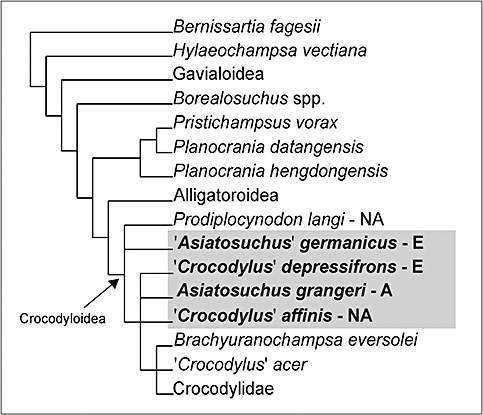
Schematic strict consensus tree summarizing the phylogenetic relationships of ‘Crocodylus’depressifronsBlainville, 1855 (see text for details and statistics). For the sake of clarity, only the name of the clade (or the genus name in the case of Borealosuchus) is shown in the cladogram for some terminal taxa. The continent of origin is specified for Prodiplocynodon and the four Asiatosuchus-like taxa (on a grey background): A, Asia; E, Europe; NA, North America. Note that the four Asiatosuchus-like taxa do not constitute a monophyletic group.
The topology of the tree resulting from this new character coding is congruent with the ones published by previous authors (Brochu, 1997, 1999, 2000, 2001b, 2003, 2007a, b; Brochu & Gingerich, 2000; Jouve, 2004; Piras et al., 2007; Vélez-Juarbe et al., 2007) and based on earlier versions of the same character matrix. The topological congruence does not reflect a few differences with the ‘Belgian crocodyloid’ character coding by Brochu (2007a): few previously unknown states have been added to the coding (characters: 18, 88, 148, 153), few states have been scored differently (characters: 73, 82, 98, 161) and others, previously scored in older matrices, have been considered as not clear enough to allow a confident assessment (characters: 3, 15, 16, 17, 45, 66, 74, 99, 115, 122, 127, 129, 152, 162, 164).
This analysis does not allow improvement of the resolution of the relationships of the considered Asiatosuchus-like taxa: ‘A.’germanicus, A. grangeri, ‘C.’depressifrons, and ‘C.’affinis do not form a monophyletic assemblage. ‘Asiatosuchus’ germanicus is polytomic with the primitive crocodyloid Prodiplocynodon langiMook, 1941, and both are basal to the polytomic group of A. grangeri, ‘C.’depressifrons, ‘C.’affinis, and the rest of the crocodyloids.
DISCUSSION
Systematics
The crocodylian remains coming from the coeval Early Eocene localities of Dormaal, Erquelinnes, Leval, and Orp-le-Grand show a rather uniform morphology, and despite some obvious differences in terms of size, they clearly belong to the same taxon. All the characters typical of ‘C.’depressifrons, as shown by the figures published by Blainville (1855) or implicit in the name, are present in the Belgian material. Not to mention the general crocodyloid appearance, noteworthy are the relatively small supratemporal fenestrae, the flattened forehead, the mandibular symphysis reaching the sixth dentary tooth, and the splenials not involved in the mandibular symphysis. Minor similarities shared by the specimens of the four localities, but not visible on the specimen figured by Blainville, are the well-marked depression on the lateral jugal surface (character visible in the remains from all the localities in which the jugal is preserved; Fig. 7), the symmetrical, shallow, and wide concavities on the palatal surfaces of pterygoids anteriorly to the internal choana (visible in the specimen from Erquelinnes and the larger skull from Orp-le-Grand; that area is not preserved in the smaller skull from Orp-le-Grand and in the Leval specimen), as well as the particular shape of the external naris (visible in the remains of three of the four localities; in Dormaal the maxillae are not preserved). The combination of all these characters supports a conspecific status of the remains from the four localities. The few differences are mostly referable to ontogenetic changes or intraspecific variability. An example is the fact that only the Leval specimen shows very small eighth and ninth dentary alveoli (nearly in contact with each other). This condition is also shown in the detail of the lower jaw of ‘C.’depressifrons published by Blainville (1855) and figured, along with a ‘normal’ condition, as a possibility for ‘A.’germanicus when the eighth tooth is ‘doubled’ (Berg, 1966: fig. 4c). This morphology seems to be best interpreted as a variable feature present in different species rather than as a diagnostic character.
The referral to ‘C.’depressifrons of the material from Erquelinnes is in agreement with Dollo (1909), Buffetaut (1985), and Vasse (1992, 1993), but referral of material from Orp-le-Grand to this species contrasts with the opinion expressed by Vasse (1993) that it could belong to a still undescribed taxon. The Leval specimen was not previously assigned to any taxon. The presence of an Asiatosuchus-like taxon at Dormaal was previously reported by Buffetaut (1985) on the basis of a few isolated distinctly ornate teeth. In fact, the members of this group seem to have, along with ‘normal’ teeth, also massive, slightly blunt or even rounded teeth whose surface is distinctly ornate; these teeth are rather different from those of some coeval European taxa (as Eosuchus or Diplocynodon) but in other cases, and mostly the posterior ones, are similar to those of other taxa (such as ‘Allognatosuchus’). In absence of a well-grounded comparative analysis of the morphology and variability of the dentition of ‘C.’depressifrons and the Asiatosuchus-like taxa, it is here proposed to avoid potential taxonomic, chronological, and biogeographical confusion in the already complex issue concerning this group and formally to refer isolated teeth, such as those from Dormaal, to Crocodylia indet. (it is also the case for ‘Asiatosuchus’ sp. from the middle Eocene of Kazakhstan; Efimov, 1988). However, the presence of an Asiatosuchus-like taxon at Dormaal as proposed by Buffetaut (and then by Lambrecht, 2003) is supported by some skull fragments and postcranial elements whose morphology matches with that of ‘C.’depressifrons remains from the three other coeval Belgian localities.
Significant congruence between the Belgian crocodyloids and ‘C.’depressifrons was already underlined by Brochu (1997) who, despite considering the holotype as uninformative, suggested that the French material from the Early Eocene of Meudon (see Fig. 4 for chronological relationships of this locality; Planté, 1870; Russell et al., 1990) could belong to this species (same splenial symphysis and frontoparietal suture–supratemporal fossae relationships; morphology of ‘C.’depressifrons assessed on the figures by Blainville), and that the Belgian crocodylians are congruent with the Meudon specimens and therefore might represent ‘C.’depressifrons (however, he hesitated to accept this identification, awaiting further material from the Paris Basin).
As for the generic allocation of ‘C.’depressifrons, its referral to the genus Crocodylus can be easily excluded on the basis of the characters available in the Belgian materials, and in particular by the absence of blind pockets on the medial wall of the caviconchal recess and by the absence of a dorsal constriction in the posterior process of the ilium (see Brochu, 2000, 2007b). The morphology of the Belgian remains is congruent with that of the Eocene Asiatosuchus-like taxa but differs from ‘A.’germanicus because of the position of the frontoparietal suture on the skull table, the partly interfingering occlusal pattern, and by the absence of a splenial symphysis; differs from ‘A.’germanicus, A. grangeri, and ‘C.’affinis because of the large medial jugal foramen, and from ‘C.’affinis because the anterior squamosal process is not dorsally shifted and therefore the postorbital is visible on the skull table's lateral margin (see Norell & Storrs, 1986; Brochu, 1997). Comparison with other species putatively referred to the genus Asiatosuchus (see Introduction) is not discussed here because their morphology, and therefore their taxonomic status, is unclear.
Remarks on the morphology of‘Crocodylus’depressifrons
The Belgian remains allow further discussion of some of the morphological features of ‘C.’depressifrons: the occlusal pattern, the size of the medial jugal foramen, and the pterygoid and jugal depressions. As already stated by Brochu (1997), the occlusion pattern of the Belgian basal crocodyloids testifies to a transitional state between a complete overbite and completely interfingering occlusion. Indeed, this character is slightly variable: in one case (specimen IRSNB R253 from Orp-le-Grand) pits are located in interalveolar positions at least between the first eight maxillary alveoli (Fig. 10), but distinctly more medially (Fig. 13A) in the region corresponding to the suborbital fenestrae as testified by the associated lower jaw (Fig. 12); in another case (specimen IRSNB R251 from Erquelinnes) the maxillary occlusal pits are more medially located in the anterior region but clearly interalveolar after the seventh tooth (Fig. 5). A deep occlusal pit centred in the interalveolar spaces before and after the seventh alveolus is also visible in the specimen IRSNB R254 from Leval. Therefore, the typology of the occlusal pattern scored for ‘C.’depressifrons is the intermediate condition between a lingual occlusion (that characterizes, among others, the alligatoroids) and an in line occlusion (that mostly characterizes the crocodylids). Such an intermediate condition is shared by ‘C.’affinisand A. grangeri and agrees with their basal position within the crocodyloid clade.
A further interesting character is represented by the size of the jugal medial foramen. This foramen is extremely large in A. depressifrons (see descriptions above and, among others, Brochu, 1997) but it is reported to be small in ‘C.’affinis and A. grangeri (Brochu, 1997; and note that the corresponding character is scored as ‘?’ for the latter in the available matrixes; see, among others, Salisbury et al., 2006; Brochu, 2007b). Because of the fact that the size of this foramen shows a considerable intraspecific variation (Massimo Delfino, pers. observ.), it would be interesting to analyse a wider sample of A. grangeri (but its remains are far from being abundant at present) and to assess its size in the several specimens of ‘A.’germanicus that are already available in European collections.
The shallow depression located anteriorly to the choana on the ventral surface of the pterygoids (feature visible in the left pterygoid fragment of specimen IRSNB R251 from Erquelinnes and in the fragmentary but better preserved pterygoids of specimen IRSNB R252 from Orp-le-Grand) is apparently wider and less defined than that reported by Salisbury & Willis (1996: figs 3B, 4B) for the mekosuchine Kambara implexidensSalisbury & Willis (1996) from the Early Eocene of Australia, but somehow similar to the depression shown by extant Osteolaemus. Salisbury & Willis (1996) interpreted the depression of Kambara as possibly housing a salt-secreting gland (see also considerations about biogeography made by Angielczyk & Gingerich, 1998). It is worth noting that the cladistic analysis performed by Salisbury & Willis (1996: fig. 15) suggested sister taxon relationships among an ‘Asiatosuchus’ species, ‘A.’germanicus and the Mekosuchinae (relationships not confirmed by other analyses; among others Brochu, 2007b). Further investigations are needed to understand if these depressions are homologous and what their function was in the fossils, also taking into consideration their (apparently unknown) function in extant Osteolaemus.
The evident depression on the lateral surface of the jugal (Fig. 7) is a character which is probably widely distributed among different taxa (apparently similar, but not so well defined, depressions are present in some specimens of extant Crocodylus spp.) and that could also be present in other Asiatosuchus-like taxa. However, the presence of this depression proved to be useful for the identification of isolated Asiatosuchus-like jugals in Palaeogene crocodylian assemblages (as in the case of the material from the Late Palaeocene of Mont de Berru / Cernay-les-Reims).
Remarks on the phylogenetic relationships of theAsiatosuchus-like taxa
The cladistic analysis here presented, even if based on a new character coding for ‘C.’depressifrons, does not succeed in retrieving a monophyletic grouping of the Asiatosuchus-like taxa. The same topology has been already obtained several times by previous authors (see Introduction).
However, close similarities among the four Asiatosuchus-like taxa, both in terms of gross morphology and fine anatomy (at least according to the information available at present), may suggest closer relationships than those emerging from the cladistic analysis. In this respect, Brochu (1997: 281) has already noticed that Prodiplocynodon and ‘A.’germanicus are linked only by the position of the frontoparietal suture on the skull table and that ‘a closer relationship between “A.”germanicus and more derived crocodyloids appears more likely’. The apparent relationships of these two taxa could be a result of the poor preservation of the former (C. A. Brochu, pers. comm.).
As for the relationships among the type species of Asiatosuchus, A. grangeri, ‘C.’affinis, and ‘C.’depressifrons, it is worth noting that their character codings available at present are nearly completely redundant. The coding of A. grangeri is 100% redundant to that of ‘C.’affinis (the latter is much more complete than the former), whereas the differences with ‘C.’depressifrons are limited to the size of the medial jugal foramen (large in ‘C.’depressifrons and small in A. grangeri but see above for further discussion of this character) and the shape of the naris. The naris of A. grangeri has been scored as it if were wider than long (Brochu, 2007b). It is not possible to verify the morphology of the naris because the snout is not preserved in the type material published by Mook (1940) and this character has probably been evaluated on unpublished material. However, it is remarkable that the same status is scored for ‘C.’affinis. This appears to be a probable miscoding because the naris of the latter is obviously not wider than long in the type specimen figured by Norell & Storrs (1986: fig. 1). Similarly, the status of A. grangeri could have been affected by a miscoding (this character should be verified on the fossils before changing the matrix and therefore it was not changed in the matrix analysed in this work).
Only an accurate analysis of the fossil remains already collected (if not the collection of new fossils), and therefore the revision of the codings of all the Asiatosuchus-like taxa (with the possible inclusion of further characters), will demonstrate if their nonmonophyly is only a result of the weak resolution power of the available matrixes in the area of the basal crocodylids, or if, despite a considerable morphological affinity, there is sound evidence for splitting the Asiatosuchus-like taxa into different genera.
CONCLUSIONS
The species ‘C.’depressifronsBlainville, 1855 is identified in the Belgian localities of Dormaal, Erquelinnes, Orp-le-Grand, and Leval, whose age, early Ypresian, Early Eocene, is approximately coeval with that of Muirancourt (Oise, France), the type locality of this species. The abundant Belgian material has allowed us to present for the first time the diagnosis of this species. It appears to be similar to the type species of Asiatosuchus, A. grangeriMook, 1940, as already supposed by several authors in the last decades. Nevertheless, the four Asiatosuchus-like taxa cladistically analysed here (with a new character coding for ‘C.’depressifrons) do not form a monophyletic group, confirming previous phylogenetic analyses. The species erected by Blainville is therefore provisionally referred to its former description genus and not to Asiatosuchus. A complete revision of the remains of all of the Asiatosuchus-like taxa and the identification of further characters are needed to increase the resolution power of the available matrixes in the area of the basal crocodyloids.
ACKNOWLEDGEMENTS
Annelise Folie (RBINS, Brussels), France de Lapparent, and Bernard Battaille (MNHN, Paris) collaborated in the literature search and assisted M. Delfino while working in their institutions. Roman Croitor (Università di Firenze) kindly provided translations from Russian. Simone Maganuco (Università di Firenze) assisted and discussed issues related to the phylogenetic analysis. Christopher A. Brochu (University of Iowa, Iowa City), Marco P. Ferretti, and Lorenzo Rook (Università di Firenze), Paolo Piras (Università di Roma Tre), T. Rossmann (Technische Universität, Darmstadt), Roberto Sindaco (IPLA Torino), and Alberto Venchi (Agrotech Roma) discussed the nomenclature of ‘C.’depressifrons or provided literature and suggestions. We thank Richard Smith (Bruxelles) for access to specimens from his collection, Stéphane Berton (RBINS, Brussels) for the restoration of the Leval skeleton, and P. Harcourt Davies (Hiddenworlds, Orvieto) for linguistic revision. C. A. Brochu and the anonymous reviewers significantly improved earlier drafts of the manuscript. This research received support from the SYNTHESYS Project (http://www.synthesys.info/), which is financed by European Community Research Infrastructure Action under the FP6 ‘Structuring the European Research Area’ Programme (BE-TAF 970 and FR-TAF-967 to M. Delfino). Work was partially supported by Research Project MO/36/020 of the Belgian Federal Science Policy Office to T. Smith.
Appendix
Character coding for ‘Crocodylus’ depressifrons realised on the basis of the remains from Dormaal, Erquelinnes, Orp-le-Grand, and Leval. Character list (166 discrete morphological characters) and definitions as in Brochu (2007b).
???000100? 111????10? ?00001111? ?00110???1 1010??0001 011??????1 11?00?0001 101?020110 2200100110 01?01?01?0 010000?001 1301?00001 0?1000?1?1 00010?1?01 1000010010 0?1??????0 0???00


