Polyphasic approach and typification of selected Phormidium strains (Cyanobacteria)
Abstract
Cyanobacteria (phylum Cyanophyta/Cyanobacteria, class Cyanophyceae) are among the most widespread organisms and are able to adapt themselves to different extreme environments. These micro-organisms have an important ecological role, given their ability to perform oxygenic photosynthesis, and are employed in different fields based on their ability to produce several bioactive compounds. Their prokaryotic nature, the presence of many cryptic species, and the coexistence of different nomenclature systems make the taxonomic identification of cyanobacteria particularly difficult. Moreover, for several species, the original reference strains (holotypes) are lacking. Increasingly, authors are using a polyphasic approach to characterize cyanobacteria, while typification is a recent trend that is being used to solve the problem of missing holotypes in other micro-organisms. Here we focus on a filamentous cyanobacterium, isolated from the Euganean Thermal District (Padova, Italy) and temporarily named strain ETS-02, using a polyphasic approach that includes morphological, ultrastructural, biochemical (pigment and fatty acid content), physiological (nitrogen fixation), and genetic (16S rRNA, 16S–23S ITS, cpcB-IGS-cpcA, rpoC1, gyrB, rbcL, nifD loci) analyses. The description of Phormidium cf. irriguum CCALA 759 as the epitype of Phormidium irriguum was also used to complete the characterization of strain ETS-02.
Cyanobacteria (phylum Cyanophyta/Cyanobacteria, class Cyanophyceae) are among the most widespread, morphologically distinct, and abundant prokaryotes (Whitton, 1992). They inhabit almost all environments, playing an important role as primary producers. In particular, they are able to adapt themselves to different extreme habitats, such as desert soils, alkaline lakes, hypersaline ponds, glaciers, and hot springs (Ward et al., 1998; Paerl et al., 2000; López-Cortés et al., 2001; Jungblut et al., 2005; Sánchez-Baracaldo et al., 2005; Taton et al., 2006).
Cyanobacteria are also known to produce several bioactive compounds, most of which have various therapeutic applications (Li et al., 2005; Singh et al., 2005; Khan et al., 2006; Eriksen, 2008; Gerwick et al., 2008; Sivonen and Börner, 2008). Nearly half of the active cyanobacterial compounds known thus far are produced by members of the order Oscillatoriales (Gerwick et al., 2008). This order, representative genera including Lyngbya, Oscillatoria, and Phormidium, includes filamentous species which are not able to form heterocysts (Komárek and Anagnostidis, 2005).
The identification of new cyanobacterial strains is a complex issue, due to small cell sizes and morphologies that can look very similar among phylogenetically distinct entities.
Cyanobacteria show the phenomenon of “cryptic species,” i.e. organisms that seem to belong to a particular species from a morphological point of view but are genetically distinct (Ward et al., 1998; Casamatta et al., 2005; Taton et al., 2006; Marquardt and Palinska, 2007). Moreover, different ecotypes of the same species can exploit separate ecological niches of the same environment and/or can be found in geographically distant areas worldwide (García-Pichel et al., 1996; Palińska et al., 1996; Ward et al., 1998; Palińska and Marquardt, 2008).
The arbitrary use of two different nomenclature codes, Botanical and Bacteriological, makes the situation even more complex. For nearly 30 years, taxonomists have been trying to find a solution that will lead to the creation of a unique nomenclature system (Oren, 2004; Komárek, 2005, 2010; Marquardt and Palinska, 2007; Oren et al., 2009).
Moreover, the description of several strains deposited in international culture collections relies only on morphological traits, without molecular characterization. Conversely, many nucleotide sequences found in public databases are attributed to strains that have not been morphologically described (Wilmotte and Herdman, 2001; Rajaniemi et al., 2005). A further problem is that, for many genera, the described type species are no longer available for comparative purposes.
This picture is particularly true for the order Oscillatoriales, whose heterogeneous and polyphyletic nature has been emphasized by many authors (Casamatta et al., 2005; Teneva et al., 2005; Premanandh et al., 2006; Palińska and Marquardt, 2008).
At present, many studies suggest the use of a polyphasic approach to properly identify new cyanobacterial strains (García-Pichel et al., 2001; Komárek, 2005, 2010; Palińska and Marquardt, 2008; Moro et al., 2010; Sciuto et al., 2011). Moreover, a recently proposed tendency to overcome the problem of unavailable holotypes in other groups of micro-organisms, such as fungi and microalgae, is to describe new epitypes (Hyde and Zhang, 2008; Jahn et al., 2008; Tuji and Williams, 2008; Maistro et al., 2009; Rybalka et al., 2009), i.e. specimens or illustrations selected to serve as interpretative types when the holotypes, or all original material associated with a validly published name, are demonstrably ambiguous and cannot be critically identified for purposes of the precise application of the name of a taxon (McNeill et al., 2006).
The Euganean Thermal District (Padova, Italy) has been known since Roman times for its therapeutic properties, and many people come from all over the world to benefit from thermal water and mud treatments. Some researchers have highlighted the important contribution provided to the therapeutic properties of the mud by cyanobacteria (Bellometti et al., 1997, 2000; Galzigna et al., 1998), which during the mud maturation process develop as conspicuous mats on its surface. In particular, a filamentous strain isolated from this environment and characterized as Phormidium sp. ETS-05 (Ceschi-Berrini et al., 2004) showed large amounts of polyunsaturated fatty acids in its membrane glycolipids, conferring anti-inflammatory properties to the mud (Lalli et al., 2004; Bruno et al., 2005; Marcolongo et al., 2006).
In spite of this, the biodiversity of the Euganean Thermal District has been poorly studied. In fact, knowledge of the Euganean cyanobacterial diversity is based largely on reports dealing with morphological identification (Andreoli and Rascio, 1975; Tolomio et al., 2004), while, only more recently, analyses have been carried out using more extensive approaches, including ultrastructural, pigment, and genetic analyses (Ceschi-Berrini et al., 2004; Moro et al., 2007a,b, 2010). We were therefore interested in studying new cyanobacterial strains growing on the thermal mud surface through more complete surveys, as well as gaining a better understanding of their contribution to the properties of the mud.
We carried out analyses using morphological, ultrastructural, biochemical, physiological, and molecular approaches to characterize strain ETS-02, another filamentous cyanobacterium isolated from the Euganean Thermal District. For the molecular analyses, a multilocus approach, as in Lineau et al. (2010), was adopted, considering seven different genomic regions: 16S rRNA, 16S–23S ITS, cpcB-IGS-cpcA, rpoC1, gyrB, rbcL and nifD. The 16S rRNA gene and 16S–23S intergenic transcribed spacer (16S–23S ITS) were employed as molecular markers for cyanobacteria and, more generally, for prokaryotes (Iteman et al., 2000; Casamatta et al., 2005; Premanandh et al., 2006; Palińska and Marquardt, 2008). The cpcB-IGS-cpcA locus (coding for a portion of the phycocyanin operon), the rpoC1 gene (coding for the γ subunit of the cyanobacterial RNA polymerase core), the gyrB gene (coding for the β subunit of the DNA gyrase, a type II topoisomerase), and the rbcL gene (coding for the large subunit of ribulose 1,5-bisphosphate carboxylase/oxygenase, the enzyme controlling CO2 fixation during the Calvin cycle) are single copy regions that have more recently been used in cyanobacterial phylogeny (Toledo and Palenik, 1997; Fergusson and Saint, 2000; Wilson et al., 2000; Watanabe et al., 2001; Seo and Yokota, 2003; Teneva et al., 2005; Premanandh et al., 2006; Tomitani et al., 2006; Ballot et al., 2008). In addition, the nifD gene, coding for two subunits of the enzyme complex nitrogenase (an enzyme involved in N2 fixation) and initially amplified as further proof of nitrogen fixation ability, was employed for phylogenetic reconstruction, as previously reported (Henson et al., 2004).
For a more precise identification of this cyanobacterium, we propose Phormidium cf. irriguum CCALA 759 as the epitype of the species P. irriguum (Kützing ex Gomont) Anagnostidis and Komárek.
We show that the combination of a polyphasic approach and the typification of cyanobacterial strains, deposited in international culture collections and corresponding to the original descriptions, provides a good “combined method” to solve the taxonomic problems associated with the phylum Cyanophyta/Cyanobacteria and to identify new cyanobacterial strains when the holotypes (i.e. the original specimens or illustrations used by the author or designated by the author as the nomenclatural types and conserved in one herbarium or other collection or institution; McNeill et al., 2006) are lacking or no longer suitable for a complete polyphasic comparison.
Materials and methods
Cyanobacterial strains and growth conditions
Samples of microbial mats were collected from the thermal mud surface in a tank of the Garden Hotel (Montegrotto Terme, Italy) in May 2005. At the moment of sampling, the water temperature was about 45 °C and the pH 6.8. From these samples, a filamentous strain was isolated, grown in axenic cultures, and named strain ETS-02 (“ETS” = “Euganean Thermal Springs”).
The following cyanobacterial strains were obtained for comparison from international culture collections: Lyngbya aestuarii PCC 7419, Lyngbya majuscula CCAP 1146/4, Oscillatoria sancta PCC 7515, Oscillatoria tenuis PCC 9107, Phormidium cf. irriguum CCALA 759, Phormidium ambiguum IAM M-71, and Phormidium autumnale CCALA 143.
All of the cyanobacterial strains were cultured in BG11 medium (Rippka et al., 1979) at 20 ± 2 °C, with a photon flux density of 16 μmol photons/m2/s and a 12/12-h light–dark cycle. For the Lyngbya strains, the culture medium comprised BG11 and ASN-III (1 : 1, v/v) (Rippka et al., 1979).
A living culture of strain ETS-02 was deposited at the Culture Collection of Autotrophic Organisms, Czech Republic (CCALA 946), where it is available to the public.
Light and fluorescent light microscopy
Light and fluorescent light microscopy observations were performed using a DMR 5000 Leica (Wetzlar, Germany) microscope, equipped with a digital image acquisition system. The viability of cyanobacterial cells was investigated through observation of chlorophyll red autofluorescence when excited with UV light.
Morphological identification of strain ETS-02 was based on the recent classification of Hoffmann et al. (2005) and on the traits proposed by Komárek and Anagnostidis (2005).
Electron microscopy
For electron microscopy [scanning (SEM) and transmission electron microscopy (TEM)], the samples were prepared as previously described (Moro et al., 2007a,b, 2010). TEM analyses were carried out with an Hitachi HS9 (Tokyo, Japan) transmission electron microscope operating at 75 kV. SEM observations were conducted with a Stereoscan 260 (Cambridge Instruments, Cambridge, UK) instrument operating at 25 kV.
Water-soluble pigment analyses
Phycobiliproteins (PBPs) were extracted from pellets of cultured cells, finely ground in a mortar with liquid nitrogen, using a phosphate buffer (0.01 m NaH2PO4, 0.15 m NaCl, pH 7). The extract absorption spectra were analysed on a DU530 Beckman Coulter spectrophotometer (Fullerton, CA, USA).
Lipid-soluble pigment analyses
Chlorophyll a and carotenoid extracts were obtained from cell pellets, ground as described above in 90% acetone and then analysed by reversed-phase high-performance liquid chromatography (HPLC).
The analyses were carried out according to Färber and Jahns (1998) and Komárek et al. (1999) with an Agilent HPLC system (Waldbronn, Germany), composed of a Rheodyne valve (Rohnert Park, CA, USA), a reversed-phase column (5-μm particle size; 25 × 0.4 cm; 250/4 RP 18 Lichrocart), a binary pump, and an Agilent 1100 series diode array detector.
Fatty acid content analyses
Dried cell pellets were disrupted with an Ultra-Turrax instrument (IKA-Werke GmbH & Co., Staufen, Germany) in a mixture of dichloromethane/methanol (2 : 1, v/v) to extract lipids. The total fatty acids were obtained as methyl esters from these extracts according to Christie (1982) and then analysed by gas-chromatography at the L.A.Z. Centre (Department of Animal Sciences, Legnaro, Padova, Italy).
Nitrogen fixation and gliding motility tests
To test for nitrogen fixation ability, some cultures of strain ETS-02 were grown under the described environmental conditions but in BG110, a medium in which NaNO3 is replaced by an equimolar concentration of NaCl (Rippka et al., 1979).
To assess the gliding ability of strain ETS-02 on a solid surface and the nature of its photomovement, some filaments were placed on a solid medium in the centre of plates, whose surface was three-quarters covered with a black sheet. The plates were exposed to a continuous photon flux density of 16 μmol photons/m2/s and observed after 1–2 weeks.
Genetic analyses
Genomic DNA was extracted from cell pellets ground in a mortar with liquid nitrogen, using the Genomic DNA Purification Kit from Fermentas (Burlington, ON, Canada).
The 16S rRNA gene, the 16S–23S ITS region, the rpoC1 gene, the gyrB gene, the rbcL gene, and the cpcB-IGS-cpcA locus were amplified from the DNA extracts by PCR, as previously described (Seo and Yokota, 2003; Ballot et al., 2004; Tomitani et al., 2006; Moro et al., 2010).
For amplification of the nifD gene, the forward primer NifD1F (5′-TYGGWGGHGACAARAARCT-3′) and the reverse primer NifD2R (5′-TARTCCCARGAGTGCATYTG-3′) were designed, based on multiple alignment of several cyanobacterial nifD gene sequences, which included those of both heterocystous and non-heterocystous filamentous forms (Table 1). Standard PCR protocols were carried out in 25-μL aliquots with Taq DNA polymerase (Fisher, Molecular Biology, Trevose, PA, USA), according to the manufacturer’s recommendations, on a Peltier Thermal Cycler PTC-200 (MJ Research, St Bruno, Quebec, Canada). Thermocycling conditions were as follows: one cycle at 94 °C for 5 min; 35 cycles of 94 °C for 45 s, 50 °C for 30 s, and 72 °C for 45 s; and a final step at 72 °C for 8 min. Approximately 80 ng of template DNA per reaction was used.
| Strain | nifD accession number |
|---|---|
| Anabaena cylindrica PCC 7122 | AF442506 |
| Anabaena sp. PCC 7120 | V01482 |
| Calothrix sp. PCC 7101 | AY196953 |
| Cylindrospermum majus PCC 7604 | AY196952 |
| Leptolyngbya boryana PCC 6306 | EF576854 |
| Leptolyngbya sp. PCC 7004 | EF576858 |
| Leptolyngbya sp. PCC 7104 | EF576859 |
| Leptolyngbya sp. PCC 73110 | EF576865 |
| Leptolyngbya sp. PCC 7375 | EF576867 |
| Lyngbya aestuarii PCC 7419 | EF576869 |
| Nodularia spumigena PCC 73104 | AF442509 |
| Nostoc sp. PCC 7120 | AF442504 |
| Nostoc sp. PCC 7423 | AF442503 |
| Oscillatoria sancta PCC 7515 | EF576870 |
| Pseudanabaena sp. PCC 6802 | EF576857 |
| Pseudanabaena sp. PCC 7403 | EF576864 |
| Pseudanabaena sp. PCC 7409 | EF576868 |
| Scytonema sp. PCC 7814 | AY196954 |
| Symploca atlantica PCC 8002 | EF576872 |
The PCR product analyses, sequencing, and final consensus sequence assembly were performed as described by Moro et al. (2010).
The following different data sets were constructed, using the obtained sequences (from the investigated organism and the strains acquired from culture collections) plus suitable sequences found in the DDBJ/GenBank/EBI Data Bank: 16S rRNA, 16S–23S ITS, rpoC1, gyrB, rbcL, cpcB-IGS-cpcA, nifD, and 16S rRNA + rpoC1 + gyrB. The last-named was produced by concatenating the sequences shared by all of the corresponding single gene data sets, as described in Maistro et al. (2007, 2009). In the construction of the data sets, we took care to insert sequences obtained both from members of the subclass Oscillatoriophycidae, family Oscillatoriaceae, and from members of the subclass Oscillatoriophycidae, family Phormidiaceae, as the investigated organism could belong to both of these groups, based on morphological and ultrastructural data. Moreover, being filamentous non-heterocystous cyanobacteria as well, members of the subclass Synechococcophycideae, family Pseudanabaenaceae, were considered in the phylogenetic analyses. To root the trees, each data set included members of the subclass Nostocophycideae, a group of filamentous heterocystous cyanobacteria, long known to be monophyletic and phylogenetically closely related to the filamentous non-heterocystous cyanobacteria (Hoffmann et al., 2005; Tomitani et al., 2006; Gupta and Mathews, 2010). A summary of all the organisms considered in the phylogenetic analyses is given in Fig. S1.
Unfortunately, we were able to concatenate only three of the five genes used in our phylogenetic surveys, as many of the sequences available for the considered markers in public databases were obtained from different strains of the same species. As we are working with prokaryotes, we consider that concatenating gene sequences of the same species but obtained from different strains is unacceptable. The effect of using different strains of the same species in prokaryotic phylogeny is shown also by Naum et al. (2008). Thus, we have concatenated only the three genes for which there were enough available sequences (in addition to those we obtained here) for the same strains of a given cyanobacterial species.
The multiple alignments for the phylogenetic analyses were generated with the ClustalW computer program (Thompson et al., 1994). Multiple alignment of the 16S rRNA gene was analysed by the Gblocks program, using default settings, and ambiguously aligned nucleotides were removed from the final data set (Castresana, 2000).
An a priori estimation of the phylogenetic signal present in the multiple alignments was performed by maximum-likelihood (ML) mapping (Strimmer and von Haeseler, 1997). The phylogenetic signal was evaluated using the Tree-Puzzle 5.2 program (Schmidt et al., 2002).
Finally, only the 16S rRNA, the rbcL, the nifD, and 16S rRNA + rpoC1 + gyrB gene data sets were used for the phylogenetic analyses.
The analyses were performed according to the maximum-parsimony (MP) method, using MEGA ver. 4.0 software (Tamura et al., 2007), and the ML method, with the PHYML 2.4.4 program (Guindon and Gascuel, 2003), by applying the GTR + I + Γ evolutionary model (Lanave et al., 1984). Non-parametric bootstrap re-sampling (Felsenstein, 1985) was performed to test the robustness of the tree topologies (1000 replicates).
Bayesian inference analyses were also carried out, using MrBayes version 3.1 (Ronquist and Huelsenbeck, 2003). The substitution model in this case was again GTR + I + Γ, and the Bayesian analyses were performed with four search chains for 1 000 000 generations, sampling trees every 100 generations. The first 2500 trees were discarded as burn-in. For the analysis of the 16S rRNA gene data set, the chains were run for 10 000 000 generations, and the first 25 000 trees were discarded as burn-in. Parameter stability was estimated by plotting log-likelihood values against generation time, and a consensus tree with posterior probabilities was then produced. The nexus files for the Bayesian analyses were generated with the Mesquite 2.71 software package (Maddison and Maddison, 2009). The resulting trees were visualized through NJplot (Perriére and Gouy, 1996).
Results
The cultured strain ETS-02 appeared to be composed of long, variously curved, bright blue–green trichomes (Fig. 1), strictly packed to form mats in older cultures, or more dispersed in liquid medium in younger cultures. The aggregated filaments were able to float at the surface of the liquid medium or grow attached to sides and bottom of the flask. Trichomes were isopolar, composed of cells that were wider (3–5 μm) than long (1–2 μm) (Fig. 2), ending with rounded apical cells, and surrounded by a thin colourless sheath, which could protrude from the filament itself (Fig. 3).
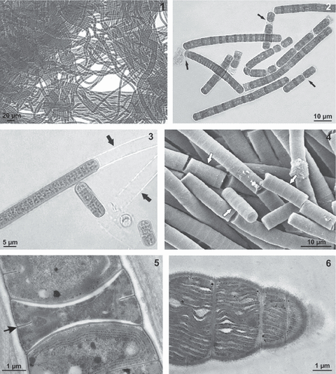
Light micrograph of strain ETS-02. Flexuous, densely packed trichomes. Fig. 2. Light micrograph of strain ETS-02. Reproduction by trichome fragmentation in short hormogonia through the formation of necridia (arrow). Fig. 3. Light micrograph of strain ETS-02. Trichomes surrounded by a colourless sheath, protruding from the filament itself (arrow). Fig. 4. Scanning electron micrograph of strain ETS-02 showing trichome fragmentation in short hormogonia (arrow). Fig. 5. Transmission electron micrograph of strain ETS-02. Necridic cell (arrow) between two neighbouring cells. Fig. 6. Transmission electron micrograph of strain ETS-02. Trichome showing thylakoids (t) parallel to the longitudinal cell walls.
Reproduction was by trichome fragmentation in short hormogonia through the formation of necridia (Figs 4 and 5). Subsequently, each hormogonium reconstituted the average length of a filament by binary fission of the component cells. The daughter cells began a new division before being completely separated.
TEM observations revealed that thylakoids did not assume a particular arrangement in the cell, but could lie parallel to the longitudinal cell walls (Fig. 6), parietal (Fig. 5), radial, or variously distributed. Some cells showed widened thylakoids, with a net-like appearance (Fig. 6). No gas vesicles were observed in the cytoplasm.
Most of the features reported for strain ETS-02 were observed also in Phormidium cf. irriguum CCALA 759, although cells of the latter isolate were larger (9–12 μm wide, 2–3 μm long) (Figs 7 and 8).
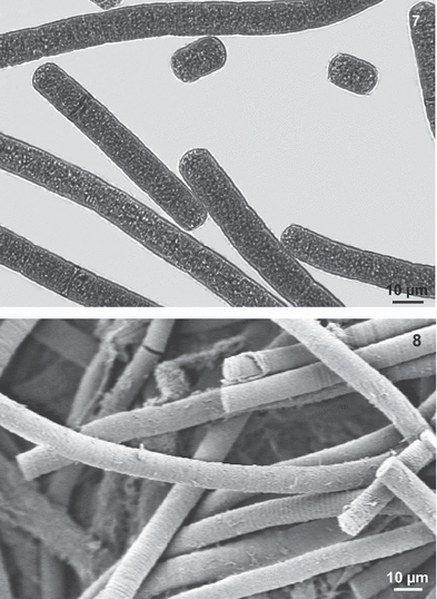
Light micrograph showing trichomes of Phormidium irriguum CCALA 759. Fig. 8. Scanning electron micrograph showing trichomes of Phormidium irriguum CCALA 759.
Strain ETS-02 was able to survive in medium without organic nitrogen, as ascertained through the observation of the chlorophyll red autofluorescence of cells maintained in this condition for 3 weeks. No heterocysts or other specialized cells were observed by light microscopy in this culture condition (not shown).
The organism was also able to move towards the light on a solid surface (data not shown).
Spectrophotometric measurements of buffered saline PBP extract of strain ETS-02 showed the presence of two peaks at 576 and 608 nm (Fig. 9), corresponding to phycoerythrocyanin (PEC) (Bryant, 1982; García-Pichel et al., 1996) and C-phycocyanin (C-PC) (Edwards et al., 1996), respectively. The same spectrum was also found for Phormidium cf. irriguum CCALA 759. Lipid-soluble pigment analyses, carried out with HPLC, showed corresponding peaks in strain ETS-02 and Phormidium cf. irriguum CCALA 759 (Table 2). Strain ETS-02 presented an additional peak that was not possible to identify.
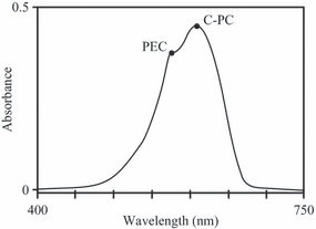
Absorption spectrum of phycobiliprotein extract of strain ETS-02 showing the presence of two peaks corresponding to phycoerythrocyanin (PEC) and C-phycocyanin (C-PC).
| Peak no. | Pigment | λmax (nm) | ETS-02 | CCALA 759 |
|---|---|---|---|---|
| 1 | Myxol 2′-dymethyl-glycoside | 296, 368, 448, 476, 506 | + | + |
| 2 | Nostoxanthin | 428, 452, 480 | + | + |
| 3 | Caloxanthin | 428, 452, 480 | + | + |
| 4 | Zeaxanthin | 428, 452, 480 | + | + |
| 5 | Unknown pigment | 304, 446, 470, 502 | + | − |
| 6 | Echinenone | 460 | + | + |
| 7 | trans-β-carotene | 428, 450, 476 | + | + |
| 8 | cis-β-carotene | 426, 444, 470 | + | + |
- Peaks are listed according to their elution order and for each the maximum wavelengths are given (“+,” presence; “−,” absence).
Preliminary fatty acid content analysis of strain ETS-02 highlighted the presence of the following molecules: C14:0 (myristic acid), C14:1 (myristoleic acid), C16:0 (palmitic acid), C16:1 (palmitoleic acid), C18:0 (stearic acid), C18:1, Δ9 (oleic acid), C18:1, Δ11 (cis-vaccenic acid), C18:2 (linoleic acid), C18:3α (α-linolenic acid), and C20:0 (arachidic acid).
The gene sequences obtained in this study for the different molecular markers were deposited in the DDBJ/GenBank/EBI Data Bank with the accession numbers reported in Table 3, which also includes the accession numbers of the reference cyanobacterial sequences used for comparison.
| Taxon | Strain designation | 16S rRNA | 16S–23S ITS | cpcAB | rpoC1 | gyrB | rbcL | nifD |
|---|---|---|---|---|---|---|---|---|
| Strain ETS-02 | ETS-02 | FN813342 | FN813339 | FN813309 | FN813320 | FN813313 | FN813327 | FN813334 |
| Phormidium cf. irriguum | CCALA 759 | FN813343 | FN813340 | FN813310 | FN813321 | FN813314 | FN813328 | FN813335 |
| Phormidium ambiguum | IAM M-71 | AB003167 | – | – | FN813322 | FN813315 | FN813329 | FN813336 |
| Phormidium autumnale | CCALA 143 | FN813344 | AM778716 | FN813311 | FN813323 | FN813316 | FN813330 | FN813337 |
| Oscillatoria sancta | PCC 7515 | AF132933 | EF178272 | AJ401185 | FN813324 | FN813317 | FN813331 | EF576870 |
| Oscillatoria tenuis | CCAP 1459/4 | FN813345 | FN813341 | FN813312 | FN813325 | FN813318 | FN813332 | FN813338 |
| Lyngbya aestuarii | PCC 7419 | AJ000714 | AY768375 | AJ401187 | FN813326 | FN813319 | AB075915 | EF576869 |
| Lyngbya majuscula | CCAP 1446/4 | AY768394 | AY768384 | – | – | – | FN813333 | DQ078751 |
- A dash indicates that the sequence was not obtained. Bold type indicates sequences obtained in this survey.
The phylogenetic signal of the different multiple alignments (both the single and the combined data sets) was calculated with the ML mapping method, and the corresponding results, given in quartet percentages, are reported in Table 4.
| Data set | QFR (%) | QPU (%) | QFU (%) |
|---|---|---|---|
| 16S rRNA | 98.5 | 1.6 | 0.0 |
| 16S–23S ITS | 69.6 | 16.4 | 14.1 |
| cpcB-IGS-cpcA | 91.7 | 3.3 | 5.1 |
| rpoC1 | 97.9 | 1.9 | 0.2 |
| gyrB | 91.7 | 5.1 | 3.1 |
| rbcL | 98.4 | 1.5 | 0.1 |
| nifD | 95.6 | 1.3 | 3.2 |
| 16S rRNA + rpoC1 + gyrB | 99.3 | 0.7 | 0.0 |
- QFR, quartets fully resolved; QPU, quartets partially unresolved; QFU, quartets fully unresolved.
- In total, 10 000 quartets were evaluated in each calculation.
The 16S rRNA, rpoC1, rbcL, and 16S rRNA + rpoC1 + gyrB gene multiple alignments showed the highest phylogenetic signals, with percentages of fully resolved quartets ranging from 97.9% to 99.3%. The nifD multiple alignment signal was lower, with 95.6% of fully resolved quartets. The 16S–23S ITS locus showed the lowest phylogenetic signal, with only 69.9% of quartets fully resolved and the highest percentage of fully unresolved quartets (14.1%). For the cpcB-IGS-cpcA and gyrB multiple alignments, the percentage of fully resolved quartets was much lower (91.7%) than for the other multiple alignments. However, for the phycocyanin operon multiple alignment, the totally unresolved quartets were 5.1%, while the gyrB multiple alignment showed this percentage for the partially unresolved quartets.
The 16S–23S ITS locus and the cpcB-IGS-cpcA operon were only used for nucleotide identity comparisons, aligning the sequences of strain ETS-02 and Phormidium cf. irriguum CCALA 759 (Figs S2 and S3).
Both ETS-02 and CCALA 759 16S–23S ITS loci presented a tRNAIle and a tRNAAla sequence inside, each with 100% nucleotide identity between the two strains. Moreover, the structurally important domains D1, D1′, D2, D3, and D4, and the anti-terminator region boxA, reported in Iteman et al. (2000), were conserved in the two organisms (100% identity). Some differences were observed in the remaining parts of the 16S–23S ITS locus. In particular, strain ETS-02 presented a 7-bp gap in the region between the tRNAIle and tRNAAla sequences, while strain CCALA 759 showed two subsequent gaps of 5 and 14 bp after the tRNAAla sequence.
In the phycocyanin operon, the IGS region was 94 bp for strain ETS-02 and 78 bp for strain CCALA 759. The alignment between the two strain operons revealed 15.2% identity for the cpcB region (25 substitutions on 164 aligned positions), 41.5% identity for the IGS region (16 insertions/deletions and 23 substitutions on 94 aligned positions), and 6.5% identity for the cpcA region (eight substitutions on 122 aligned positions).
16S rRNA, rbcL, and nifD gene data sets were used for phylogenetic reconstructions, as well as the data set joining the 16S rRNA, rpoC1 and gyrB gene sequences (16S rRNA + rpoC1 + gyrB).
In the 16S rRNA gene trees (10, 11), several clades were clearly distinguishable. Clade A, including strains belonging to the genera Leptolyngbya and Pseudanabaena, was a sister taxon to clade B, comprising two Pseudanabaena strains. Within clade A, Leptolyngbya sp. IAM M-99 and Leptolyngbya sp. PCC 73110 were joined by a node with bootstrap values of 100% in both the MP and the ML trees (100% BTMP and BTML, respectively) and a posterior probability (PP) of 100%. The same was observed for Leptolyngbya sp. PCC 7375 and Pseudanabaena persicina CCMP 638. The remaining named clades (C–G), plus Planktothrix agardhii IAM M-244, represented other Oscillatoriales, spanning the families Oscillatoriaceae and Phormidiaceae. Clade C, with 69/59/59% BTMP/BTML/PP, included strain ETS-02, Phormidium cf. irriguum CCALA 759, and Phormidium ambiguum IAM M-71. Strain ETS-02 and Phormidium cf. irriguum CCALA 759 were sister taxa with 95/79/78% BTMP/BTML/PP. Clade D (57/58/79% BTMP/BTML/PP) comprised two more Phormidium strains: Phormidium uncinatum SAG 81.79 and Phormidium tergestinum CCALA 155. Clade E, supported by 99/100/100% BTMP/BTML/PP, included three members of the species Phormidium autumnale. In particular, Phormidium autumnale Arct-Ph5 and Phormidium autumnale SAG 78.79 were joined by a node with 92/94/100% BTMP/BTML/PP, while Phormidium autumnale CCALA 143 was associated with Oscillatoria nigro-viridis PCC 7112 with a node showing 78/80% BTMP/BTML support. Finally, Arthrospira platensis IAM M-135 and Lyngbya aestuarii PCC 7419 constituted clade F (97/99/100% BTMP/BTML/PP) and Oscillatoria sancta PCC 7515 and Trichodesmium erythraeum IMS-101 formed clade G (100/100/100% BTMP/BTML/PP).
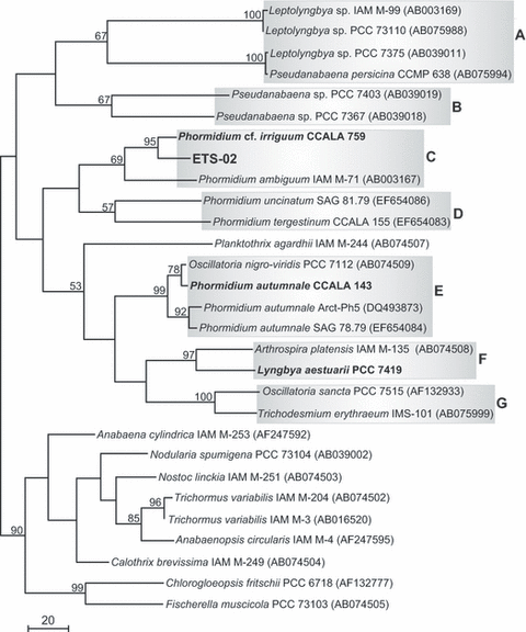
Phylogenetic tree based on 16S rRNA gene sequences and reconstructed using the maximum-parsimony (MP) analysis. Bootstrap values (≥ 50%) are provided for each node. Sequences determined in this work are indicated in bold. GenBank accession numbers are indicated in parentheses. Bar represents 20 nucleotide substitutions per site.
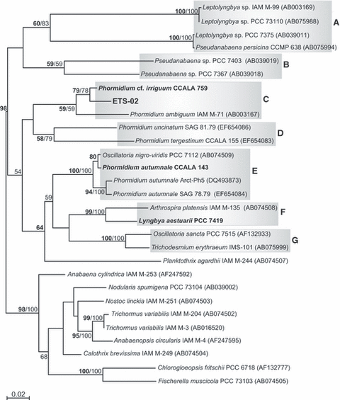
Phylogenetic tree based on 16S rRNA gene sequences and reconstructed using the maximum-likelihood (ML) analysis of evolutionary distances determined by the GTR + I + Γ model. Bootstrap values (≥ 50%) and Bayesian posterior probabilities are provided for each node (ML in bold and BI in normal font). Sequences determined in this work are indicated in bold. GenBank accession numbers are indicated in parentheses. Bar represents 0.02 nucleotide substitutions per site.
For members of clades C–E, levels of 16S rRNA gene sequence similarity were also calculated (Table 5).
| Taxon | 1 | 2 | 3 | 8 | 9 | 4 | 5 | 6 | 7 |
|---|---|---|---|---|---|---|---|---|---|
| Clade C | |||||||||
| 1. Strain ETS-02 | – | 2.4 | 6.7 | 9.2 | 9.8 | 9.4 | 9.6 | 8.9 | 8.4 |
| 2. Phormidium cf. irriguum CCALA 759 | 97.6 | – | 6.4 | 8.8 | 9.6 | 9.3 | 9.5 | 8.8 | 8.5 |
| 3. Phormidium ambiguum IAM M-71 | 93.3 | 93.4 | – | 9.9 | 10.1 | 10.3 | 10.5 | 9.9 | 9.6 |
| Clade D | |||||||||
| 8. Phormidium uncinatum SAG 81.79 | 90.8 | 91.2 | 90.2 | – | 8.8 | 8.7 | 9.2 | 8.3 | 8.3 |
| 9. Phormidium tergestinum CCALA 155 | 90.2 | 90.4 | 89.9 | 91.2 | – | 10.3 | 10.7 | 10.7 | 10.5 |
| Clade E | |||||||||
| 4. Oscillatoria nigro-viridis PCC 7112 | 90.6 | 90.7 | 89.7 | 91.3 | 89.7 | – | 0.9 | 2.7 | 3.0 |
| 5. Phormidium autumnale CCALA 143 | 90.4 | 90.5 | 89.5 | 90.9 | 89.3 | 99.1 | – | 2.9 | 3.2 |
| 6. Phormidium autumnale Arct-Ph5 | 91.1 | 91.3 | 90.1 | 91.8 | 89.3 | 97.3 | 97.1 | – | 1.2 |
| 7. Phormidium autumnale SAG 78.79 | 91.6 | 91.5 | 90.4 | 91.7 | 89.5 | 97.0 | 96.8 | 98.8 | – |
- Each strain considered is associated with a number in the first column, reported in the first line of the table. Values refer to a 1383-bp alignment.
Heterocystous cyanobacteria were found to be monophyletic (90/98/100 BTMP/BTML/PP) and constituted the outgroup of the whole tree.
In January 2010, sequences for the 16S rRNA gene (EU196638) and the 16S–23S ITS locus (EU196671) of Phormidium cf. irriguum CCALA 759 were deposited in the DDBJ/GenBank/EBI Data Bank. The alignment of each of these new online sequences with the corresponding sequences obtained during this study revealed that they were completely different. In particular, the 16S–23S ITS sequence of strain CCALA 759 (EU196671) did not contain any tRNA sequence, in contrast to ours. Moreover, accession EU196638 and the 16S rRNA gene sequence obtained in this survey for strain CCALA 759 showed only 90.50% identity (1084 aligned positions). A tree based on the 16S rRNA gene was also made adding the EU196638 sequence to the data set. This clearly showed that the online sequence was phylogenetically distant from that obtained here (data not shown).
In the phylogenetic reconstruction based on the 16S rRNA + rpoC1 + gyrB multiple alignment (Fig. 12), four clades were observed. Clade A (97/97/100% BTMP/BTML/PP), including three different members of the family Pseudanabaenaceae, could be recognized. Within this group, Leptolyngbya sp. IAM M-99 was a sister taxon to Leptolyngbya sp. PCC 7375 with 80/92/100% BTMP/BTML/PP, and the two Pseudanabaena strains (PCC 7376 and PCC 7403) were joined by a node with 95/100/100% BTMP/BTML/PP.
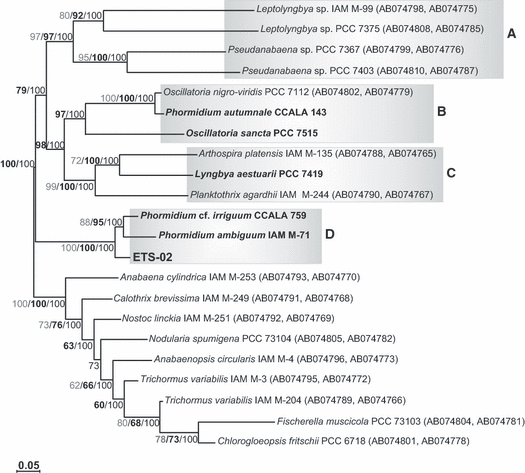
Phylogenetic tree based on the 16S rRNA + rpoC1 + gyrB multiple alignment, reconstructed using the maximum-likelihood analysis of evolutionary distances determined by the GTR + I + Γ model. MP and ML bootstrap values (≥ 50%) and Bayesian posterior probabilities are provided for each node (MP in grey, ML in bold, BI in normal font). Sequences determined in this work are indicated in bold. GenBank accession numbers are indicated in parentheses. Bar represents 0.05 nucleotide substitutions per site.
Clade B (97/100% BTML/PP), consisted of Oscillatoria nigro-viridis PCC 7112, Phormidium autumnale CCALA 143, and Oscillatoria sancta PCC 7515, and clade C (99/100/100% BTMP/BTML/PP), including Arthrospira platensis IAM M-135, Lyngbya aestuarii PCC 7419, and Planktothrix agardhii IAM M-244, represented a sister taxon to clade A (98/100% BTML/PP).
Strain ETS-02, Phormidium cf. irriguum CCALA 759, and Phormidium ambiguum IAM M-71 grouped in clade D with 100/100/100% BTMP/BTML/PP. Within this clade, Phormidium cf. irriguum CCALA 759 was a sister taxon to Phormidium ambiguum IAM M-71 (88MP/95ML/100% BT/BT/PP).
Also in this case, the outgroup, represented by heterocystous cyanobacteria, was well supported (100/100/100% BTMP/BTML/PP).
The phylogenetic relatedness between strain ETS-02 and Phormidium cf. irriguum CCALA 759 was confirmed in the phylogenetic tree obtained with the nifD gene multiple alignment (Fig. 13), where they clustered with 100/100/100% BTMP/BTML/PP. Leptolyngbya sp. PCC 7004 and Symploca atlantica PCC 8002 grouped with them in clade A (80/84/100% BTMP/BTML/PP).
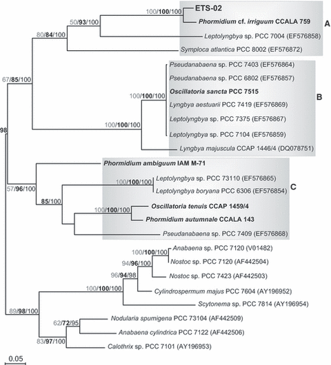
Phylogenetic tree based on nifD gene sequences, reconstructed using the maximum-likelihood analysis of evolutionary distances determined by the GTR + I + Γ model. MP and ML bootstrap values (≥ 50%) and Bayesian posterior probabilities are provided for each node (MP in grey, ML in bold, and BI in normal font). Sequences determined in this work are indicated in bold. GenBank accession numbers are indicated in parentheses. Bar represents 0.05 nucleotide substitutions per site.
Clade B (100/100/100% BTMP/BTML/PP), comprising members of the genera Pseudanabaena, Oscillatoria, Lyngbya, and Leptolyngbya, was joined to clade A by a node with 67/85/100% BTMP/BTML/PP.
In this case, Phormidium ambiguum IAM M-71 was placed distantly to strains ETS-02 and CCALA 759, being included in clade C (57/96/100% BTMP/ BTML/PP) with other oscillatoriacean cyanobacteria. In particular, within this last group, Leptolyngbya boryana PCC 6306 and Leptolyngbya sp. PCC 73110 clustered together with 100/100/100% BTMP/BTML/PP. The same was observed for Phormidium autumnale CCALA 143 and Oscillatoria tenuis PCC 9107.
Finally, in the tree based on the rbcL gene (Fig. 14), strain ETS-02 grouped with Phormidium ambiguum IAM M-71 and Phormidium autumnale CCALA 143 in clade A (99/100/100% BTMP/BTML/PP). Phormidium cf. irriguum CCALA 759 and Oscillatoria tenuis PCC 9107 clustered in clade B (94/74/59% BTMP/BTML/PP). Lyngbya aestuarii PCC 7419 and Lyngbya majuscula CCAP 1146/4 grouped in clade C (60/82/100% BTMP/BTML/PP). Symploca atlantica PCC 8002 and Pseudanabaena sp. PCC 7403 were joined by a node with 52/84% BTML/PP (clade D), while Oscillatoria sancta PCC 7515 and Trichodesmium erythraeum IMS-101 were joined by a node with 100/100/100% BTMP/BTML/PP (clade E). Clade F (85/69/85% BTMP/BTML/PP) included Leptolyngbya sp. PCC 73110, Pseudanabaena persicina CCMP 638, and Geitlerinema sp. PCC 8501. The outgroup was again represented by heterocystous cyanobacteria, grouped with 85/100% BTML/PP support.
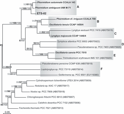
Phylogenetic tree based on rbcL gene sequences, reconstructed using the maximum-likelihood analysis of evolutionary distances determined by the GTR + I + Γ model. MP and ML bootstrap values (≥ 50%) and Bayesian posterior probabilities are provided for each node (MP in grey, ML in bold, and BI in normal font). Sequences determined in this work are indicated in bold. GenBank accession numbers are indicated in parentheses. Bar represents 0.05 nucleotide substitutions per site.
Discussion
The correct identification of cyanobacteria is an important issue for microbiologists and ecologists, but the nature of these micro-organisms makes this complicated. The situation is made even more difficult by the arbitrary use of different systems of nomenclature, the unavailability of many described type species for comparison, and disagreements relating to the use of classical and molecular techniques. All this has often led to confusion and erroneous identification of cyanobacteria.
The Euganean Thermal District has been exploited for therapeutic purposes since ancient times. Recent studies have underlined how the curative properties of the muds are linked to the colonization of its surface by cyanobacteria. In particular, the filamentous strain Phormidium sp. ETS-05 has been shown to be rich in polyunsaturated fatty acids with anti-inflammatory activity (Lalli et al., 2004; Bruno et al., 2005). During samplings to assay the biodiversity of the Euganean Thermal District, another filamentous cyanobacterium similar to strain ETS-05 was found. Comparison of their 16S rRNA gene sequences and more extensive morphological analyses revealed that the new organism was distinct from strain ETS-05. The finding of distinct but morphologically very similar cyanobacteria sharing the same habitat is a common one, and cryptic species are a widespread phenomenon in the phylum Cyanophyta/Cyanobacteria (Ward et al., 1998; Casamatta et al., 2005; Taton et al., 2006; Marquardt and Palinska, 2007). Therefore, strain ETS-02 was subjected to a polyphasic study in order to better identify it.
As a filamentous non-heterocystous cyanobacterium, and given its thylakoid arrangement, strain ETS-02 was assigned to the subclass Oscillatoriophycidae and to the order Oscillatoriales. Features such as the presence of a thin sheath surrounding each filament, cell width, and the reproduction through the formation of necridic cells led us to hypothesize that the organism could be attributed to the families Oscillatoriaceae or Phormidiaceae as well, in agreement with Komárek and Anagnostidis (2005) and Hoffmann et al. (2005). As, except for rRNA operons, very few nucleotide sequences were available in public databases for cyanobacteria belonging to these families, we used strains of Lyngbya, Phormidium, and Oscillatoria for molecular comparisons, choosing those morphologically more similar to ETS-02 and/or more representative of their genera.
Of the molecular markers considered, the 16S–23S ITS and cpcB-IGS-cpcA data sets gave the poorest phylogenetic signal, indicating that the sequences included in the analyses were too divergent and could not be used for a reasonable phylogenetic reconstruction. This is not surprising, as the 16S–23S ITS and the IGS of the phycocyanin operon non-coding genomic regions are potentially more likely to accumulate mutations during evolution. This also confirms previous suggestions by many authors of using these markers to discriminate cyanobacteria at lower taxonomic ranks, such as at the strain level (Robertson et al., 2001; Papke et al., 2003; Teneva et al., 2005; Premanandh et al., 2006).
Almost all of the phylogenetic reconstructions clearly revealed relatedness between strain ETS-02 and Phormidium cf. irriguum CCALA 759. Phormidium irriguum (Kützing ex Gomont) Anagnostidis and Komárek belongs to the group VIII of the genus Phormidium. In fact, given the taxonomic complexity of this taxon, the genus Phormidium has been divided into eight groups based mainly on apical cell morphology (Komárek and Anagnostidis, 2005). Only in the rbcL phylogenetic tree strain ETS-02 is not associated with strain CCALA 759, but is in a group including two Phormidium strains: P. autumnale CCALA 143 and P. ambiguum IAM M-71. The latter, belonging to group VIII as well, is phylogenetically related to both strains ETS-02 and CCALA 759 in all the trees obtained, apart from the reconstruction based on the nifD gene. Thus, based on our phylogenetic analyses, strain ETS-02 belongs to group VIII of the genus Phormidium.
The contrasting results obtained using different molecular markers are probably due to their cellular function. In fact, the molecular markers employed in the phylogenetic analyses can be divided into two categories: the first, represented by the 16S rRNA, rpoC1 and gyrB genes, is involved in replication and translation reactions of the genetic material; and the second one, consisting of the rbcL and nifD genes, is involved in carbon and nitrogen fixation processes, respectively. The two categories of genes, in all probability, followed distinct evolutionary pathways, which are reflected by the different phylogenies obtained. Perhaps, because the replication and translation reactions are more basic for the functionality of the cell, the genes involved in these functions better reflect cyanobacterial history at the phylum level, while the others are more suitable to understand the relationships within certain taxa. In support of this interpretation, besides the known evidence that not all cyanobacteria are able to fix nitrogen and so lack the enzyme complex nitrogenase, some unicellular cyanobacteria unable to perform oxygenic photosynthesis, and lacking the genes coding for the Calvin–Benson cycle and for photosystem II, have recently been found (Bothe et al., 2010).
Nucleotide identity analyses carried out on the 16S rRNA gene, 16S–23S ITS locus, and cpcB-IGS-cpcA operon strengthen the relatedness between strain ETS-02 and Phormidium cf. irriguum CCALA 759. In particular, the conserved and structurally important domains of the 16S–23S ITS and the tRNA sequences found within this locus are identical in the two organisms. Moreover, the level of divergence (6.5%) of the phycocyanin operon cpcA region between the two strains is similar to those previously observed by Teneva et al. (2005) among different strains of P. autumnale. In the same study, the authors found a larger level of divergence (44%) among different Phormidium species.
Following the proposed threshold levels of 16S rRNA gene sequence similarity used to assign two cyanobacteria to the same genus (95%) or to the same species (97.5%) (Stackebrandt and Goebel, 1994), the calculated similarity values confirm some previous findings, i.e. that Oscillatoria nigro-viridis PCC 7112 belongs to group VII of the genus Phormidium (Komárek and Anagnostidis, 2005). However, focusing on strain ETS-02 and those grouping with it in the 16S rRNA and 16S rRNA + rpoC1 + gyrB phylogenetic trees and according to the above threshold values, strain ETS-02 is considered to belong both to the same genus and to the same species as strain CCALA 759, while P. ambiguum IAM M-71, besides representing a distinct species, belongs to a different genus from both ETS-02 and CCALA 759. Given all of the phylogenetic reconstructions and the morphologies of the three strains, this seems highly improbable. Evidently, 16S rRNA gene sequence similarity threshold values can be taken into account to identify cyanobacteria, but cannot constitute sufficient indication alone.
The nucleotide differences found between the 16S rRNA gene and 16S–23S ITS sequences published for strain CCALA 759 and the corresponding ones obtained here confirm the existence of multiple copies of rrn operons in cyanobacteria. The fact that both in our survey and in Lokmer’s (2007), only one of these operons was amplified may be due to the different primers used in the amplification reactions and to the previously documented differential efficiency and specificity of amplification by diverse primer pairs (Reysenbach et al., 1992; Suzuki and Giovannoni, 1996; Nübel et al., 1997). Thus there is the need to complement a more classical 16S rRNA gene phylogenetic analysis with phylogenetic reconstructions based on other single-copy molecular markers, such as the rpoC1 and gyrB genes.
These last two loci, together, proved meaningful to attribute strain ETS-02, Phormidium cf. irriguum CCALA 759, and P. ambiguum IAM M-71 (90.2–90.8% sequence identities among the rpoC1 + gyrB gene fragments of these taxa) to the same genus in our analyses, and thus they may prove important for the identification of Phormidium strains at the genus level.
The level of phylogenetic relatedness found between strain ETS-02 and strain CCALA 759 is also supported by the biochemical, morphological, and ultrastructural analyses. Indeed, the lipid-soluble pigment patterns are highly similar, while the water-soluble pigment patterns are identical, with both the organisms containing phycoerythrocyanin, a rare PBP found only in a limited number of cyanobacteria (Bryant, 1982; García-Pichel et al., 1996). Moreover, the 608-nm peak, corresponding to C-PC and present in both organisms, is of note, having been reported, to our knowledge, only in Synechococcus lividus, a cyanobacterium found in Yellowstone National Park (Edwards et al., 1996).
The widened thylakoid aspect, called keritomy, is present both in strain ETS-02 and in CCALA 759. Keritomy is observed throughout the whole spectrum of cyanobacterial taxonomic groups, but it appears to be specific, genetically determined, although differentially occurring during the life cycle and/or according to environmental conditions (Komárek and Anagnostidis, 2005).
Table 6 summarizes the morphological, ultrastructural, physiological, biochemical, and molecular features between strain ETS-02 and strain CCALA 759.
| Characteristic | Strain ETS-02 | Phormidium cf. irriguum CCALA 759 |
|---|---|---|
| Cell morphology | Wider than long | Wider than long |
| Cell width (μm) | 3–5 | 9–12 |
| Thylakoid arrangement | Irregular | Irregular |
| Sheath | + | + |
| Keritomy | + | + |
| Necridic cells | + | + |
| Heterocyst formation | − | − |
| PEC production | + | + |
| Isolation source | Thermal mud surface | Littoral zone of sand pit lake |
| t-RNAIle | + | + |
| t-RNAAla | + | + |
- PEC, phycoerythrocyanin.
In conclusion, we can attribute strain ETS-02 to group VIII of the genus Phormidium (subclass Oscillatoriophycidae, order Oscillatoriales, family Phormidiaceae). It seems also to belong to the same species as strain CCALA 759, i.e. P. irriguum (Kützing ex Gomont) Anagnostidis and Komárek. This species is a new combination by Anagnostidis and Komárek (1988, p. 405) based on the description and illustration of O. irrigua by Gomont (1892). As the holotype strain is not a living culture, it cannot be used as a reference strain according to our polyphasic method, which focuses not only on external morphology, but also on aspects such as ultrastructure, pigment composition, and genetic features. When the original material associated with a validly published name is demonstrably ambiguous and/or cannot be used for the precise application of the name of a taxon, as in this case, the International Code of Botanic Nomenclature allows the institution of an epitype (McNeill et al., 2006). We designate strain CCALA 759 as this epitype, as it resembles the original description of P. irriguum according both to Lokmer’s (2007) observations and ours.
Nevertheless, cell size clearly distinguishes strain ETS-02 from P. irriguum CCALA 759. Considering the original description of P. irriguum and both Lokmer’s analyses and ours, this species shows a range of cell width that varies between 6 and 12 μm, while cells of strain ETS-02 are smaller, with a width ranging between 3 and 5 μm, particularly if culture parameters, such as organic nitrogen availability, temperature, and light intensity, are changed (data not shown). However, in our opinion the cell sizes are not sufficient alone to consider strain ETS-02 as representing a novel species, especially evaluating the whole spectrum of our observations. Some authors suggest, in certain well-documented cases, the creation of the subspecific taxonomic categories of variety (“the category variety could be used in the cases of differentiation in one remarkable or more features, usually combined with ecological or phytogeographical deviation”) or form (“the category form could be applied for a stable, recognizable deviation without ecological specificity”) (Komárek and Golubić, 2005). For this reason we propose the name Phormidium irriguum f. minor for the Euganean strain (where “f.” stands for forma, form).
Phormidium irriguum f. minor Sciuto et Moro
Thallus blue–green. Trichomes bright blue–green, long, variously curved, strictly packed to form mats. Isopolar trichomes, composed by cells wider (3–5 μm) than long (1–2 μm), ending with rounded apical cells, and surrounded by a thin colourless sheath, sometimes protruding from the filament itself. Reproduction by trichome fragmentation in short hormogonia through the formation of necridia. During cell division, daughter cells can begin a new division before growing to the size of the mother cell. Thylakoids with irregular arrangement, sometimes keritomized. Able to produce phycoeritrocyanin.
-
Type material: A living culture is deposited at the Culture Collection of Autotrophic Organisms (CCALA) (Czech Republic) as strain CCALA 946.
-
Type locality: Phormidium irriguum f. minor was isolated from cyanobacterial mats covering the thermal muds of the Garden Hotel of Montegrotto Terme (Padova, Italy).
-
Etymology: The specific form name refers to the cell size, which is smaller than those of the species Phormidium irriguum (Kützing ex Gomont) Anagnostidis et Komarek.
Another finding that results from the present survey is the heterogeneity of the genus Phormidium Kützing ex Gomont, as attested mainly by the phylogenetic reconstructions based on 16S rRNA gene sequences (10, 11) in which more clades were detected within this taxon. In particular, clade D, comprising P. uncinatum (group VIII) and P. tergestinum (group V), and clade E, including four Phormidium strains of group VII, placed far from the Phormidium entities grouped in clade C (group VIII). These clades should probably be ascribed to different genera, but further investigation is required. The genus Phormidium thus needs revision; several studies by other authors have highlighted the polyphyletic and heterogeneous nature of this group, as it is presently intended (Teneva et al., 2005; Palińska and Marquardt, 2008; Komárek, 2010).
We thus make the following proposal. As the type species of the genus Phormidium is P. lucidum Kützing ex Gomont, a member of group VIII, the name Phormidium should be maintained only for members of group VIII. Moreover, as P. lucidum is based on an illustration only, which is not suitable for a modern approach including ultrastructural and molecular analyses, and we were not able to find a culture of P. lucidum that could be subjected to a polyphasic analysis and then typified, we suggest another type species for the future new genus Phormidium. The new type species could be P. irriguum, belonging to group VIII of the current genus Phormidium (and thus probably being also phylogenetically related to P. lucidum), and the designed type strain could be P. irriguum CCALA 759, on which a polyphasic characterization has been carried out in this study. If our proposal is accepted it could help other researchers regarding the complex systematics of Phormidium.
The proposed “combined approach” consists of the following steps:
- 1
Preliminary morphological and ultrastructural observations of the cyanobacterial strain of interest, focusing on those characteristics (presence/absence of thylakoids, thylakoid arrangement, presence of differentiated cells) that allow its immediate assignation to one of the four subclasses of the phylum (Hoffmann et al., 2005): Gloeobacterophycidae, Synechococcophycidae, Oscillatoriophycidae and Nostochophycidae.
- 2
More detailed morphological, ultrastructural, biochemical, and physiological analyses to allocate the cyanobacterial strain of interest to more precise sub-taxa (orders, families, genera, and/or species) within the individual subclass or, at least, to reduce the number of possible related taxa.
- 3
Phylogenetic multilocus analyses, based on the classical 16S rRNA gene and other single-copy markers (e.g. the rpoC1 gene). The data set reconstructions should be determined based on morphological, biochemical, and physiological analyses made to this point, amplifying the chosen molecular markers from selected cyanobacterial strains, deposited in international culture collections, if the corresponding sequences are not already available in public databases.
- 4
Focus on the cyanobacterial strain (or strains) that are phylogenetically related to the organism of interest, extending the morphological, biochemical and physiological analyses.
- 5
If necessary, typify the considered cyanobacterial strain (or strains) in order to provide a reference for future studies. The form of typification should be based on the specific case.
- 6
The conclusions and identification of the studied cyanobacterium should take account of all available information, detecting possible autapomorphic characters, and including a nomenclatural analysis.
At present, a polyphasic approach to characterize cyanobacteria seems essential. This approach should not only include different types of analyses, ranging from the more classical (morphological, biochemical, and physiological) to the molecular, but also comparisons with deposited type species. Where the holotypes of the described reference species are lacking or not suitable for a detailed comparison, a typification with cultured strains can be useful.
Therefore, far from proposing completely new analytical methods, the described “combined approach” joins the more classical analyses to the more recent molecular ones, making them complementary and equally important to the correct identification of new strains belonging to the phylum Cyanophyta/Cyanobacteria, avoiding the incongruities between only classical-based and only molecular-based identifications. The amount of information produced by such an approach should facilitate subsequent studies on cyanobacteria, contributing to solve the taxonomic and classification problems of this complex and interesting phylum.
Acknowledgements
This work was supported by grants provided by Centro Studi Termali “Pietro d’Abano,” Abano Terme (Padova, Italy). We thank Dr Nancy Jenkins for help with the English text.


