First record of Philometra ovata (Nematoda) infection in Gobio lozanoi in Portugal
Abstract
Gobio lozanoi, from the River Febros northern Portugal, contained female Philometra ovata in the body cavity causing abdominal swelling. A mild chronic inflammation and some tissue necrosis were observed in infected fish. Significant correlations were found between occurrence of infection and host length, and gonado-somatic index; and between intensity of infection and condition factor.
The genus Philometra comprises several species of nematodes of the abdominal cavity and tissues of fishes (Moravec et al., 1998; Anderson, 2000). In recent years, there has been a growing awareness of the importance of philometrid nematodes because they may cause serious damage to the ovaries and also parasitic castration (Moravec, 2006). Philometra ovata (Zeder, 1803) Skryabin, 1923, referred to frequently as its junior synonym Philometra abdominalis Nybelin, 1928, is a parasite of cyprinids of the genera Gobio, Phoxinus and Leuciscus (Moravec, 2004). It has been recorded in several European countries but never in the Iberian Peninsula.
During ichthyological surveys carried out in several tributaries of the River Douro system, northern Portugal, during January 2007, more than half of the sampled Gobio lozanoi Doadrio & Madeira, the gudgeon species that occurs in Iberian Peninsula and southern France, were infected with P. ovata. In order to characterize the G. lozanoi–Philometra infection, including the associated pathology in the host population during its reproductive period (spring), a parasitological survey was conducted in April 2007, in River Febros, a tributary of River Douro system (41°07″ N; 8°35′ W).
A total of 97 fish were captured by electrofishing, measured (fork length, LF) to the nearest mm, weighed (total mass, M) to the nearest g, sexed, gonad removed and weighed (gonad mass, MG) to the nearest g and examined for parasites. Fulton’s condition factor (K) was calculated from  and the gonado-somatic index (IG) from IG= 100 MGM−1. Parasites were removed, counted, fixed in hot 70% ethanol and the prevalence, intensity and abundance of infection determined according to Bush et al. (1997). Differences in occurrence and intensity of infection among males, females and immature specimens were analysed by χ2 and Kruskal–Wallis tests. Spearman’s test (rS; Siegel & Castellan, 1989) was used to identify correlations between occurrence and intensity of infection, and LF, K and IG.
For histological studies, some fish were fixed in 10% neutral buffered formalin, routinely processed and stained with Mallory.
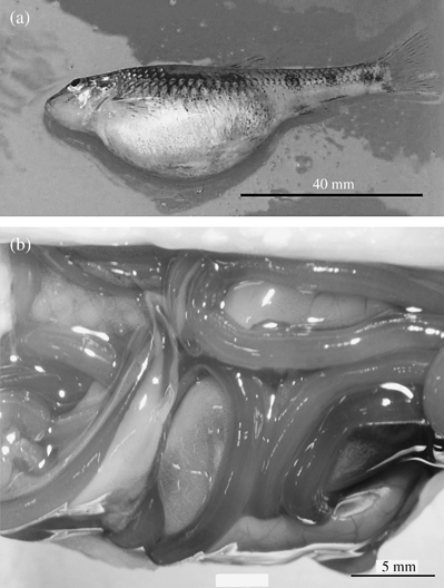
Specimens of P. ovata females were located in the body cavity causing abdominal swelling [Fig. 1(a), (b)]. When a small incision was made in the body wall, the worms would emerge from the incision. Infected fish had impaired mobility that reduced their ability to escape. In some females, 30 large nematodes were detected amounting to 20% of M.

Gobio lozanoi heavily infected with Philometra ovata showing: (a) a swollen abdomen and (b) female worms coiled among visceral organs.
Data on LF, K, IG and the parasitological variables prevalence, intensity and abundance are presented in Table I.
| Index | Fish examined (n) | |||
|---|---|---|---|---|
| Total (97)* | Immature (23) | Male (43) | Female (27) | |
| L F (mm) | 76 ± 26 (33–128) | 45 ± 10 (33–68) | 84 ± 20 (47–113) | 83 ± 19 (52–125) |
| K | 1·32 ± 0·21 (0·83–2·25) | 1·38 ± 0·30 (0·83–2·25) | 1·29 ± 0·16 (0·99–1·77) | 1·33 ± 0·17 (1·06–1·68) |
| I G | 4·2 ± 3·9 (0·1–14·9) | — | 2·0 ± 0·8 (0·1–3·9) | 8·4 ± 3·5 (2·4–14·9) |
| Prevalence (%) | 53·6 | 34·8 | 51·2 | 74·1 |
| Intensity | 7·4 ± 7·6 (1–30) | 5·2 ± 6·2 (1–19) | 6·5 ± 7·3 (1–25) | 9·3 ± 8·6 (1–30) |
| Abundance | 4·0 ± 6·7 (0–30) | 1·8 ± 4·3 (0–19) | 3·3 ± 6·1 (0–25) | 6·9 ± 8·5 (0–30) |
- * Sex was not determined in four fish.
Occurrence of infection was significantly different among male, female and immature specimens (χ2, P < 0·05). Female fish showed the highest prevalence (74·1%). Intensity of infection was not significantly different among sex and maturity groups (Kruskal–Wallis, P > 0·05), but the highest value was observed in females.
Significant positive correlations were found between occurrence of infection and LF (n= 97, P < 0·05) and IG (n= 74, P < 0·05) and between intensity and K (n= 52, P < 0·05).
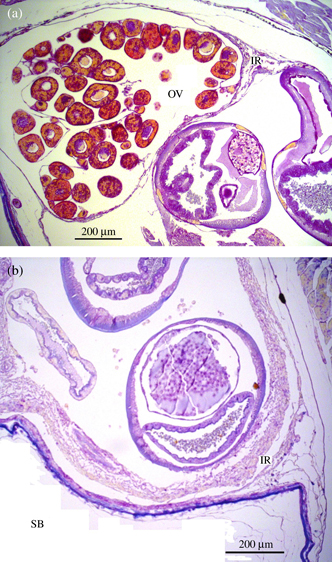
Histological examination of infected fish showed a mild chronic inflammatory response and, in some cases, some necrotic tissue [Fig. 2(a), (b)].

Body cavity of Gobio lozanoi infected with Philometra ovata showing (a) a mild inflammatory response (IR) near the ovary (OV) caused by the nematodes and (b) necrosis of the swimbladder (SB) and an inflammatory host response (IR) around the nematodes.
Although prevalence was high in both sexes, females were more often infected, as also reported by Hesp et al. (2002) for Philometra lateolabracis in west Australian dhufish Glaucosoma hebraicum Richardson.
Occurrence of infection was positively related to host LF and to sexual development (IG). Philometrids utilize copepods as intermediate hosts so higher infection rates can be expected in fish that have fed on a larger number of prey items. This relationship also seemed to be confirmed by the positive correlation between intensity of infection and K.
The most visible effects of infection were the abdominal swelling and increase of M caused by nematode load that decreased swimming ability.
The low number of females captured in the present study may reflect a higher mortality rate due to high infection levels that cause lower escape efficiency. This is supported by data from Miñano et al. (2003) from south-east Spain, who showed that the sex ratio of G. lozanoi is slightly biased in favour of females.
Acknowledgments
The authors thank the technician M. H. Moreira for processing the material for histology.


