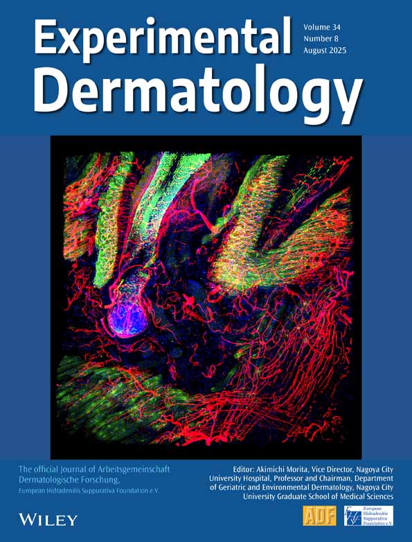
Processing of POMC peptides by dermal microvascular endo-thelial cells: implication for EC biologic functions and skin inflammation
Abstract
Dermal microvascular endothelial cells (ECs) are both source and target of the pro-opiomelanocortin (POMC) peptides ACTH and α-melanocyte-stimulating hormone (α-MSH). The availability of neuropeptides as important modulators of innate and adaptive immune responses is controlled by neuropeptide-specific peptidases such as neutral endopeptidase (NEP) or angiotensin-converting enzyme (ACE). In this study, we have tested the possibility that NEP or ACE expressed by ECs may influence the local bioavailability of POMC peptides. Incubation of ACTH1−39 with cell membranes prepared from the high NEP-/low ACE-expressing microvascular EC line 1 (HMEC-1) or from low NEP-/high ACE-expressing primary human dermal ECs (HDMECs) for 30–480 min resulted in a decrease in ACTH immunoreactivity (IR) over time in membrane supernatants that could be partially blocked with NEP inhibitors as detected by radioimmunoassay. In parallel, α-MSH IR increased peaking after 60 min. Fragments generated by incubation of HMEC-1 or HDMEC membranes with ACTH1−39, ACHT1−24 or α-MSH for 1–120 min were further analysed by mass spectroscopy. HMEC-1 membranes generated peptide products which could be altered by inhibition of NEP, but not ACE. Likewise, HDMEC membranes fragmented ACTH similar to HMEC-1 membranes in the presence of NEP inhibitors. Some of the proteins can be assigned to regular proteolytic cleavage, while others seem to be modified. Importantly, HMEC-1 and HDMEC membranes also slowly degraded α-MSH, suggesting that EC proteolytic peptidases locally control ACTH/α-MSH bioavailability, which may be important in controlling cutaneous inflammation.


