The clinical picture of the Börjeson–Forssman–Lehmann syndrome in males and heterozygous females with PHF6 mutations
Abstract
The usual description of the Börjeson–Forssman–Lehmann syndrome (BFLS) is that of a rare, X-linked, partially dominant condition with severe intellectual disability, epilepsy, microcephaly, coarse facial features, long ears, short stature, obesity, gynecomastia, tapering fingers, and shortened toes. Recently, mutations have been identified in the PHF6 gene in nine families with this syndrome. The clinical history and physical findings in the affected males reveal that the phenotype is milder and more variable than previously described and evolves with age. Generally, in the first year, the babies are floppy, with failure to thrive, big ears, and small external genitalia. As schoolboys, the picture is one of learning problems, moderate short stature, with emerging truncal obesity and gynecomastia. Head circumferences are usually normal, and macrocephaly may be seen. Big ears and small genitalia remain. The toes are short and fingers tapered and malleable. In late adolescence and adult life, the classically described heavy facial appearance emerges. Some heterozygous females show milder clinical features such as tapering fingers and shortened toes. Twenty percent have significant learning problems, and 95% have skewed X inactivation. We conclude that this syndrome may be underdiagnosed in males in their early years and missed altogether in isolated heterozygous females.
The striking constellation of clinical features known as the Börjeson–Forssman–Lehmann syndrome (BFLS) (OMIM 301900) was first described in 1962 in two adult brothers and their uncle (1). All three had severe intellectual handicap, and two were epileptic. All had a similar appearance, with thickening of the skin of the face, deep-set eyes, and large, fleshy ears. They had short stature with gynecomastia, small external genitalia, and no body hair. X-Linked inheritance was suggested and confirmed in 1989 when two large families with BFLS exhibited linkage to Xq27 (2, 3). Recently, we identified mutations in the PHD-like zinc-finger gene, PHF6, in both of these families, in five other unpublished families, and in two sporadic cases (4). Here, we describe the clinical features of the affected males, the changes with age, and heterozygous expression in the female.
Clinical report
The series consisted of 25 affected males in nine families designated F1 to F9 to correspond with the numbering used by Lower et al. (4). Ages ranged from 10 months to 59 years. At the time of the last or their only assessment, seven were children under 10 years of age, four were in the 10–19 age group, nine in the 20–39 age group, and five in the 40–59 age group. The amount and kind of clinical information varied between subjects. Information was gathered from published papers (2, 3), reports from hospitals, and institutions, parents, and caregivers. Many of the affected individuals had been seen in clinical consultation on one occasion only, but five subjects from F1, with present ages of 17–59 years, had also been assessed 15–17 years earlier (2). The family pedigrees are shown in Fig. 1.
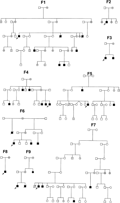
Pedigrees of the nine families, slightly modified from Lower et al. (4) but with similar family numbering. Solid symbols = affected; symbols with dot = obligate or DNA-positive hetero-zygotes; and symbols with cross = DNA tested negative.
Results
Males
Infancy. Babies were born normally at term after uneventful pregnancies, with only one mother recalling reduced fetal movements. The mean birth weight (n = 8) was 3.12 kg, with a range from 2.7 to 3.6 kg. The penis was small with undescended testes and a small scrotum, but one baby had completely normal genitalia. Strikingly large ears were obvious from birth (Fig. 2). The only congenital abnormalities were a cleft lip and palate in one baby – the mother had been taking anticonvulsant drugs during the pregnancy. Mild generalized hypotonia was the norm with poor feeding. Three infants were treated in hospital for failure to thrive. Two brothers (F9) developed hypoglycemia in the newborn period and were found to have hypopituitarism.
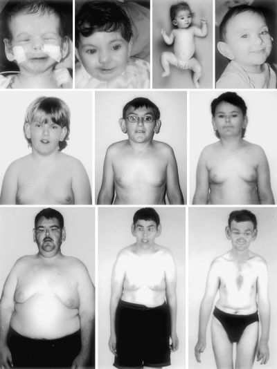
Börjeson–Forssman–Lehmann syndrome at different ages. From left to right. Top row: newborn; 1 year with repaired cleft lip (F6); 10 months (F7); and 2 years (F9). Middle row – adolescence: 12 years (F8); 14- and 13-year-old brothers (F1). Bottom row – adults: 26 (F1) and 48 and 41 years (F7).
Childhood. There were two deaths. One with hypopituitarism died at the age of 4 years with a respiratory illness (F9). The other (not DNA tested but probably had BFLS) developed seizures at the age of 6 weeks, had profoundly retarded growth, and died at the age of 2 years (F1). In all the children, delayed development in both speech and motor skills was recognized early and it was accepted, before entering school, that special education would be needed. The degree of intellectual handicap was moderate or mild. Hearing impairment requiring aids was recognized in two male cousins (F1). Overall growth in height and head circumference (HC) is shown in Fig. 3. Moderate short stature was the norm, but some subjects were above average in height. One boy had marked short stature – this individual had an affected brother with normal height (F1) (Fig. 2, middle row). Truncal obesity developed in late childhood in nine of 15 boys. HCs were mostly within 2 standard deviations of the means for age with more above than below the mean. However, there were two with microcephaly and three with macrocephaly, all from different families (F1, F4, and F8). The facies were relatively normal, but large ears with long, fleshy ear lobes were evident. The external genitalia remained underdeveloped, with impalpable or small, soft testes.
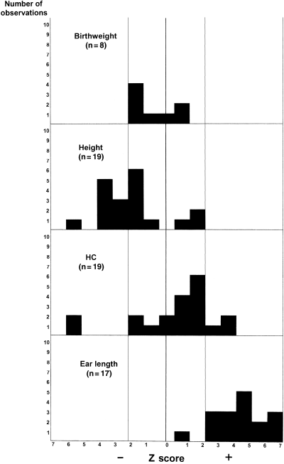
Histograms of physical growth data of males with Börjeson–Forssman–Lehmann syndrome. All measures are in Z scores. For each parameter, only one observation was used for each subject; n = number of subjects; HC = head circumference. Means are indicated by vertical heavy lines and plus and minus 2 standard deviations or Z scores are indicated by faint vertical lines.
Adolescence. Global learning difficulties persisted. Most were talkative and cheerful and interacted well with family and peers. There was little participation in sporting activities. Gynecomastia was recognized in one child at age 4 years. In others, it was perceptible before puberty but became obvious and almost universal during adolescence (Fig. 2). Obesity contributed to breast enlargement but increased underlying breast tissue was present in all.
Adults. All required some degree of supervision, but this varied. Most of the men lived in their parent's home or in group homes. However, four had been placed in institutions because of their socially unacceptable behavior. None was reported to be sexually active, but one man had a steady partner. The average height in nine adults was 169 cm, with a range from 154 to 185 cm. There was moderate obesity with gynecomastia in all but one individual (Fig. 2). The genitalia were small. Deep-set eyes became noticeable, with some coarsening of the facial features (Fig. 2).
Fingers and toes
The fingers were tapered, hypotonic, and malleable, with lax interphalangeal joints similar to those seen in the Coffin–Lowry syndrome. The feet were broad with shortening of the toes, sometimes most marked in toes 4 and 5 or in toe 4 alone (Fig. 4). Flexed toes were common with or without extension of the metatarsophalangeal joints.
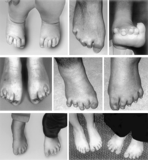
Toes in Börjeson–Forssman–Lehmann syndrome. In the upper and middle rows, all are males. From left to right. Top row: newborn (F6), 59 years old with fixed hammer toes, and 28 years old with fixed extended toes and weight-bearing on the metatarsal heads. Middle row: adults from F7 and F1. Bottom row – left: mother and her affected 16-year-old son (F1). Bottom row – right: obligate-carrier mother with short, broad toes and her 14-year-old daughter (shown to have the mutation) with normal toes.
Occasional findings
Unilateral Perthes disease of the hip occurred in two cousins (F1) in late childhood. One child had hyperopia, and another child had mild nystagmus. An adult in his thirties was found to have a cataract. Another man developed a small keloid scar after surgical removal of a nevus. Two subjects had well-controlled epilepsy.
Female heterozygotes
There was some clinical information on 14 heterozygous females from five of the eight families. All the mothers of the affected males were functioning in the community and coping with bringing up their intellectually handicapped sons. One mother was epileptic with an intelligence quotient of 73. Two teenage heterozygous sisters were doing well at school, with one topping her class. One maternal aunt had hypothyroidism with moderate retardation, epilepsy, short stature (150 cm), obesity, oligomenorrhea, and totally skewed X inactivation (F9).
Physical manifestations of BFLS were mild and variable. Two women had some coarsening of their facial features, with more pronounced supraorbital ridges and deeper set eyes than their non-carrier sisters. Thickened, fleshy ear lobes were noted in 11. Four had tapered fingers, and six had shortened toes.
Skewed X inactivation using a number of different loci (FMR1, AR, and PGK-1) was examined in 21 obligate carriers. In all, skewing was marked except for one who had only slight skewing (4).
Discussion
This study shows that the phenotype of BFLS evolves over time and is seldom as severe as that seen in the original patients. Clinical manifestations are quite variable, in that both microcephaly and macrocephaly and short and tall stature may be encountered. Most have clearevidence of hypogonadism and some of hypo-pituitarism, but the basic endocrinologic abnormalities are yet to be elucidated.
The most constant clinical manifestations are mild-to-moderate mental retardation, big ears, underdeveloped genitalia, gynecomastia, truncal obesity, tapering fingers, and short toes. Help in the diagnosis may come from an X-linked pattern of inheritance and the presence of minor clinical features in the mother who may well have skewed X inactivation. In addition, the diagnosis of BFLS should be considered in any male infant with hypopituitarism or with delayed development and big ears. Differential diagnoses in later childhood include the Prader–Willi, Wilson–Turner, Cohen, and Klinefelter syndromes.
Mutation testing of the PHF6 gene in these families revealed seven with missense mutations, all different, except in F4 and F8, and two truncation mutations in exons 10 and 2 in F1 and F9, respectively. Although the two brothers in one family (F9) were the most severely affected, in other families there was marked intrafamilial variation, and no direct correlation between the mutation and the clinical phenotype could be recognized. Phenocopies may exist, because many males have been referred for mutation analysis with the clinical diagnosis of BFLS in whom no mutation could be found.
BFLS may prove to be a significant cause of intellectual handicap in the female. In the present study, there was bias in the selection of the heterozygotes, because most were mothers who had functioned well socially. However, there were three unmarried women with significant intellectual handicap. In addition, there are two known BFLS families (without recorded mutation studies) with intellectually handicapped females who also had short stature, amenorrhea, and scanty head and body hair. In one of these, there was an affected brother (5); in the other, the sisters also had macrocephaly and epilepsy (6). Female carriers of BFLS might be mistaken for the Coffin–Lowry syndrome or pseudohypoparathyroidism.
BFLS has been considered a rare entity. It may prove to be more common when the clinical presentation in the pediatric age group is more widely recognized and the diagnosis considered in females presenting with developmental delay.
Acknowledgements
The authors express their thanks to the families for their patience and co-operation, in particular F1. This work was supported in part by a grant from the Australian National Health and Medical Research Council.


