Clinical efficacy of blood derivatives on wound healing: A systematic review and network meta-analysis
Abstract
This study aims to evaluate the clinical effects of different blood derivatives on wound healing using network meta-analysis. PubMed, Embase, OVID, Web of Science, SCOPUS and Cochrane Central were searched to obtain studies about blood derivatives on wound healing until October 2023. R 4.2.0 and Stata 15.0 softwares were used for data analysis. Forty-four studies comprising 5164 patients were included. The results of network meta-analysis showed that the healing area from high to low was GF + ORCCB, ORCCB, GF, PRF, Unnas paste dressing, APG, PRP injection, PRP, PRP + thrombin gel, PPP, HPL, CT. The healing time from low to high was PRP + thrombin gel, GF, PRP, PC + K, PC, APG, PRF, CT, Silver sulfadiazine ointment. The number of patients cured from high to low was APG, PRP injection, PRP, Aurix, PRF, Leucopatch, HPL, Antimicrobial Ointment Dressing, CT, 60 μg/cm2 repifermin, 120 μg/cm2 repifermin, AFG, PPP. The order of analgesic effect from high to low was AFG, Aminogam gel, PRF, PRP, Oxidised oil, APG, GF, CT. The order of the number of wound infection cases from low to high is APG, 20 μg/cm2 repifermin, 60 μg/cm2 repifermin, PRP, LeucoPatch, CT, PPP, Antiseptic ointment dressing. Healing area: GF + ORCCB had the best effect; Healing time: PRP + thrombin gel took the shortest time. The number of cured patients and the reduction of wound infection: APG has the best effect. Analgesic effect: AFG has the best effect. More studies with large sample sizes are needed to confirm the above findings.
Abbreviations
-
- AFG
-
- autologous fibrin gel
-
- APG
-
- autologous platelet gel
-
- APL
-
- autologous platelet lysate
-
- Aurix
-
- aurix gel
-
- CT
-
- conventional treatment
-
- GF
-
- growth factor
-
- HPL
-
- autologous platelet lysate
-
- MCMC
-
- Markov chain Monte Carlo
-
- MD
-
- mean difference
-
- NOS
-
- Newcastle Ottawa scale
-
- OR
-
- odds ratio
-
- ORCCB
-
- oxidised regenerated cellulose and collagen biomaterial
-
- Ozonated oil
-
- ozone gas dissolved in olive oil
-
- PC + K
-
- keratinocytes suspended in platelet concentrate
-
- PC
-
- platelet concentrate
-
- PG
-
- platelet gel
-
- PPP
-
- platelet-poor plasma
-
- PRF injection
-
- injectable platelet-rich fibrin
-
- PRF
-
- platelet-rich fibrin
-
- PRG
-
- platelet rich gel
-
- PRP
-
- platelet-rich plasma
-
- PSRF
-
- potential scale reduction factor
-
- RCT
-
- randomised controlled trial
1 INTRODUCTION
With the acceleration of China's aging population, the number of patients with acute and chronic trauma caused by burns, surgery, diabetes, ulcers and other reasons is increasing. The increasing types and refractory coefficients of wounds have brought unprecedented challenges to clinical researchers.1 The long-standing wounds not only affect the quality of life of patients and increase the difficulty of nursing, but also may lead to a variety of complications (pain, electrolyte disorders, osteomyelitis, cancer, etc.), and the serious ones may face amputation or even life-threatening risks.
Wound healing is a complex biological process that is highly regulated by a variety of growth factors, cytokines, cells and cell matrix.2 It involves four stages (haemostasis, inflammation, proliferation and remodelling) that occur gradually and overlap with each other.3 In addition to surgical repair, exogenous growth factors are also one of the effective ways to promote wound healing. Plasma derivatives are rich in high concentrations of platelets, which produce a large number of growth factors after activation. The main products of plasma derivatives in the clinic are platelet-rich plasma (PRP), platelet-gel (PG), platelet rich gel (PRG), platelet-rich fibrin (PRF), growth factor rich plasma (GF), autologous platelet lysate (APL), platelet concentrate (PC), autologous platelet gel (APG) and autologous fibrin gel (AFG), etc. There are many kinds of blood derivatives for the treatment of wound healing, and there is a lack of efficacy comparison between them, which is not conducive to clinical promotion and the selection of the best scheme. This study intends to use a network meta-analysis to compare the clinical efficacy of different plasma derivatives in the treatment of wound healing, to provide references and evidence for the selection of clinical drugs.
2 MATERIALS AND METHODS
2.1 Literature search
We searched PubMed, EMBASE, OVID, Web of Science, SCOPUS, and Cochrane Central, and manually included references of the retrieved literature as supplements. The retrieval time was from the establishment of each database to October 2023. The search was performed by combining subject words and free words. The retrieval strategy were as follows (‘blood product’ OR ‘plasma derivatives’ OR ‘platelet-rich plasma’ OR ‘PRP’ OR ‘platelet-rich in growth factors’ OR ‘GF’ OR ‘platelet rich fabric’ OR ‘PRF’ OR ‘concentrated growth factor’ OR ‘CGF’) AND (‘healing of wound’ OR ‘wound healing’ OR ‘wound’ OR ‘surgery’ OR ‘burn’).
2.2 Document selection criteria
2.2.1 Inclusion criteria
(1) The types of studies were RCT and cohort study; (2) The language is limited to English; (3) The study subjects were wounds caused by ulcers, burns, surgery, tooth extraction and other reasons, without limiting the age and gender of patients; (4) The intervention type was that the experimental group was mostly treated with blood derivatives alone or combined with other measures, without limiting the intervention time, and the control group was mostly treated with conventional/standard nursing. The experimental group and the control group should be consistent except for the inconsistent intervention measures.
2.2.2 Exclusion criteria
(1) Repeatedly published literature, reviews, meta-analyses, treatises and conferences; (2) Literature on relevant outcomes could not be extracted; (3) Literature with errors; (4) The literature with ≤5 patients in the treatment group or the control group.
2.3 Literature quality evaluation
2.3.1 Cochrane risk of bias assessment
The Cochrane bias risk assessment tool was used for evaluation,4 including random allocation method, allocation scheme concealment, whether the research object and scheme implementer were blinded, whether the outcome evaluator was blinded, data integrity, selective reporting and other sources of bias. Each RCT included was evaluated as ‘low-risk’, ‘unclear’ and ‘high-risk’ from the above seven aspects.
2.3.2 Newcastle Ottawa scale bias risk assessment
Cohort study: the Newcastle Ottawa scale (NOS)5 was used to evaluate the quality of the five included cohort studies, including patient selection (four items, full score 4), comparability between groups (one item, full score 2) and exposure factors (three items, full score 3). A total score of ≥6 points was considered to be of high quality. Two researchers independently evaluated the literature. In case of disagreement, the third researcher ruled.
2.4 Literature screening and data extraction
Two researchers independently screened the literature according to the inclusion and exclusion criteria, extracted the data and cross-checked them. According to the included literature, the corresponding data were extracted, mainly including the basic information of the study (research title, first author, publication time, country), the information needed for Cochrane and NOS risk of bias assessment, the characteristics of the study subjects (type of trauma, age, gender), grouping, intervention measures, follow-up time and various outcome indicators: ① healing area; ② wound healing time; ③ number of wound healing cases; ④ pain score; ⑤ number of wound infection cases. In case of different opinions, discuss with each other to reach an agreement. If no consensus can be reached, the third investigator will be consulted.
2.5 Statistical analysis
Review Manager (version 5.3) was used to draw the risk bias chart, and the graph package of R (version 4.03) software was used to draw the network evidence chart of intervention measures. Dichotomous variables were expressed by odds ratio (OR); Mean difference (MD) was used for continuous variables. If heterogeneity was low (I2 < 50%), the fixed-effects model was used. Otherwise, the random-effects model was employed. The gemtc package of R 4.1.0 software was used for Bayesian network analysis, and the probability ranking of the interventions was performed. Markov chain Monte Carlo (MCMC) random/fixed effects model was used for analysis, and the parameters were set; The initial value is set to 2.5, and the number of simulated annealing and iteration for four chains is 20 000 and 50 000. Evaluate the potential scale reduction factor (PSRF). When the PSRF is close to 1 (1.00–1.05), it indicates that the convergence of the iteration is good. Otherwise, it needs to increase the number of simulations and re-evaluate. Finally, the comparison correction funnel plot was drawn with Stata SE (version 15.0) software to identify whether there was a small sample effect and publication bias in the results.
3 RESULTS
3.1 Results of literature search
The computer system searched PubMed (n = 120), Embase (n = 6561), OVID (n = 2232), Web of science (n = 5184), SCOPUS (n = 3011) and Cochrane CENTRAL (n = 2142) databases. After removing duplicates, 12 025 records remained. Twenty-six references were retrieved from the references as supplements. According to the inclusion and exclusion criteria, 11 898 records were preliminarily excluded by browsing the titles and abstracts, and 153 records were obtained. After reading the full text, 42 records were finally screened, as shown in Figure 1.

3.2 Basic characteristics of the included literature
Among the 42 records, 38 were RCTs and four were cohort studies, including 5164 patients. In two studies, 55 patients were randomly treated with PRF or PRP on one side and conventional treatment on the remaining side. Among them, seven were from Italy, five each from Egypt and the United States, four from China, three each from Iran, India, and Spain, two from Turkey and one each from the remaining countries (Denmark, Czech Republic, Korea, Lithuania, United Kingdom, France, Germany, Switzerland, Greece and Australia), as shown in Table 1.
| Study | Year | Study type | Country | The type of disease | Case | Median age/years | Intervention measures | Follow up (day) | ||
|---|---|---|---|---|---|---|---|---|---|---|
| (T/C) | (Male/female) | T | C | |||||||
| Amse6 | 2021 | RCT | Egypt | Palatal wound healing | 13/13/13 | NA | 18–60 | T1: PRF, T2: ozonated oil | CT | 28 |
| Capion7 | 2021 | RCT | Denmark | Total hip arthroplasty | 16/17 | 22/11 | T: 65.6 ± 8.5; C: 68.9 ± 7.1 | PRP | CT | 28 |
| Malekpour Alamdari8 | 2021 | RCT | Iran | Diabetic foot ulcers | 43/47 | 56/34 | T: 56.3 ± 7.1; C: 56.7 ± 7.2 | PRP | Silver sulfadiazine ointment | 180 |
| Vaheb9 | 2021 | RCT | Iran | Burn injury | 33/33 | 17/16 | 33.10 ± 2.60 | PRF | CT | 15 |
| Elbarbary10 | 2020 | RCT | Egypt | Chronic venous leg ulcer | 30/30/30 | 72/18 | T1: 45.4 ± 9.35 (22–60); T2: 43.4 ± 13 (25–61); C: 41.80 ± 13.3 (23–66) | T1: PRP, T2: PRP injections | CT | 365 |
| Elsaid11 | 2020 | RCT | Egypt | Non-healing diabetic foot ulcers | 12/12 | 14/10 | T: 54.7 ± 6.6; C: 55.6 ± 6.5 | PRP | CT | 140 |
| Kiziltoprak12 | 2020 | RCT | Turkey | Palatal wound healing | 12/12/12 | 9/27 | T1: 28.92 ± 9.66; T2: 33.25 ± 10.97; C: 32.08 ± 9.46 | T1: PRF, T2: AFG | CT | 90 |
| Lektemur Alpan13 | 2020 | RCT | Turkey | Subepithelial connective tissue wound healing | 20/20 | 19/21 | T: 30.6 ± 6.45; C: 30.89 ± 6.92 | PRF | CT | 7 |
| Slaninka14 | 2020 | RCT | Czech Republic | Trophic (vascular, diabetes) 17; Trauma 3; Burn injury 1 | 20/20 | 9/15 | Male: 60.44 (18–91), Female: 70.13 (55–84) | PRP | CT | 33 |
| Xie15 | 2020 | RCT | China | Diabetic sinus tract wounds | 25/23 | 27/21 | T: 60.50 ± 8.27 (49–81); C: 61.10 ± 7.90 (48–80) | APG | CT | 56 |
| Yuvasri16 | 2020 | RCT | India | Chronic venous leg ulcers | 10/10 | NA | NA | PRF | Unna's paste dressing | 28 |
| De Angelis17 | 2019 | Prospective study | Italy | Chronic ulcers | 182/182 | NA | 20–89 | PRP | CT | 120 |
| Goda18 | 2019 | RCT | Egypt | Diabetic foot ulcers | 25/25 | 30/20 | T: 56.88; C: 55.8 | PRP | PPP | 140 |
| Gude19 | 2019 | RCT | US | Chronic diabetic foot ulcers | 66/63 | 100/29 | T: 64.7; C: 66.9 | Aurix | CT | 84 |
| Jeong20 | 2019 | RCT | Korea | Ulcers | 7/7 | 12/2 | T: 72.57 ± 7.74; C: 71.57 ± 5.41 | PRP | CT | 28 |
| Rainys21 | 2019 | RCT | Lithuania | Leg ulcers | 35/34 | 35/34 | T: 62.23 ± 14.72; C: 68.01 ± 14.89 | PRP | CT | 56 |
| Game22 | 2018 | RCT | UK | Diabetic foot ulcers | 137/132 | 217/52 | T: 61.9 ± 11.4; C: 62.0 ± 11.9 | LeucoPatch | CT | 140 |
| Guazzo23 | 2018 | RCT | Italy | Wound healing after mandibular third molar extraction | 62/66 | 46/82 | T: 46.9 ± 6.4; C: 48.5 ± 6.1 | Aminogam gel | CT | 14 |
| Singh24 | 2018 | Prospective study | India | Diabetic foot ulcers | 29/26 | 34/21 | T: 53.76 ± 10.38 (30–82); C: 55.69 ± 10.35 (35–75) | PRP | CT | 28 |
| Yeung25 | 2018 | RCT | China | Burn injury | 15/12 | 18/9 | T: 45.27 ± 15.82; C: 46.00 ± 14.03 | PRP | CT | 21 |
| Ahmed26 | 2017 | RCT | Egypt | Diabetic foot ulcers | 28/28 | 38/28 | T: 43.2 ± 18.2; C: 49.8 ± 15.4 | PRP | Antiseptic ointment dressing | 90 |
| Escamilla Cardenosa27 | 2017 | RCT | Spain | Venous ulcers | 55/47 | 31/71 | T: 64.09 ± 13.72; C: 64.20 ± 16.26 | GF | CT | 168 |
| Hersant28 | 2017 | RCT | France | Necrotizing soft tissue infections | 14/13 | 13/14 | T: 54.8 ± 19.8; C: 57.7 ± 15.8 | PRP + thrombin gel | CT | 8 |
| Samani29 | 2017 | RCT | Iran | Free gingival graft's donor site wound healing | 10/10 | NA | 20–45 | PRP | CT | 42 |
| Somani30 | 2017 | RCT | India | Chronic venous leg ulcers | 9/6 | NA | NA | PRF | CT | 28 |
| Volpe31 | 2017 | RCT | Italy | Diabetic foot ulcers | 10/10 | 13/7 | T: 72.8 ± 8.7; C: 72.5 ± 10.3 | APG | CT | 30 |
| Yang32 | 2017 | RCT | China | Lower-extremity ischemic ulcers | 38/38 | 36/40 | T: 40.1 ± 10.2; C: 43.7 ± 9.8 | APG | CT | 30 |
| Raposio33 | 2016 | RCT | Italy | Chronic skin ulcer | 16/21 | 21/19 | T: 74.5; C: 70.75 | PRP | CT | 540 |
| Aguirre Anda34 | 2015 | RCT | Spain | Venous ulcers | 12/11 | 9/14 | T: 68.7 ± 8.9; C: 74.3 ± 9.3 | GF | CT | 56 |
| Li35 | 2015 | RCT | China | Cutaneous ulcers | 59/58 | 75/42 | T: 61.4 ± 13.1; C: 64.1 ± 9.4 | PRF | CT | 84 |
| Serraino36 | 2015 | Prospective study | Italy | Sternotomy wound | 422/671 | 694/399 | NA | PRP | CT | 365 |
| Dorge37 | 2013 | RCT | Germany | Deep sternal wound | 97/99 | 142/54 | T: 68 ± 8.6; C: 67 ± 9.5 | PRP | CT | 30 |
| Guerid38 | 2013 | RCT | Switzerland | Trauma 13, burn 12, ulcer 6, cutaneous tumours 4, others 10 | 15/15/15 | 25/20 | T1: 46.9 ± 5.3; T2: 45.5 ± 3.9; C: 42.5 ± 3.1 | T1: PC + K; T2: PC | CT | 20 |
| Serra39 | 2013 | Prospective study | Italy | Transmetatarsal amputation | 26/32 | 46/12 | T: 6 7.5 (43–85); C: 6 3.5 (41–79) | APG | CT | 30 |
| Anitua40 | 2008 | RCT | Spain | Chronic cutaneous ulcers | 8/7 | 8/7 | T: 45 ± 20; C: 61 ± 16 | GF | PPP | 56 |
| Kakagia41 | 2007 | RCT | Greece | Diabetic foot ulcers | 17/17/17 | T1: 57 ± 12; T2: 61 ± 9; C: 58 ± 10 | T1: GF + ORCCB; T2: ORCCB | GF | 56 | |
| Driver42 | 2006 | RCT | US | Diabetic foot ulcers | 40/32 | 59/13 | T: 56.4 ± 10.2; C: 57.5 ± 9.1 | PRP | CT | 84 |
| Robson43 | 2004 | RCT | US | Chronic venous ulcers | 123/112/117 | 216/136 | T1: 60.9 ± 13.7; T2: 61.8 ± 15.6; C: 61.0 ± 15.4 | T1: 60 μg/cm2 repifermin; T1: 120 μg/cm2 repifermin | CT | 182 |
| Saldalamacchia44 | 2004 | RCT | Italy | Diabetic foot ulcers | 7/7 | 6/8 | T: 61.1 ± 9.4; C: 58.14 ± 7.8 | APG | CT | 35 |
| Robson45 | 2001 | RCT | US | Venous ulcers | 31/32/31 | 61/33 | T1: 61 ± 13 (39–86); T2: 59 ± 14 (33–83); C: 59 ± 13 (35–91) | T1: 20 mg/cm2 repifermin; T2: 60 mg/cm2 repifermin | CT | 84 |
| Stacey46 | 2000 | RCT | Australia | Venous ulcer | 44/42 | 36/50 | T: 72 (35–90); C: 70 (26–92) | HPL | CT | 140 |
| Steed47 | 1992 | RCT | US | Chronic cutaneous ulcers | 7/6 | 9/4 | T: 58.7 ± 12.4; C: 54.2 ± 12.9 | HPL | CT | 140 |
- Abbreviations: AFG, autologous fibrin gel; APG, autologous platelet gel; Aurix, aurix gel; C, control group; CT, conventional treatment; GF, growth factor; HPL, autologous platelet lysate; NA, not applicable; ORCCB, oxidised regenerated cellulose and collagen biomaterial; PC, platelet concentrate; PC + K, keratinocytes suspended in platelet concentrate; PPP, platelet-poor plasma; PRF, platelet-rich fibrin; PRP, platelet-rich plasma; RCT, randomised controlled trial; T, treatment group.
3.3 Quality evaluation of literature
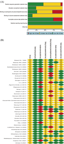
A total of 38 RCTs were included. One literature27 used the date of birth for numbering and grouping, and one20 indicated nonrandom grouping, rated as ‘high risk of bias’. Nineteen records8, 10, 12, 14-16, 21, 25, 26, 29-31, 33, 37, 43-47 only mentioned the use of random grouping, but did not specify the method of random grouping, and was rated as ‘unclear risk of bias’. Seventeen records6, 7, 9, 11, 13, 18, 19, 22, 23, 28, 32, 34, 35, 38, 40-42 mentioned specific methods of randomisation, such as computer-generated random table or number generators, which were rated as ‘low risk of bias’. Nine records6, 7, 9, 11, 13, 18, 19, 22, 23, 28, 32, 34, 35, 38, 40-42 explicitly mentioned the use of envelope concealment, which was rated as ‘low risk of bias’, and 298, 12-16, 18-20, 23, 25-34, 37, 38, 40-45, 47 did not mention whether it was hidden, which was rated as ‘unclear risk of bias’. Ten records6, 9, 18, 23, 38, 42, 43, 45-47 were blinded to subjects and researchers, rated as ‘low risk of bias’, 17 records6, 9, 18, 23, 38, 42, 43, 45-47 did not mention whether to use blinding to subjects and researchers, rated as ‘unclear risk of bias’, and 11 records6, 9, 18, 23, 38, 42, 43, 45-47 were open-label, rated as ‘high risk of bias’. Five records7, 9, 13, 22, 23 clearly adopted the blind method for the evaluation results, and were rated as ‘low risk of bias’. Thirty-three records6, 8, 10-12, 14-16, 18-21, 25-35, 37, 38, 40-47 did not indicate whether the results were evaluated in a blinded manner, and were rated as ‘unclear risk of bias’. Twenty-eight records6, 8-14, 16, 18-20, 25, 26, 28-32, 34, 37, 38, 40, 41, 44-47 with no data loss were rated as ‘low risk of bias’, and 10 records7, 15, 21-23, 27, 33, 35, 42, 43 with data loss were rated as ‘high risk of bias’. None of the 38 records7, 15, 21-23, 27, 33, 35, 42, 43 had selective reports and were rated as ‘low risk of bias’. None of the 38 records7, 15, 21-23, 27, 33, 35, 42, 43 indicated whether there were other biases and were rated as ‘unclear risk of bias’, as shown in Figure 2A,B.

The quality of four cohort studies was evaluated by the NOS scoring standard, including 1 with 8 points,17 2 with 7 points,24, 36 and 1 with 6 points.39 The quality of these four records is relatively good, indicating that the risk of bias is small, as shown in Table S1.
3.4 Healing area
3.4.1 Evidence network
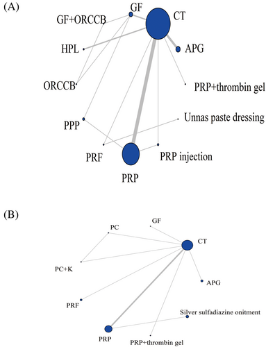
A total of 21 records reported the area of healing, involving 1077 patients, involving 12 dosing regimens, and the network evidence is shown in Figure 3A. The line between the two points represents the evidence of direct comparison between the two drugs, and no line indicates that there is no direct comparison, and the results can be obtained through indirect comparison. The thickness of the line indicates the number of studies using the two drugs in all included studies, and the size of the dot indicates the sample size of the included cases using the drug. The results showed that three closed loops were formed. CT had the largest sample size (n = 447), while PRP and CT had the largest number of studies (n = 8).

3.4.2 Results of network meta-analysis
A total of 21 studies reported on the healing area, involving 12 intervention measures, forming a total of 14 pairwise comparisons. The inconsistency test and node splitting method showed good consistency (I2 < 50%), and there was no heterogeneity between studies (p > 0.05).
The results of the network meta-analysis showed that APG, GF, GF + Oxidised regenerated cellulose and collagen biomaterial (ORCCB), ORCCB, PRF and PRP significantly increased the area of wound healing compared to the CT group. GF + ORCCB was significantly larger than Autologous Platelet Lysate (HPL), Platelet-poor Plasma (PPP), PRP, PRP injection and Unnas paste dressing. GF was significantly higher than HPL, PPP, PRP and PRP injection. ORCCB was significantly greater than PPP, PRP and PRP injection. PRF is significantly greater than PRP. PRP + thrombin gel was significantly smaller than GF, GF + ORCCB and ORCCB. APG was significantly smaller than GF, GF + ORCCB and ORCCB. HPL was significantly smaller than ORCCB and PRF, and there was no statistically significant difference (p > 0.05) compared to other intervention measures, as shown in Table 2.
| Intervention measure | PRP + thrombin gel | APG | GF | GF + ORCCB | HPL | ORCCB | PPP | PRF | PRP | PRP injection | Unnas paste dressing | CT |
|---|---|---|---|---|---|---|---|---|---|---|---|---|
| PRP + thrombin gel | 0 | |||||||||||
| APG | −4.32 (−30.75, 19.85) | 0 | ||||||||||
| GF | −48.13 (−75.4, −21.56) | −43.77 (−62.26, −23.98) | 0 | |||||||||
| GF + ORCCB | −67.62 (−102.5, −33.55) | −63.33 (−91.61, −33.73) | −19.48 (−41.23, 2.25) | 0 | ||||||||
| HPL | 10.68 (−16.06, 37.29) | 14.93 (−2.54, 35.03) | 58.78 (38.18, 80.13) | 78.29 (48.54, 108.9) | 0 | |||||||
| ORCCB | −52.43 (−87.27, −18.72) | −48.14 (−76.06, −18.87) | −4.34 (−25.67, 16.9) | 15.17 (−6.82, 37.21) | −63.07 (−93.4, −33.81) | 0 | ||||||
| PPP | 2.54 (−26.89, 30.97) | 6.82 (−14.36, 29.45) | 50.61 (30.56, 70.68) | 70.11 (40.54, 99.68) | −8.16 (−32.09, 14.92) | 54.94 (25.88, 84.12) | 0 | |||||
| PRF | −29.85 (−65.83, 5.97) | −25.5 (−55.27, 6.01) | 18.25 (−13.51, 50.56) | 37.74 (−0.49, 76.58) | −40.56 (−72.4, −8.78) | 22.5 (−15.44, 61.37) | −32.37 (−65.69, 1.69) | 0 | ||||
| PRP | 0.32 (−24.05, 23.63) | 4.66 (−9.11, 19.67) | 48.43 (31.93, 64.76) | 67.97 (40.57, 95.02) | −10.29 (−27.62, 5.89) | 52.81 (25.93, 79.57) | −2.16 (−19.77, 15.2) | 30.19 (0.32, 59.33) | 0 | |||
| PRP injection | −0.58 (−29.93, 27.97) | 3.7 (−17.33, 26.44) | 47.48 (24.13, 71.07) | 66.99 (35.27, 98.95) | −11.26 (−34.99, 11.77) | 51.82 (20.48, 83.63) | −3.15 (−27.96, 22.03) | 29.27 (−4.7, 62.65) | −0.96 (−19.05, 17.59) | 0 | ||
| Unnas paste dressing | −15.76 (−62.29, 31.06) | −11.31 (−53.18, 31.97) | 32.42 (−10.92, 76.37) | 51.93 (3.6, 100.92) | −26.41 (−69.82, 16.99) | 36.68 (−11.06, 85.67) | −18.22 (−62.68, 26.99) | 14.12 (−15.3, 43.93) | −16.03 (−57.39, 26.06) | −15.11 (−59.72, 29.82) | 0 | |
| CT | 12.84 (−9.57, 35.2) | 17.13 (6.38, 30.31) | 60.93 (46.35, 76.51) | 80.44 (54.47, 107.34) | 2.14 (−12.34, 16.8) | 65.25 (39.77, 91.89) | 10.3 (−7.65, 29.22) | 42.72 (14.51, 70.96) | 12.48 (4.68, 21.41) | 13.41 (−4.57, 32.27) | 28.58 (−12.36, 69.38) | CT |
- Abbreviations: APG, autologous platelet gel; CT, conventional treatment; GF, growth factor; HPL, autologous platelet lysate; ORCCB, oxidised regenerated cellulose and collagen biomaterial; PC, platelet concentrate; PPP, platelet-poor plasma; PRF, platelet-rich fibrin; PRF injection, injectable platelet-rich fibrin; PRP, platelet-rich plasma.
3.4.3 Ranking of intervention efficacy
The order of healing area from high to low is GF + ORCCB > ORCCB > GF > PRF > Unnas paste dressing > APG > PRP injection > PRP > PRP + thrombin gel > PPP > HPL > CT, see Table S2.
3.4.4 Publication bias assessment
The area of healing was assessed for publication bias. The funnel plot suggested that all studies were roughly symmetrically distributed on both sides of the vertical line with x = 0, indicating that there was less possibility of significant publication bias. Not all the dots are located inside the triangle, indicating that there may be a small sample effect, as shown in Figure S1.
3.5 Wound healing time
3.5.1 Evidence network
A total of ten records have reported the healing time of patients, a total of 481 patients, involving nine dosing regimens. The network evidence is shown in Figure 3B. The results showed that a closed loop was formed. CT had the largest sample size (n = 185), while PRP and CT had the largest number of studies (n = 4).
3.5.2 Results of network meta-analysis
Ten records reported patient healing times involving nine interventions, forming a total of nine pairwise comparisons. The results of the inconsistency test and node splitting method showed good consistency (I2 < 50%), with no heterogeneity among the studies (p > 0.05). The results of the network meta-analysis showed that there was no statistical difference among all interventions (p > 0.05), as shown in Table 3.
| Intervention measures | APG | GF | PC | PC + K | PRF | PRP | PRP + thrombin gel | Silver sulfadiazine onitment | CT |
|---|---|---|---|---|---|---|---|---|---|
| APG | 0 | ||||||||
| GF | 9.16 (−41.29, 59.18) | 0 | |||||||
| PC | 1.78 (−48.31, 52.08) | −7.41 (−57.18, 42.96) | 0 | ||||||
| PC + K | 3.35 (−46.63, 53.33) | −5.84 (−55.81, 44.45) | 1.51 (−34.03, 37.09) | 0 | |||||
| PRF | −0.43 (−50.31, 49.9) | −9.54 (−59.46, 40.9) | −2.15 (−52.66, 47.96) | −3.63 (−53.99, 46.4) | 0 | ||||
| PRP | 4.44 (−35.6, 43.56) | −4.74 (−44.72, 34.95) | 2.67 (−37.57, 42.33) | 1.16 (−39.09, 40.51) | 4.8 (−34.98, 44.23) | 0 | |||
| PRP + thrombin gel | 30.42 (−25.66, 87.2) | 21.23 (−34.74, 77.93) | 28.62 (−27.86, 85.38) | 27.06 (−29.47, 83.9) | 30.8 (−25.75, 87.13) | 26.09 (−21.4, 73.99) | 0 | ||
| Silver sulfadiazine onitment | −20.56 (−74.2, 32.66) | −29.67 (−83.46, 23.59) | −22.29 (−76.14, 30.83) | −23.78 (−77.54, 28.94) | −20.18 (−73.78, 32.98) | −24.98 (−60.47, 10.64) | −50.96 (−110.1, 7.91) | 0 | |
| CT | −4.9 (−40.38, 30.47) | −14.06 (−49.41, 21.38) | −6.64 (−42.27, 28.79) | −8.19 (−43.74, 27.22) | −4.53 (−39.89, 30.86) | −9.34 (−26.93, 8.94) | −35.32 (−79.81, 8.89) | 15.63 (−23.88, 55.66) | 0 |
- Abbreviations: APG, autologous platelet gel; CT, conventional treatment; GF, growth factor; PC, platelet concentrate; PC + K, keratinocytes suspended in platelet concentrate; PRF, platelet-rich fibrin; PRP, platelet-rich plasma.
3.5.3 Ranking of intervention efficacy
The order of healing time from low to high is PRP + thrombin gel <GF < PRP < Keratinocytes Suspended in Platelet Concentrate (PC + K) < PC < APG < PRF < CT < Silver sulfadiazine ointment, as shown in Table S3.
3.5.4 Publication bias assessment
Evaluate publication bias on healing time and use Stata SE15.0 for comparison correction funnel plot. The funnel plot shows that all records are roughly symmetrically distributed on both sides of the x = 0 vertical line, indicating a small possibility of significant publication bias. Some dots are scattered outside the triangle, indicating a possible small sample effect, as shown in Figure S1.
3.6 Number of wound healing cases
3.6.1 Evidence network
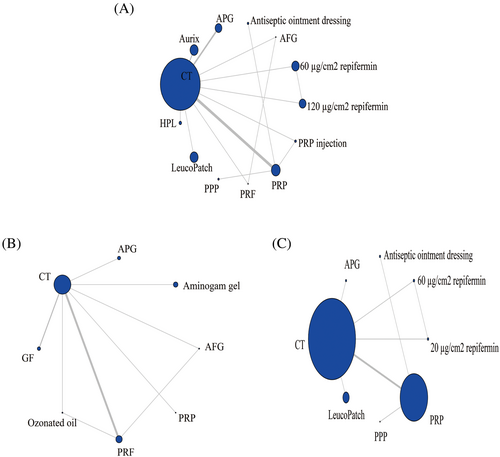
A total of 15 records have reported the number of patients who have fully recovered, with a total of 1553 patients involved in 13 medication regimens. The network evidence is shown in Figure 4A. The results showed the formation of three closed loops. CT had the largest sample size (n = 650), while PRP and CT had the largest number of studies (n = 5).

3.6.2 Results of network meta-analysis
Fifteen records have reported the number of patients who have recovered, involving 13 intervention measures, forming a total of 15 pairwise comparisons. The inconsistency test and node splitting method showed good consistency (I2 < 50%), and there was no heterogeneity between studies (p > 0.05). The results of the network meta-analysis showed that the therapeutic effect of APG was significantly greater than that of 120 μg/cm2 replifermin and CT, and the PRP was significantly greater than that of CT (p < 0.05). There was no statistically significant difference between other intervention measures (p > 0.05), as shown in Table 4.
| Intervention measures | 120 μg/cm2 repifermin | 60 μg/cm2 repifermin | AFG | Antiseptic ointment dressing | APG | Aurix | HPL | LeucoPatch | PPP | PRF | PRP | PRP injection | CT |
|---|---|---|---|---|---|---|---|---|---|---|---|---|---|
| 120 μg/cm2 repifermin | 0 | ||||||||||||
| 60 μg/cm2 repifermin | −0.29 (−2.13, 1.52) | 0 | |||||||||||
| AFG | 16.22 (−40.08, 73.89) | 16.52 (−39.73, 74.2) | 0 | ||||||||||
| Antiseptic ointment dressing | −0.34 (−3.41, 2.53) | −0.05 (−3.12, 2.86) | −16.57 (−74.3, 39.73) | 0 | |||||||||
| APG | −2.08 (−4.85, −0.19) | −1.78 (−4.55, 0.1) | −18.42 (−76.18, 37.89) | −1.76 (−4.79, 0.73) | 0 | ||||||||
| Aurix | −1.16 (−3.39, 1.1) | −0.86 (−3.1, 1.39) | −17.31 (−75, 38.83) | −0.81 (−3.46, 1.97) | 0.93 (−0.63, 3.31) | 0 | |||||||
| HPL | −0.46 (−3.14, 2.24) | −0.17 (−2.82, 2.53) | −16.65 (−74.32, 39.63) | −0.11 (−3.13, 3.08) | 1.64 (−0.43, 4.5) | 0.7 (−1.66, 3.08) | 0 | ||||||
| LeucoPatch | −1.04 (−3.61, 1.55) | −0.75 (−3.29, 1.83) | −17.22 (−74.9, 39) | −0.68 (−3.58, 2.38) | 1.05 (−0.83, 3.8) | 0.13 (−2.12, 2.37) | −0.57 (−3.26, 2.09) | 0 | |||||
| PPP | 0.23 (−2.85, 3.11) | 0.52 (−2.54, 3.42) | −16.04 (−73.78, 40.37) | 0.57 (−2.47, 3.61) | 2.34 (−0.16, 5.39) | 1.39 (−1.43, 4.03) | 0.69 (−2.51, 3.69) | 1.27 (−1.8, 4.16) | 0 | ||||
| PRF | −6.44 (−70.84, 49.94) | −6.14 (−70.53, 50.2) | −21.91 (−116.97, 72.28) | −6.07 (−70.63, 50.22) | −4.18 (−68.55, 52.1) | −5.29 (−69.71, 50.9) | −6.03 (−70.44, 50.22) | −5.42 (−69.81, 50.89) | −6.68 (−71.18, 49.71) | 0 | |||
| PRP | −1.44 (−3.66, 0.5) | −1.15 (−3.36, 0.82) | −17.69 (−75.41, 38.5) | −1.11 (−3.29, 1) | 0.66 (−0.86, 2.67) | −0.29 (−2.07, 1.24) | −0.98 (−3.33, 1.11) | −0.4 (−2.63, 1.52) | −1.67 (−3.88, 0.44) | 4.98 (−51.24, 69.38) | 0 | ||
| PRP injection | −2.07 (−4.74, 0.47) | −1.76 (−4.42, 0.76) | −18.28 (−75.93, 38) | −1.71 (−4.55, 1.13) | 0.05 (−2.07, 2.7) | −0.9 (−3.25, 1.3) | −1.59 (−4.41, 1.03) | −1.02 (−3.69, 1.49) | −2.28 (−5.16, 0.58) | 4.33 (−51.83, 68.64) | −0.6 (−2.45, 1.3) | 0 | |
| CT | −0.37 (−2.19, 1.45) | −0.08 (−1.89, 1.77) | −16.56 (−74.2, 39.64) | −0.02 (−2.34, 2.42) | 1.73 (0.61, 3.6) | 0.79 (−0.53, 2.12) | 0.09 (−1.87, 2.05) | 0.66 (−1.14, 2.48) | −0.6 (−2.92, 1.87) | 6.11 (−50.12, 70.46) | 1.08 (0.17, 2.21) | 1.69 (−0.09, 3.64) | 0 |
- Abbreviations: AFG, autologous fibrin gel; APG, autologous platelet gel; Aurix, aurix gel; CT, conventional treatment; GF, growth factor; HPL, autologous platelet lysate; PPP, platelet-poor plasma; PRF, platelet-rich fibrin; PRF injection, injectable platelet-rich fibrin; PRP, platelet-rich plasma.
3.6.3 Ranking of intervention efficacy
The order of number of wound healing cases from high to low is APG > PRP injection > PRP > Aurix > PRF > LeucoPatch > HPL > Antiseptic Ointment Dressing > CT > 60 μg/cm2 replifermin > 120 μg/cm2 replifermin > AFG > PPP, as shown in Table S4.
3.6.4 Publication bias assessment
Evaluate publication bias and small sample effects on the number of recovered cases, and use Stata SE 15.0 for comparison correction funnel plots. The funnel plot indicates that all studies are roughly symmetrically distributed on both sides of the x = 0 vertical line, indicating a small possibility of significant publication bias. All the dots are inside the triangle, indicating that there cannot be a small sample effect, as shown in Figure S1.
3.7 Pain score
3.7.1 Evidence network
There are a total of nine articles reporting VAS pain scores, with a total of 457 patients involved in eight medication regimens. The network evidence is shown in Figure 4B. The results show the formation of two closed loops. CT has the largest sample size (n = 211), while PRF and CT have the highest number of studies (n = 4).
3.7.2 Results of network meta-analysis
Nine records reported pain scores involving eight intervention measures, resulting in a total of nine pairwise comparisons. The inconsistency test and node splitting method showed good consistency (I2 < 50%), and there was no heterogeneity between studies (p > 0.05). The results of the network meta-analysis showed that the AFG score was significantly lower than that of CT (p < 0.05), and there was no statistically significant difference (p > 0.05) compared to other intervention measures, as shown in Table 5.
| Intervention measures | AFG | Aminogam_gel | APG | GF | Ozonated oil | PRF | PRP | CT |
|---|---|---|---|---|---|---|---|---|
| AFG | 0 | |||||||
| Aminogam gel | −2.35 (−21.73, 15.57) | 0 | ||||||
| APG | −11.42 (−30.89, 6.49) | −9.08 (−27.94, 9.72) | 0 | |||||
| GF | −11.68 (−28.47, 3.76) | −9.3 (−25.78, 7.07) | −0.2 (−16.43, 16.01) | 0 | ||||
| Ozonated oil | −9.28 (−26.52, 7.1) | −6.97 (−24.58, 11.34) | 2.1 (−15.37, 20.16) | 2.33 (−12.54, 17.85) | 0 | |||
| PRF | −6.83 (−19.83, 5.82) | −4.5 (−19.03, 11.16) | 4.55 (−9.76, 20.25) | 4.78 (−6.28, 17.17) | 2.44 (−9.16, 14.72) | 0 | ||
| PRP | −9.34 (−28.52, 8.5) | −6.99 (−25.64, 11.79) | 2.09 (−16.49, 20.83) | 2.3 (−13.85, 18.57) | −0.02 (−18.21, 17.39) | −2.46 (−18.02, 11.77) | 0 | |
| CT | −12.56 (−26.42, −0.14) | −10.17 (−23.61, 3.14) | −1.11 (−14.27, 12.13) | −0.88 (−10.36, 8.55) | −3.22 (−15.51, 8.37) | −5.69 (−13.26, 0.61) | −3.18 (−16.45, 9.94) | 0 |
- Abbreviations: AFG, autologous fibrin gel; APG, autologous platelet gel; CT, conventional treatment; GF, growth factor; ozonated oil, ozone gas dissolved in olive oil; PG, platelet gel; PRF, platelet-rich fibrin; PRG, platelet-rich gel; PRP, platelet-rich plasma.
3.7.3 Ranking of intervention efficacy
The order of pain scores from low to high is AFG < Aminogem gel < PRF < PRP < Ozoned oil < APG < GF < CT (Table S5).
3.7.4 Publication bias assessment
The pain score was evaluated using Stata SE 15.0 for publication bias and small sample effects. The funnel plot indicates that all studies are roughly symmetrically distributed on both sides of the x = 0 vertical line, indicating a small possibility of significant publication bias. The two dots are scattered outside the triangle, indicating a possible small sample effect, as shown in Figure S1.
3.8 Number of wound infection cases
3.8.1 Evidence network
There are a total of eight records reports on the number of wound infection cases during treatment, with a total of 1818 patients involved in eight medication regimens. The network evidence is shown in Figure 4C. The results show the formation of a closed loop. CT has the largest sample size (n = 983), while PRP and CT have the largest number of studies (n = 4).
3.8.2 Results of network meta-analysis
Eight records have reported the number of wound infection cases, involving eight intervention measures, forming a total of eight pairwise comparisons. The inconsistency test and node splitting method showed good consistency (I2 < 50%), and there was no heterogeneity between studies (p > 0.05). The results of the network meta-analysis showed that APG had the fewest number of infection cases and was significantly lower than other intervention measures (p < 0.05). There was no statistically significant difference (p > 0.05) between other intervention measures, as shown in Table 6.
| Intervention measures | 20 μg/cm2 repifermin | 60 μg/cm2 repifermin | Antiseptic ointment dressing | APG | LeucoPatch | PPP | PRP | CT |
|---|---|---|---|---|---|---|---|---|
| 20 μg/cm2 repifermin | 0 | |||||||
| 60 μg/cm2 repifermin | −0.85 (−5.5, 3.71) | 0 | ||||||
| Antiseptic ointment dressing | −2.71 (−9.7, 4.04) | −1.86 (−8.78, 4.84) | 0 | |||||
| APG | 32.59 (2.42, 77.68) | 33.5 (3.31, 78.54) | 35.44 (5.03, 80.43) | 0 | ||||
| LeucoPatch | −1.5 (−7.71, 4.71) | −0.63 (−6.77, 5.4) | 1.21 (−5.4, 8.02) | −34.18 (−79.14, −3.98) | 0 | |||
| PPP | −2.34 (−9.81, 4.7) | −1.48 (−8.86, 5.51) | 0.38 (−6.52, 7.12) | −35.09 (−80.19, −4.5) | −0.82 (−8.13, 6.08) | 0 | ||
| PRP | −1.27 (−6.59, 3.82) | −0.41 (−5.61, 4.55) | 1.43 (−3.04, 5.99) | −33.98 (−78.85, −4.04) | 0.23 (−4.8, 5.1) | 1.03 (−3.91, 6.34) | 0 | |
| CT | −1.8 (−6.4, 2.66) | −0.95 (−5.38, 3.42) | 0.91 (−4.19, 6.19) | −34.53 (−79.24, −4.68) | −0.3 (−4.55, 3.9) | 0.52 (−4.98, 6.43) | −0.53 (−3.02, 2.08) | 0 |
- Abbreviations: APG, autologous platelet gel; CT, conventional treatment; PPP, platelet-poor plasma; PRP, platelet-rich plasma.
3.8.3 Ranking of intervention efficacy
The order of the number of wound infection cases from low to high is APG < 20 μg/cm2 replifermin < 60 μg/cm2 replifermin < PR < LeucoPatc < CT < PPP < Antiseptic Ointment Dressing, as shown in Table S6.
3.8.4 Publication bias assessment
Evaluate publication bias and small sample effects on wound infections. The funnel plot indicates that all studies are roughly symmetrically distributed on both sides of the x = 0 vertical line, indicating a small possibility of significant publication bias. All the dots are inside the triangle, indicating that there cannot be a small sample effect, as shown in Figure S1.
4 DISCUSSION
Plasma derivatives are substances prepared from healthy blood using separation and purification techniques such as chromatography and centrifugation. The mechanisms by which plasma derivatives promote wound healing are as follows: ① Platelets in plasma derivatives are activated to release powerful cell/growth factors (TGF-β, PDGF, IGF-1 and EGF, etc.), which act on a variety of target cells to promote cell proliferation, matrix synthesis, collagen deposition, and ultimately achieving tissue repair.48 ② The anti-inflammatory cytokines in plasma derivatives can regulate the process of inflammatory response.49 ③ Adhesion factors (fibrin, fibronectin and hyalonin) in plasma derivatives construct a three-dimensional structure locally (required for tissue repair), repairing cell movement and facilitating wound repair.50 ④ Plasma derivatives induce the release of local growth factors, cytokines, or microRNAs through autocrine and paracrine pathways, promoting wound healing.51 ⑤ Platelet activation in plasma derivatives releases bioactive substances (chemokines, histamines and adenosine) that directly induce platelet aggregation, alleviate pain and indirectly chemotactic leukocytes to exert bactericidal effects.52 In recent years, with the rapid development of regenerative medicine, the use of plasma derivatives for wound treatment has achieved remarkable results,53 but the lack of direct or indirect comparison of efficacy among derivatives is not conducive to clinical promotion and the selection of the best program. In this study, network meta-analysis was used to analyse the healing area, healing time, healing number, late infection number and analgesia score, in order to provide evidence for the selection of clinical drugs.
The healing area shows that GF + ORCCB has the largest healing area, and the combination of wound environment regulator (ORCCB) and GF can significantly accelerate the healing area of the wound. This may be due to: ① ORCCB can change the external environment of the wound by binding and inactivating proteases and gelatinases that seep out of the wound, creating a protective environment for growth factor healing.54 ② ORCCB can physically bind proteases. When the proteases are fully controlled, this material has the ability to bind and protect growth factors, and can send 70% of the growth factors back to the wound surface, which is beneficial for wound healing.55 Zhang et al.56 confirmed through meta-analysis that ORCCB is beneficial in improving the wound healing rate and relative percentage reduction compared to other traditional nursing materials (non-MMP, inhibitory biomaterials and modern excipients).
PRP + thrombobin gel has the shortest healing time. PRP increases the rate of wound healing by increasing haemostasis, releasing growth factors, re-epithelialization, inducing fibroblast proliferation of extracellular matrix and promoting angiogenesis. Thrombin is another important component of wound healing.57 The combination of the two can convert fibrin in PRP into a network structure, embed platelets, white blood cells, and growth factors, and produce a certain adhesive effect, retaining the sample at the delivery site and stimulating the release of growth factors, accelerating wound healing time.58
APG has the best effect on healing and reducing post-infection. APG can accelerate wound healing and promote tissue regeneration by increasing the production of extracellular matrix and granulation tissue,59 which is consistent with the results of this analysis. The activated platelets in APG contain a series of microbicidal proteins with antibacterial activity, which exert strong antibacterial effects mainly by altering the permeability of cell membranes and inhibiting the synthesis of large molecules.60 This may be a key factor in reducing the number of infections in the later stages of surgery.
Postoperative pain assessment is an important component of wound healing and plays a decisive role in wound healing. Segal et al.61 found through a randomised trial that there was no significant difference in the analgesic effect of AFG compared to not using AFG (p = 0.988). Contrary to Segal N's research, this article found that AFG has a significantly higher postoperative analgesic effect than CT, which may be due to the release of mediators such as histamine, 5-hydroxytryptamine and dopamine from concentrated platelets, thereby reducing the occurrence of pain.12 This indicates that there are relatively few included literature on some blood derivatives with low quality, and more high-quality studies with larger sample sizes are needed to confirm them in the later stage.
Limitations of this study: ① Most of the 38 RCT studies included were of low quality, and the vast majority did not indicate specific randomised, hidden, or blind methods, which poses a certain risk of bias and affects the authenticity and reliability of the results.② Some studies include a small sample size, which may reduce the credibility of the results.③ The inclusion of research implemented in multiple countries around the world has a certain impact on the results due to differences in medical standards and standard protocols. ④ Inconsistent inclusion of causes of trauma (surgical wounds, ulcers, burns, etc.) may affect the accuracy of the results. ⑤ The lack of direct comparison between different blood derivatives affects the credibility of the results. ⑥ This study included four retrospective cohort studies, which may have a certain impact on the results. Given the above limitations, clinical practice should maintain a rigorous and cautious attitude, conduct more multi-centre, large-sample clinical studies and provide more sources of evidence for further verifying its efficacy.
5 CONCLUSIONS
GF + ORCCB is the best in reducing wounds, and PRP + thrombin gel has the shortest treatment time. The number of people who have recovered from APG and the reduction of wound infections are the most prominent, and the analgesic effect of APG is the best. Clinical medical staff can choose appropriate treatment plans based on the needs of patients.
ACKNOWLEDGEMENTS
The authors thank all the participants of this work.
FUNDING INFORMATION
This research was supported by the Yucai Foundation of General Hospital of Southern Theater Command (Grant No. 2022NZB002).
CONFLICT OF INTEREST STATEMENT
The authors declare that they have no competing interests.
Open Research
DATA AVAILABILITY STATEMENT
The data used in this network meta-analysis are available to the authors upon reasonable request.


