Prevention efficacy of dentin tubule sealing with Nd:YAG laser against tooth discoloration induced by vital pulp treatment
Abstract
Background
The discoloration potential of dental materials and applications such as vital pulp therapy also cause discoloration due to the presence of blood. Dentin tubule sealing methods could be used for the prevention of discoloration.
Aim
The purpose of this study was to evaluate the effect of sealing pulp chamber walls with Nd:YAG laser against discoloration caused by tricalcium silicate-based materials in the presence and absence of blood contamination.
Design
Eighty-one extracted human maxillary incisors were prepared and randomly divided into nine groups according to the Nd:YAG laser application, the cement to be used (NeoMTA Plus/Biodentine), and the presence or absence of blood. The color measurements were obtained with a spectrophotometer at baseline and then at the sixth (T1) and 12th (T2) months thereafter.
Results
Sealing with Nd:YAG laser and placing NeoMTA Plus or Biodentine on human blood significantly increased discoloration at T1 and T2 (p < .05). Similarly, without sealing the cavity and placing NeoMTA Plus or Biodentine on human blood significantly increased discoloration at T1 and T2 (p < .05).
Conclusion
Regardless of sealing the dentin tubules with Nd:YAG laser, NeoMTA Plus and Biodentine caused more discoloration in the presence of blood contamination.
Why this paper is important to paediatric dentists
- Regardless of sealing the dentin tubules with Nd:YAG laser, NeoMTA Plus and Biodentine caused more discoloration in the presence of blood contamination.
- In vital pulp therapies, sealing the dentinal tubules with a Nd:YAG laser application before placing calcium silicate-based materials could reduce discoloration.
- Nevertheless, in the presence of blood contamination, Nd:YAG laser application could not prevent discoloration.
1 INTRODUCTION
Vital pulp therapy (VPT) is defined as a treatment aimed at preserving the vitality of dental pulp affected by extensive dental caries, dental trauma, or iatrogenic reasons.1 Vital pulp therapies expect to simulate the remaining pulp tissue to regenerate the dental pulp complex. Clinically, vital pulp treatment includes indirect pulp capping, direct pulp capping, partial pulpotomy, and complete pulpotomy.2 The treatment options for cariously exposed pulp are shifting to avoid pulpectomy and apply biologically based VPTs. An appropriate dental biomaterial application for VPTs is highly crucial for favorable outcomes. The literature suggests the use of a wide range of materials as pulp-protective dressing materials.3
Mineral trioxide aggregate (MTA) was first introduced as an experimental calcium silicate-based material in 1993.4 Mineral trioxide aggregate has been reported to be an excellent biocompatible material, which has supported apatite formation and stimulates dentinogenesis. It has superior sealing properties and antibacterial effects due to highly alkaline pH and calcium hydroxide release.5-7 Hence, MTA has been suggested and used in many clinical applications such as direct and indirect pulp capping, pulpotomies, perforation repairs, apexification procedures, and revascularization treatment. Although MTA has several advantages, numerous drawbacks such as difficulty in handling, long setting time, and discoloration potential have also been clinically reported.2, 8 It has also been reported that discoloration is caused by bismuth oxide in MTA.9, 10 NeoMTA Plus (Avalon Biomed Inc., Houston, TX, USA) is a tricalcium-based material developed for indications similar to MTA and contains tantalum oxide as a radiopacifier to prevent potential discoloration.11
In 2011, Biodentine (BD; Septodont, France), a relatively new calcium silicate-based cement, was introduced to eliminate the disadvantages of MTA. Biodentine is a bioactive and biocompatible material composed of tricalcium silicate, dicalcium silicate, zirconium oxide, calcium carbonate, calcium oxide, and iron oxide. The interactions between BD with dentin and pulp induce remineralization with tertiary dentin formation and allow its use in VPTs. Shorter setting time and improvements in mechanical properties compared with MTA have facilitated its widespread use.1, 3 In addition, BD has also been reported to cause less tooth discoloration than MTA in many studies.12-14
The prevention of potential tooth discoloration caused by materials used during endodontic procedures has become very important due to increasing esthetic demands in recent years. Tooth discoloration has been attributed to the penetration of dental material contents into the dentin tubules or their inclusion into the pulp chambers.15 Various methods have been studied to prevent tooth discoloration.15-18 It has been reported that sealing the dentinal tubules of the pulp chamber with a bonding agent may be a suitable alternative to minimize tooth discoloration.15 Another treatment option for sealing dentinal tubules is laser application; favorable results have been reported.16 Nd:YAG laser obliterates dentinal tubules with its melting and recrystallization potential in the hard tissue. It has been stated that the Nd:YAG laser can reduce dentin permeability when the dentinal tubules are occluded.19 Thus, preventing or reducing the discoloration potential of the materials applied to the pulp chamber may be possible. In addition to the discoloration potential of dental materials, applications such as VPT also cause discoloration due to the presence of blood. Blood has been attributed to induce tooth discoloration. The presence of blood has been shown to modify the discoloration potential of calcium silicate-containing materials in an ex vivo model.15
The purpose of this in vitro study was to evaluate the preventive efficacy of sealing pulp chamber walls with Nd:YAG laser against discoloration caused by tricalcium silicate-based materials in the presence and absence of blood contamination.
2 MATERIALS AND METHODS
2.1 Preparation of tooth specimens
The sample size calculation was performed with a significance level of 5% and a test power of 80% using Shokouhinejad et al.'s study.15 The minimum sample size required was determined to be nine samples per group. The teeth were collected under the approval of the Institutional Review Board of Bezmialem Vakif University (protocol no. 02/293), Istanbul, Turkey.
Eighty-one recently extracted human maxillary central and lateral incisors that were without caries or restorations and developmental defects were used. The teeth were disinfected with 0.5% chloramine T solution for 48 h. Each specimen was separated 2 mm below the cementoenamel junction using a high-speed diamond disk under water cooling. Standardized cavities (3 mm in diameter and 3 mm in depth) were prepared for each specimen on the lingual surface of the crowns without removing the roof chamber using a cylindrical bur with a diameter of 3 mm. The height of the tooth structure between the labial surface of the crowns and the cavity floor was obtained using an electronic caliper of 2 mm for each specimen. After the preparation, all the specimens were irrigated with 1% sodium hypochlorite for 30 min, followed by 20% EDTA solution for 5 min to remove the smear layer. The specimens were then stored in normal saline solution.
2.2 Experimental design
The study groups were designed according to the Nd:YAG laser application, the tricalcium silicate-based cements to be used, and the presence or absence of blood contamination. A direct pulp capping model was created in groups with blood contamination. The tricalcium silicate-based cements (NeoMTA Plus and Biodentine) were applied with 2 mm thickness. The material compositions and applications are presented in Table 1. For this study, fresh human blood was collected from a volunteer member of the research group.
| Materials | Manufacturer | Composition | Application procedure |
|---|---|---|---|
| Nd:YAG Laser | LightWalker, Fotona, Slovenia | NA | The inner surfaces were irradiated with a pulse 10 Hz–1 W, with a total irradiation time of 60 s |
| NeoMTA Plus | Avalon Biomed Inc. Bradenton, FL, USA |
Powder: Tricalcium silicate, dicalcium silicate, tantalum oxide, tricalcium aluminate, and calcium sulfate. Liquid: Water-based gel with thickener agents and water-soluble polymers |
One scoop of powder and three drops of water-based gel are mixed on a glass slab. |
| Biodentine | Septodont, Saint Maur des Fosses, France |
Powder: Tricalcium silicate, dicalcium silicate, calcium carbonate, calcium oxide, and zirconium oxide (in a capsule) Liquid: Water, calcium chloride, and hydrosoluble polymer |
Five drops of liquid was poured into the capsule and mixed for 30 s on a mixing device. |
The specimens were randomly divided into nine groups (n = 9) as follows (Table 2):
| Groups | Sealed with Nd:YAG laser | Cement | Presence of blood |
|---|---|---|---|
| Control | No | No | Yes |
| Group 1 | No | NeoMTA Plus | No |
| Group 2 | Yes | NeoMTA Plus | No |
| Group 3 | No | NeoMTA Plus | Yes |
| Group 4 | Yes | NeoMTA Plus | Yes |
| Group 5 | No | Biodentine | No |
| Group 6 | Yes | Biodentine | No |
| Group 7 | No | Biodentine | Yes |
| Group 8 | Yes | Biodentine | Yes |
Group 1: The interior surfaces of the cavity were not sealed, and NeoMTA Plus (Avalon Biomed Inc. Bradenton, FL, USA) was placed on the cavity floor.
Group 2: The interior surfaces of the cavity were sealed with Nd:YAG laser, and NeoMTA Plus was placed on the cavity floor.
Group 3: The interior surfaces of the cavity were not sealed, and NeoMTA Plus was placed on 1.5 mL of human blood applied to the cavity floor.
Group 4: The interior surfaces of the cavity were sealed with Nd:YAG laser, and NeoMTA Plus was placed on 1.5 mL of human blood applied to the cavity floor.
Group 5: The interior surfaces of the cavity were not sealed, and Biodentine (Septodont, Saint Maur des Fosses, France) was placed on the cavity floor.
Group 6: The interior surfaces of the cavity were sealed with Nd:YAG laser, and Biodentine was placed on the cavity floor.
Group 7: The interior surfaces of the cavity were not sealed, and Biodentine was placed on 1.5 mL of human blood applied to the cavity floor.
Group 8: The interior surfaces of the cavity were sealed with Nd:YAG laser, and Biodentine was placed on 1.5 mL of human blood applied to the cavity floor.
Control: Only 1.5 mL of human blood was applied to the cavity floor; the interior surfaces of the cavity were not sealed, and no material was placed.
The cavities were then restored with resin-modified glass ionomer cement (Equia, GC, Tokyo, Japan). Each specimen was placed into distilled water-filled single tubes and then stored in the dark at room temperature. The distilled water was refreshed every 10 days to avoid a pH drop.
2.3 Color assessment
2.4 Statistical analysis
Means and standard deviations for the color differences were calculated using the SPSS 25.0 software (SPSS, Chicago, Ill., USA) at a significance level of α equals 0.05. Shapiro–Wilk test was used for confirming the normal distribution of the data. The color changes over time and were analyzed using repeated-measures ANOVA, and adjustments for multiple comparisons were assessed using the Bonferroni tests.
3 RESULTS
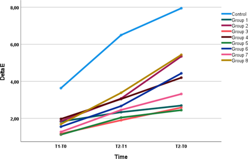
The mean ΔE values for each group at different measurement times are shown in Table 3 and Figure 1. The results of this study showed that sealing the interior surfaces of the cavity with Nd:YAG laser and placing NeoMTA Plus (Group 4) or Biodentine (Group 8) on human blood significantly increased discoloration at the end of the sixth (T1) and 12th (T2) month (p < .05). Similarly, without sealing the interior surfaces of the cavity with Nd:YAG laser and placing NeoMTA Plus (Group 3) or Biodentine (Group 7) on human blood significantly increased discoloration at T1 and T2 (p < .05). In addition, significantly increased discoloration at T1 and T2 was observed in the control group (p < .01). There was no significant difference between NeoMTA Plus and Biodentine regardless of sealing the interior surfaces of the cavity with Nd:YAG laser applied without blood (p > .05).
| T1-T0 | T2-T1 | T2-T0 | |
|---|---|---|---|
| (mean ± SD) | (mean ± SD) | (mean ± SD) | |
| Control | 1.69 ± 0.72Aa | 3.38 ± 1.32Ab | 5.43 ± 1.31Ac |
| Group 1 | 3.63 ± 2.82B | 6.49 ± 5.09B | 7.94 ± 6.7B |
| Group 2 | 1.82 ± 1.05 | 2.33 ± 1.08C | 2.69 ± 1.12C |
| Group 3 | 1.85 ± 0.83a | 3.07 ± 1.73Db | 5.34 ± 1.57c |
| Group 4 | 1.17 ± 0.4a | 1.89 ± 0.41b | 2.58 ± 0.58c |
| Group 5 | 1.96 ± 1.31a | 3.04 ± 1.89Eb | 4.19 ± 2.67c |
| Group 6 | 1.12 ± 0.61C | 2.04 ± 0.43F | 2.44 ± 0.56D |
| Group 7 | 1.55 ± 0.5a | 2.66 ± 1.21Gb | 4.43 ± 1.37c |
| Group 8 | 1.26 ± 0.38Da | 2.45 ± 0.93Hb | 3.31 ± 1.4Ec |
| **p | <.01 | <.01 | <.01 |
- Note: Different lowercase letters within each row and different uppercase letters within each column represent statistical differences according to Bonferroni post hoc comparisons.

At the sixth-month measurement, the color change value of the control group was significantly higher than that of the groups whose cavity surfaces were not sealed and applied NeoMTA Plus without human blood (Group 1) and sealed with Nd:YAG laser and applied Biodentine with (Group 8) and without (Group 6) human blood (p < .05).
At the 12th-month measurement, the highest color change value was also observed in the control group. The color change value of the control group was significantly higher than that of the groups whose cavity surfaces were sealed with Nd:YAG laser (Group 2) and not sealed (Group 1) and applied NeoMTA Plus without human blood. Similarly, the color change value of the control group was significantly higher than that of the groups whose cavity surfaces were sealed with Nd:YAG laser and applied Biodentine with (Group 8) and without (Group 6) human blood (p < .05). Photographs of a specimen in each group at baseline and at the end of the 12th month are shown in Figure 2.

4 DISCUSSION
Tooth discoloration is an unfavorable outcome that can occur after VPTs. The materials used in VPTs and the presence of blood are the most important factors of tooth discoloration. In our study, the conditions that may cause tooth discoloration and the sealing of the dentinal tubules with Nd:YAG laser as a preventive procedure were evaluated. Calcium silicate-containing biomaterials caused significant discoloration in the presence of blood contamination despite the sealing of dentinal tubules with Nd:YAG laser application.
The present study was conducted using human teeth to stimulate the clinical conditions required by VPTs. The height of the tooth structure between the labial surface of the crowns and the cavity floor was standardized in all specimens according to research by Yoldaş et al.20 Thus, the discoloration occurring in each sample could be observed from the tooth tissue of the same thickness. Color measurement was performed with the spectrophotometer, which has been used in many previous studies and provides reliable color determination.14-16, 20 A spectrophotometer provides color determination according to internationally accepted ISO standards by measuring according to the CIE color model. The ΔE value was calculated to observe the color change, and a value above 3.5 is the clinically visible difference. In this study, the control group and the groups whose cavity surfaces were not sealed and applied NeoMTA Plus with (Group 3) and without (Group 1) blood contamination caused changes in ΔE values that were greater than 3.5.
It is known that infiltration of blood components into dentinal tubules or the gaps of cement materials causes discoloration. In particular, blood products such as hemoglobin and hematin molecules, which are released by the lysis of red blood cells, are responsible for discoloration.21, 22 Tooth discoloration induced by blood has been shown in many studies.20-23 In the present study, more discoloration occurred in the presence of blood contamination, although new-generation tricalcium silicate-based cements and sealed inner surfaces of the cavity with Nd:YAG laser were used. In addition, the content of the materials is of great importance in the color change potential. The development of discoloration is the result of metal components such as bismuth and iron. Felman and Parashos reported that the iron content of MTA may cause a color change with the penetration of the dentinal tubules and its oxidation to the calcium aluminoferrite phase during the setting of MTA.21 Bismuth oxide, used as a radiopacifier in MTA, has been shown to cause significant chromogenic changes in the tooth crown when exposed to light in an oxygen-free environment.24
Alternative tricalcium silicate-based materials have been developed for preventing discoloration. It has been stated that the discoloration potential of tricalcium silicate materials without bismuth oxide is lower than that of MTA. NeoMTA Plus contains tantalum oxide instead of bismuth oxide as a radiopacifier, whereas Biodentine contains zirconium oxide.13, 25-27 Camilleri reported that NeoMTA Plus and Biodentine are materials that do not cause discoloration and can be alternatives to MTA.27 Similarly, in the current study, NeoMTA Plus and Biodentine did not cause significant discoloration in the absence of blood contamination. NeoMTA Plus and Biodentine, however, include nonstaining components and are known to not cause discoloration; NeoMTA Plus induced higher discoloration than the control group. In addition, NeoMTA Plus caused significantly more color change than Biodentine at the end of 12 months, although there was no significant difference in their coloring potential at the end of 6 months in the absence of blood contamination. Although the most likely cause of tooth discoloration is blood, it has been shown in our study that materials may also influence discoloration.
There are a number of tooth-bleaching techniques that clinicians can use on vital teeth. Home bleaching and in-office bleaching are widely used in dental practice. The combination of in-office and home bleaching has been suggested to strengthen the bleaching effect and improve color stability.28 In addition, persistent discoloration cases may require additional bleaching sessions, resulting in increased cost, time spent, and risk of complications. Therefore, the application of preventive discoloration procedures is clinically important. The experimental method in the present study was designed to avoid discoloration with the dentin tubule occlusion approach. Only a few studies assess the effectiveness of dentin tubule occlusion applications.15-18 In their case, Kim et al.18 used an adhesive dentin bonding agent to prevent discoloration caused by triple antibiotic paste and reported decreased crown discoloration. In addition to dentin bonding agent applications, Nd:YAG laser is also used to reduce or prevent discoloration. It has been reported that the Nd:YAG laser can obliterate dentin tubules and produce a sealing depth of approximately 4 μm.19
Our study showed that Nd:YAG laser application decreased the potential discoloration in the absence of blood contamination, although it could not prevent or reduce discoloration in the presence of blood contamination. Although the results cannot be compared directly, our result is in accordance with that of Fundaoğlu Küçükekenci et al.16 who reported that the use of dentin bonding agent, Nd:YAG laser, or teethmate desensitizer reduced crown color change caused by triple-antibiotic paste. They also stated that there was no statistically significant difference between the dentin tubule occlusion approaches. Another study using a dentin bonding agent to prevent discoloration reported that coronal discoloration caused by triple-antibiotic paste is reduced but not completely prevented.15 Razzak and Mahdee reported that the discoloration caused by gray MTA and triple-antibiotic paste decreased in their study using a dentin bonding agent and Er,Cr:YSGG laser as dentin tubule occlusion methods.17 They, however, also stated that the effectiveness of dentin tubule occlusion methods decreased after 4 months.
In a clinical setting, blood and residual calcium silicate-based cement can remain in the cavity access. In the present study, first, the dentin tubule occlusion method was applied, and then blood and cement were placed in the cavity. This factor is a limitation of the study that may contribute to tooth discoloration. Further research with combined dentin tubule occlusion methods is needed to evaluate the prevention of discoloration.
Blood is one of the main factors that cause tooth discoloration. There is a high probability of discoloration in treatments performed in the presence of blood, such as VPTs. Although the sealing of dentin tubules with Nd:YAG laser has a reducing effect on discoloration, the use of NeoMTA or Biodentine cements, which are known to have less tooth discoloration potential, cannot completely prevent tooth discoloration in the presence of blood.
AUTHOR CONTRIBUTIONS
Pınar Kınay Taran conceived the idea. Pınar Kınay Taran and Özlem Kara collected the data, reviewed the literature and analyzed the data. Pınar Kınay Taran led the manuscript writing.
FUNDING INFORMATION
No funding supported this research.
CONFLICT OF INTEREST STATEMENT
The authors have no conflict of interest to declare.
Open Research
DATA AVAILABILITY STATEMENT
The data that support the findings of this study are available from the corresponding author upon reasonable request.


