Multiple cerebral hemorrhages associated with Friend leukemia integration 1 (FLI1) positive cardiac angiosarcoma and left atrial thrombi
Multiple simultaneous cerebral hemorrhages can be rare manifestations of the thrombogenic conditions in the heart. Here we report a case of cardiac angiosarcoma that coexisted with a large thrombus in the left atrium presented as multiple hemorrhages in the cerebrum.
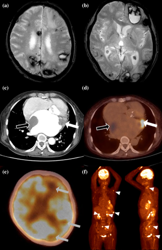
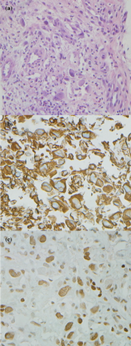
A 65-year-old, previously healthy woman complained of headache developed a month ago. Neurologic examination revealed right hemianopia. In brain magnetic resonance imaging (MRI), multiple scattered hemorrhages were found in both cerebral hemispheres (Fig. 1a,b). Most of them contained cerebromalatic change without enhancement, suggesting simultaneous hemorrhagic embolisms. On the chest computed tomography, two discrete large masses in the dilated left atrium were found; a large hypo-attenuated, nonenhancing mass along the right wall, and another hypo-attenuated, heterogeneously enhancing mass in the appendage (Fig. 1c). Transthoracic echocardiograph revealed left atrial wall mass as a thrombi, and the distinct mass with spontaneous echo-contrast in the appendage. On whole-body positron emission tomography (PET), the mass in the appendage was hypermetabolic with maximal standardized uptake value (SUV) 7·5, suggesting a malignant tumor. The left atrial thrombus and multiple cerebral lesions homogeneously appeared as metabolic defects. Additionally, there were multiple hyper-uptakes in bilateral adrenal glands, vertebras, left femur, scapula, and ninth-rib with maximum SUV 10·6, suggesting metastasis (Fig. 1d–e). Biopsy performed at the rib was confirmed as a high-grade angiosarcoma with positive Friend leukemia integration 1 (FLI1) (Fig. 2).

Images of the multiple cerebral hemorrhages and cardiac angiosarcoma. Gradient-echo images (a–b) show multiple hemorrhages of similar stages, scattered in bilateral cerebral cortexes, which were hypometabolic (dashed arrows, e). Chest computed tomography and whole-body positron emission tomography reveals a large thrombus (hollow arrows), and another heterogeneously enhancing, hypermetabolic tumor (white arrows, c–d). There are hypermetabolic bone uptakes with maximal standardized uptake value (SUV) 10·6 in the sacrum (arrowheads, f).

Photomicrographs of the tumor. Photomicrographs revealed pleomorphism, hyperchromasia, and irregular vascular spaces lined by malignant endothelial cells (a, H&E 10 × 20). Cytoplasm is stained with vimentin (b) and nucleus with Friend leukemia integration 1 (c).
Although rare, cardiac angiosarcoma is mostly located in the right atrium presenting as multiple pulmonary hemorrhages 1. However, in this case, angiosarcoma situated in the left atrium might have provoked multiple cerebral hemorrhagic embolisms. Furthermore, FLI1, a marker of angiosarcoma 2, might have a pathophysiologic role. FLI1 promotes angiogenesis, proliferation/differentiation of megakaryocytes 3. Therefore, FLI1 overexpression in the left atrium might have facilitated formation of a large thrombus and consecutive multiple embolisms. Accompanying hemorrhagic transformation might have resulted from ischemia and aberrant local hemato-angiogenesis by the tumor component of the emboli.


