Radial sclerosing lesions found on core needle biopsy: excision can be safely avoided
Abstract
Aims
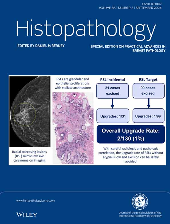
Radial sclerosing lesions (RSLs) are benign breast lesions composed of glandular and epithelial proliferations with stellate architecture and fibro-elastotic stroma, which can mimic invasive carcinoma on imaging. Surgical management following a core biopsy diagnosis of RSLs remains controversial.
Methods and results
We retrospectively identified core biopsies with RSLs without atypia who underwent subsequent surgical excision between 2015 and 2021. All core biopsy slides were reviewed to confirm the diagnosis. Imaging was reviewed to determine radiological–pathological concordance. An upgrade was defined as invasive carcinoma or ductal carcinoma in situ (DCIS) in the excision. The final cohort consisted of 130 core biopsies from 124 women (median age = 52 years, range = 27–76). The imaging modality was mammogram in 52 (40%) cases, MRI in 52 (40%) and ultrasound in 26 (20%). One hundred and seven (82%) core biopsies were vacuum-assisted and 23 (18%) were ultrasound-guided without vacuum assistance. The median lesion size on imaging was 9 mm (range = 2–41). Overall, two (1%) cases were upgraded at excision, including one microinvasive lobular carcinoma and one 2 mm focus of invasive mammary carcinoma with associated DCIS. In both cases, the upgraded foci of carcinoma were not closely associated with the biopsy site and were considered incidental upgrades.
Conclusions
This study adds to the body of literature supporting observation, rather than routine excision of radial sclerosing lesions without atypia.
Graphical Abstract
Radial sclerosing lesions (RSLs) are benign breast lesions which can mimic invasive carcinoma on imaging. Surgical management following a biopsy diagnosis of RSL remains controversial. We show that the likelihood of upgrade to carcinoma is low (1%), and thus close imaging surveillance is appropriate management for these patients.

Conflicts of interest
AG consulted for Paige; AI; MM receives honoraria from Roche and Exact Sciences. All other authors declare no relevant financial or non-financial relationships.
Open Research
Data availability statement
Data sharing not applicable to this article as no datasets were generated or analysed during the current study.