Volume 55, Issue 12 pp. 1926-1933
Full-Length Original Research

“Subtotal” hemispherectomy in children with intractable focal epilepsy

Harry T. Chugani

Corresponding Author

Harry T. Chugani

Department of Pediatrics, Wayne State University School of Medicine, Children's Hospital of Michigan, Detroit Medical Center, Detroit, Michigan, U.S.A

Department of Neurology, Wayne State University School of Medicine, Children's Hospital of Michigan, Detroit Medical Center, Detroit, Michigan, U.S.A

Address correspondence to Harry T. Chugani, Division of Pediatric Neurology, Children's Hospital of Michigan, 3901 Beaubien Blvd, Detroit, MI 48201, U.S.A. E-mail: [email protected]Search for more papers by this author
Eishi Asano

Eishi Asano

Department of Pediatrics, Wayne State University School of Medicine, Children's Hospital of Michigan, Detroit Medical Center, Detroit, Michigan, U.S.A

Department of Neurology, Wayne State University School of Medicine, Children's Hospital of Michigan, Detroit Medical Center, Detroit, Michigan, U.S.A

Search for more papers by this author
Csaba Juhász

Csaba Juhász

Department of Pediatrics, Wayne State University School of Medicine, Children's Hospital of Michigan, Detroit Medical Center, Detroit, Michigan, U.S.A

Department of Neurology, Wayne State University School of Medicine, Children's Hospital of Michigan, Detroit Medical Center, Detroit, Michigan, U.S.A

Search for more papers by this author
Ajay Kumar

Ajay Kumar

Department of Pediatrics, Wayne State University School of Medicine, Children's Hospital of Michigan, Detroit Medical Center, Detroit, Michigan, U.S.A

Department of Neurology, Wayne State University School of Medicine, Children's Hospital of Michigan, Detroit Medical Center, Detroit, Michigan, U.S.A

Department of Radiology, Wayne State University School of Medicine, Children's Hospital of Michigan, Detroit Medical Center, Detroit, Michigan, U.S.A

Search for more papers by this author
William J. Kupsky

William J. Kupsky

Department of Pathology, Wayne State University School of Medicine, Children's Hospital of Michigan, Detroit Medical Center, Detroit, Michigan, U.S.A

Search for more papers by this author
Sandeep Sood

Sandeep Sood

Department of Neurosurgery, Wayne State University School of Medicine, Children's Hospital of Michigan, Detroit Medical Center, Detroit, Michigan, U.S.A

Search for more papers by this author
First published: 03 November 2014
Citations: 34

Summary

Objective

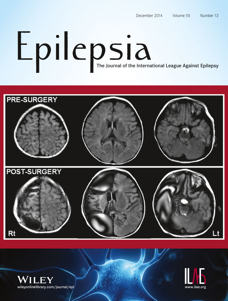
Cortical resections in epilepsy surgery tend to be larger in children, compared to adults, partly due to underlying pathology. Some children show unilateral multifocal seizure onsets involving much of the hemisphere. If there were a significant hemiparesis present, hemispherectomy would be the procedure of choice. Otherwise, it is preferable to spare the primary sensorimotor cortex. We report the results of “subtotal” hemispherectomy in 23 children.

Methods

All children (ages 1 year and 4 months to 14 years and 2 months) were operated on between 2001 and 2013 at Children's Hospital of Michigan (Detroit). Patients were evaluated with scalp video–electroencephalography (EEG), magnetic resonance imaging (MRI), 18F-fluorodeoxyglucose–positron emission tomography (FDG-PET) scans, and neuropsychological assessments when applicable. Subsequently, each case was discussed in a multidisciplinary epilepsy surgery conference, and a consensus was reached pertaining to candidacy for surgery and optimum surgical approach. The actual extent of resection was based on the results from subdural electrocorticography (ECoG) monitoring. The surgical outcome is based on International League Against Epilepsy (ILAE) classification (class 1–6).

Results

Among the 23 patients, 11 had epileptic spasms as their major seizure type; these were associated with focal seizures in 3 children. MRI showed focal abnormalities in 12 children. FDG-PET was abnormal in all but one subject. All except two children underwent chronic subdural ECoG. Multiple subpial transections were performed over the sensorimotor cortex in three subjects. On histopathology, various malformations were seen in 9 subjects; the remainder showed gliosis alone (n = 12), porencephaly (n = 1), and gliosis with microglial activation (n = 1). Follow-up ranged from 13 to 157 months (mean = 65 months). Outcomes consisted of class 1 (n = 17, 74%), class 2 (n = 2), class 3 (n = 1), class 4 (n = 1), and class 5 (n = 2).

Significance

Extensive unilateral resections sparing only sensorimotor cortex can be performed with excellent results in seizure control. Even with the presence of widespread unilateral epileptogenicity or anatomic/functional imaging abnormalities, complete hemispherectomy can often be avoided, particularly when there is little hemiparesis.

The full text of this article hosted at iucr.org is unavailable due to technical difficulties.

click me