Oligomeric α-synuclein and β-amyloid variants as potential biomarkers for Parkinson's and Alzheimer's diseases
Abstract
Oligomeric forms of α-synuclein and β-amyloid are toxic protein variants that are thought to contribute to the onset and progression of Parkinson's disease (PD) and Alzheimer's disease (AD), respectively. The detection of toxic variants in human cerebrospinal fluid (CSF) and blood has great promise for facilitating early and accurate diagnoses of these devastating diseases. Two hurdles that have impeded the use of these protein variants as biomarkers are the availability of reagents that can bind the different variants and a sensitive assay to detect their very low concentrations. We previously isolated antibody-based reagents that selectively bind two different oligomeric variants of α-synuclein and two of β-amyloid, and developed a phage-based capture enzyme-linked immunosorbent assay (ELISA) with subfemtomolar sensitivity to quantify their presence. Here, we used these reagents to show that these oligomeric α-synuclein variants are preferentially present in PD brain tissue, CSF and serum, and that the oligomeric β-amyloid variants are preferentially present in AD brain tissue, CSF, and serum. Some AD samples also had α-synuclein pathology and some PD samples also had β-amyloid pathology, and, very intriguingly, these PD cases also had a history of dementia. Detection of different oligomeric α-synuclein and β-amyloid species is an effective method for identifying tissue, CSF and sera from PD and AD samples, respectively, and samples that also contained early stages of other protein pathologies, indicating their potential value as blood-based biomarkers for neurodegenerative diseases.
Introduction
Protein aggregation is a common feature in the progression of many neurological disorders, including Alzheimer's disease (AD) and Parkinson's disease (PD) (Tsigelny et al., 2008; Crews & Masliah, 2010; Guerrero-Muñoz et al., 2014). In diseased brain, monomeric proteins can assemble into different aggregates, including smaller oligomers and larger protofibrillar and fibrillar assemblies (Guerrero-Muñoz et al., 2014). Although fibrils constitute the primary pathological diagnostic feature, recent research suggests that oligomers are the most toxic species (Guerrero-Muñoz et al., 2014). Various α-synuclein oligomers were shown to play toxic roles in PD (Emadi et al., 2007, 2009; Halliday & McCann, 2008; Lynch et al., 2008; Wang et al., 2010; Sierks et al., 2011; Chaari et al., 2013; Cox et al., 2014), a disorder resulting in resting tremors, posture instability, and rigidity (Dauer & Przedborski, 2003). α-Synuclein is produced in healthy neurons as a monomeric 14-kDa protein; however, in diseased tissue, it assembles into large fibrillar aggregates that are the primary constituents of Lewy bodies (LBs) and Lewy neurites, the pathological hallmarks of PD (Spillantini et al., 1997, 1998). There is also a reduction in the number of dopaminergic neurons in the substantia nigra pars compacta in PD brains (Martin & Teismann, 2009). Alternatively, aggregation of β-amyloid and tau is implicated in AD, a neurodegenerative disease that causes cognitive deficits and altered behaviour (Reitz & Mayeux, 2014). Although β-amyloid and tau fibrils are, respectively, the primary constituents of the amyloid plaques and neurofibrillary tangles (Hoozemans et al., 2006), various small oligomers of both have been implicated in the onset and progression of AD (Klein et al., 2001; Sinjoanu et al., 2008; Zhou et al., 2012; Crimins et al., 2013; Park et al., 2013; Tian et al., 2013; Yang et al., 2013; Pooler et al., 2014; Salvadores et al., 2014; Wischik et al., 2014). The relative concentrations of β-amyloid 42 and 40, and of phosphorylated and total tau, are currently used as AD biomarkers. The β-amyloid 42 form and hyperphosphorylated tau variants are both more prone to aggregation (Di Carlo et al., 2012; Moreth et al., 2013). The selective detection of specific toxic oligomeric protein variants may improve the diagnosis of these neurodegenerative diseases.
We developed an in vitro atomic force microscopy-based biopanning protocol that enables us to generate single-chain variable fragments (scFvs) from phage display libraries that selectively bind disease-related protein variants (Kasturirangan et al., 2013; Williams et al., 2015b). We previously isolated the 10H and D5 scFvs, which recognize two morphologically distinct α-synuclein oligomers and selectively bind postmortem human PD brain tissue (Emadi et al., 2007, 2009). We also previously isolated the A4 and C6T scFvs, which recognize morphologically distinct β-amyloid oligomers and selectively bind postmortem human AD brain tissue (Zameer et al., 2008; Kasturirangan et al., 2012, 2013). Here, we used these scFvs in conjunction with a previously developed phage capture enzyme-linked immunosorbent assay (ELISA) to assess oligomeric protein contents in human samples (Williams et al., 2015a). The protein variant-selective scFvs are used to capture the target antigen, and a phage-displayed version of a second scFv that is protein-specific, but not morphology-specific, is used as the detection antibody. This assay provides two separate methods with which to validate the identity of the target antigen: first, the specificity of the capture scFv, which binds only a specific variant of the target antigen; and second, the detection antibody, which binds a different epitope of the target antigen. In our study, we employed this capture ELISA to characterize postmortem brain tissue, cerebrospinal fluid (CSF) and serum samples from patients with pathologically confirmed AD and PD, and age-matched cognitively normal controls (NDs), to demonstrate the potential value of specific oligomeric protein variants as blood-based biomarkers for neurodegenerative diseases.
Materials and methods
Human samples
Postmortem human brain tissue, serum and CSF samples from patients with pathologically confirmed PD or AD, as well as age-matched NDs, were provided by T. Beach (Brain and Body Donation Program at Banner/Sun Health Research Institute) (Beach et al., 2008, 2015). Samples were obtained from deceased elderly subjects who had volunteered for the Banner Sun Health Research Institute Brain and Body Donation Program. All enrolled subjects signed an Institutional Review Board-approved informed consent form, allowing both clinical assessments during life and several options for brain and/or bodily organ donation after death. Brain tissue samples from nine different PD, six AD and five ND cases were obtained. Table 1 summarizes the characteristics of the utilized human subjects. The classification of PD samples is based on the unified staging system for LB disorders (Beach et al., 2009). Serum samples from eight of the nine PD cases, all six AD cases, and four of the five ND cases, and CSF samples from six of the nine PD cases, all six AD cases, and four of the five ND cases, were also available for analysis.
| Samples | Gender | Age (years) | PMI (h) | Neurological diagnosis (years) | Dementia (years) | Plaque density | Braak score | Unified LB stage | Pathology summary |
|---|---|---|---|---|---|---|---|---|---|
| PD 1 | F | 80 | 7.5 | 11 | – | Moderate | III | III. Brainstem/limbic | PD; microscopic changes of AD but insufficient for diagnosis |
| PD 2 | F | 82 | 2.75 | 16 | Moderate | IV | III. Brainstem/limbic | PD; moderate microscopic lesions of AD (moderate plaques) | |
| PD 3 | M | 85 | 2.16 | 6 | Moderate | IV | IIb. Limbic predominant | PD; microscopic changes of AD | |
| PD 4 | M | 72 | 10 | 17 | Sparse | II | IIb. Limbic predominant | PD | |
| PD 5 | M | 70 | 1.83 | 12 | 3 | Zero | III | III. Brainstem/limbic | PD; dementia (history) |
| PD 6 | F | 73 | 2.16 | 29 | 6 | Sparse | III | IV. Neocortical | PD; dementia (history) |
| PD 7 | M | 75 | 2.25 | 21 | 1 | Zero | III | IV. Neocortical | PD; dementia (history) |
| PD 8 | M | 77 | 4 | 11 | Sparse | III | IV. Neocortical | PD | |
| PD 9 | F | 78 | 3.5 | 16 | 6 | Sparse | III | IV. Neocortical | PD; dementia (clinical history) |
| AD 1 | F | 82 | 4.5 | 9 | 9 | Moderate | IV | 0. No LBs | AD |
| AD 2 | F | 93 | 1.4 | 4 | 4 | Moderate | IV | 0. No LBs | AD |
| AD 3 | M | 94 | 3 | 14 | 14 | Moderate | IV | 0. No LBs | AD |
| AD 4 | M | 79 | 3.92 | 7 | 7 | Frequent | V | 0. No LB | AD |
| AD 5 | F | 96 | 3 | 21 | 5 | Frequent | VI | 0. No LBs | AD |
| AD 6 | F | 87 | 3 | 5 | 5 | Frequent | V | 0. No LBs | AD |
| ND 1 | F | 87 | 2.83 | Zero | II | 0. No LBs | Control | ||
| ND 2 | F | 91 | 2 | Zero | II | 0. No LBs | Control; entorhinal cortex neurofibrillary tangles consistent with normal aging | ||
| ND 3 | F | 83 | 4.83 | Moderate | II | 0. No LBs | Control; microscopic changes of AD but insufficient for diagnosis | ||
| ND 4 | M | 89 | 2.5 | Moderate | II | 0. No LBs | Control; microscopic changes of AD but insufficient for diagnosis | ||
| ND 5 | M | 86 | 2.75 | Moderate | II | 0. No LBs | Control; microscopic changes of AD but insufficient for diagnosis |
- F, female; M, male; PMI, postmortem interval.
Brain tissue homogenization
Brain tissue from the middle temporal gyrus was homogenized for immunoassay assessment as previously described (Williams et al., 2015a). Briefly, each sample was chopped and resuspended in homogenization buffer containing 50 mm Tris and 5 mm EDTA at pH 7.0. Sonication was then completed at 50% amplitude with 10-s pulse on and 15-s pulse off for a total of 5 min. After centrifugation at ∼15 500 g for 20 min, the supernatants were stored at −80 °C for future use.
ScFvs
The 10H and D5 scFvs were previously isolated against two morphologically distinct α-synuclein oligomeric variants (Emadi et al., 2007, 2009), whereby D5 bound in vitro generated α-synuclein aggregates with molecular masses of 29 kDa and 56 kDa, corresponding to dimers and tetramers (Emadi et al., 2007), and 10H bound in vitro generated aggregates with molecular masses of 42 kDa and ~ 80 kDa, corresponding to trimers and hexamers (Emadi et al., 2009). The corresponding molecular masses of the oligomeric α-synuclein variants bound by 10H and D5 are shown in Fig. S1A [adapted from Emadi et al. (2007, 2009), Kasturirangan et al. (2013), and Xin et al. (2015)]. These scFvs did not show cross-reactivity with in vitro-generated oligomeric β-amyloid aggregates (Emadi et al., 2009), but do selectively block the cytotoxicity of PD brain homogenates as compared with control brain homogenates (Xin et al., 2015). A third anti-α-synuclein scFv, D10, was previously shown to bind multiple morphological types of α-synuclein, including monomeric, oligomeric and fibrillar forms (Zhou et al., 2004; Emadi et al., 2009), and was used here as the detection antibody in the capture ELISA (Fig. S1A). As 10H and D5 recognize different oligomeric α-synuclein species, the α-synuclein complexes formed by the use of 10H in conjunction with D10 and D5 in conjunction with D10 should differ. Therefore, analysis of each human sample with both scFvs increases the likelihood of identifying patients with oligomeric α-synuclein pathology, especially as the level of each oligomeric α-synuclein species at the time of testing may vary from person to person and with the stage of the disease.
The A4 and C6T scFvs were previously isolated against morphologically distinct oligomeric variants of β-amyloid, A4 binding an in vitro-generated aggregate, and C6T binding an AD brain-derived aggregate (Zameer et al., 2008; Kasturirangan et al., 2012, 2013). In indirect ELISAs, both A4 phage and scFv bind oligomeric β-amyloid but not monomeric or fibrillar structures (Zameer et al., 2008). A4 also protected human neuroblastoma cells against neuronal toxicity induced by oligomeric β-amyloid (Zameer et al., 2008). Western blot analysis with supernatant from hAPP-overexpressing 7PA2 cells showed C6T binding to an ~ 11-kDa protein. When brain tissues from control and triple transgenic AD mice were probed with C6T, it preferentially reacted with the tissue from triple transgenic AD mice (Kasturirangan et al., 2013). Comparison of the binding specificities of A4 and C6T via AFM show that A4 selectively binds synthetically derived β-amyloid oligomers, whereas C6T selectively binds AD brain-derived β-amyloid oligomers [Fig. S1B; adapted from Kasturirangan et al. (2013)]. These results highlighted the difference in the target antigens of A4 and C6T. The H1v2 scFv, which recognizes all morphological types of β-amyloid (Yuan et al., 2006), was used as the detection reagent. As A4 and C6T bind different oligomeric β-amyloid variants (Kasturirangan et al., 2013), the use of both scFvs with H1v2 should increase the likelihood of identifying samples containing oligomeric β-amyloid, similarly to above rationale for identifying oligomeric α-synuclein with 10H and D5.
ScFv production
To produce the scFvs, HB2151 cells containing the scFv plasmids were cultured overnight in 2xYT, 0.1% glucose and ampicillin at 37 °C with shaking. Ten millilitres of this overnight culture was added to 1 L of fresh medium, and incubated at 37 °C with shaking until the optical density at 600 nm was 0.8. After the addition of isopropyl β-d-1-thiogalactopyranoside, the flask was transferred to 30 °C. On the following day, the supernatant was concentrated with a tangential flow filter (10-kDa filter; Millipore). The purified scFv was then isolated by means of fast protein liquid chromatography with a protein A–Sepharose column (GE Healthcare, Piscataway, NJ, USA). C6T was purified with nickel nitrilotriacetic acid Sepharose beads (Qiagen, Valencia, CA, USA) and imidazole elution as previously described (Kasturirangan et al., 2013). Following dialysis, the scFvs were stored at −20 °C. Sodium dodecylsulphate polyacrylamide gel electrophoresis and western blot analysis were used to confirm the purity and presence of the antibodies. Antibody concentrations were determined with the bicinchoninic acid assay (Pierce, Rockford, IL, USA).
Phage production
The capture ELISA protocol employed here utilizes the morphology-specific scFvs as the capture antibodies and a protein-specific scFv as a detection antibody. The protein-specific scFv is expressed on a phage particle, where the phage coat proteins can be biotinylated to greatly amplify the signal (Williams et al., 2015a). Here, we used a phage-displayed version of D10, which recognizes all morphological types of α-synuclein (Zhou et al., 2004; Emadi et al., 2009), for detection of α-synuclein aggregates, and H1v2, which recognizes all morphological types of β-amyloid (Yuan et al., 2006), for detection of β-amyloid aggregates. The D10 and H1v2 phage particles were produced as previously described (MRC Laboratory of Molecular Biology and the MRC Centre for Protein Engineering, Cambridge, UK; http://www.lifesciences.sourcebioscience.com/media/143421/tomlinsonij.pdf). Basically, an overnight culture of the TG1 cells containing the scFv plasmid of interest was diluted 1 : 100 and cultured in 2xYT containing 100 μg/mL ampicillin and 1% glucose until the optical density at 600 nm was between 0.4 and 0.6. After a 30-min incubation with 2 × 10¹¹ KM13 helper phage or hyperphage (Progen, Germany), the culture was centrifuged at 3000 g for 10 min and resuspended in 2xYT containing 100 μg/mL ampicillin, 50 μg/mL kanamycin, and 0.1% glucose. The cells were cultured at 30 °C overnight, and on the following day they were centrifuged for 30 min at 3000 g. The supernatant was combined with PEG/NaCl, and incubated for 1 h on ice. The solution was again centrifuged at 3000 g for 30 min, and the pellet was saved. The pellet was resuspended in phosphate-buffered saline (PBS), and incubated on ice for 1 h. Following this incubation step, the solution was centrifuged at 11 600 g for 10 min, and the supernatant was stored at −80 °C.
The concentration of the phage was estimated with the bicinchoninic acid assay. Once produced, the phages were biotinylated by use of the EZ-Link Pentylamine-Biotinylation kit (Thermo Scientific, Rockford, IL, USA), essentially as described previously (Williams et al., 2015a).
Phage capture ELISA
The phage capture ELISA was performed as previously described (Williams et al., 2015a). Basically, the capture morphology-specific scFvs (D5 and 10H for α-synuclein, and A4 and C6T for β-amyloid) (Emadi et al., 2007, 2009; Zameer et al., 2008; Kasturirangan et al., 2012, 2013) were bound to the wells of a high-binding ELISA plate (Costar) for 30–60 min at 37 °C. After three washes with PBS with 0.1% Tween-20, the remaining unbound sites were blocked with 2% milk. Following three washes, the antigen was added. Brain tissue was diluted to 100 μg/mL, and serum and CSF samples were diluted 1 : 100 v/v. The plates were again washed three times, and incubated with 200 ng/mL of 40 mmol carboxyl biotinylated detection phage. The plates were then washed four times, and incubated with a 1 : 1000 dilution of avidin–horseradish peroxidase (Sigma-Aldrich, St Louis, MO, USA). After four more washes, the binding intensities were detected with the SuperSignal ELISA Femto Maximum Sensitivity Substrate (Thermo Scientific) kit. The signal intensities were quantified with a Wallac Victor2 microplate reader following development periods of 1 and 20 min.
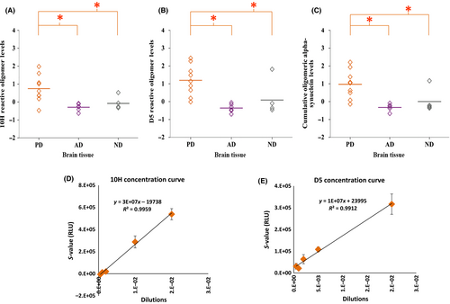
As oligomeric proteins are generally very ‘sticky’, sample collection and storage conditions need to be controlled, because this can influence the reproducibility of results. Most of the ELISA trials were conducted more than once, depending on sample availability. A representative example of sample reproducibility is shown in Fig. S2, which displays 10H's reactivity with brain tissue. The 10H and D5 calibration curves in Fig. 1D and E, respectively, are also based on the average of three trials. The R2 value is fairly high, at 0.9959 for 10H and 0.9912 for D5, and the error bars are quite small at most data points.
Statistical analysis
The raw ELISA data for each sample were divided by the PBS control data to obtain a signal ratio. The mean plus two standard deviations (SDs) was calculated for the control group, and this value was subtracted from each sample to determine positive cases (except for the CSF tested with A4 and C6T, where 1.5 SD was utilized). These results were plotted on graphs generated with the IBM spss statistics 22 program. Significant differences between the PD, AD and ND groups for the four test scFvs were evaluated with one-way anova and LSD post hoc analyses made available through spss. Results were considered to be significant at P < 0.05. The mean and SDs for each group were also reported. The other graphs and tables were created with Microsoft Excel 2010. Figures were prepared for publication using the Adobe Reader XI and Adobe Photoshop Elements 8.0 programs. The results in Fig. S2 are based on dividing the test samples by the mean plus two SDs of the control group.
For the data presented in Tables 2 and 3, each sample signal ratio was divided by the mean plus two SDs of the controls. Sample signals greater than two SDs from the controls are indicated by a ‘+’ sign, with additional ‘+’ signs being used for each additional two SD increase in the signal from controls. The sensitivity and specificity for the PD vs. ND cases and AD vs. ND cases were calculated with the use of medcalc for the brain tissue, CSF and serum results. Power analyses was carried out with the G*Power 3.1.4 analysis tool to determine the number of samples required in future studies to obtain 80% power. A one-tail a priori t-test examining the difference between two independent means was calculated for each scFv, with an alpha level of 0.05, power of 0.8, and an allocation ratio of 1, and the effect size was computed by using the mean and SDs of the test and control groups from the current study.
| Samples | Brain tissue | CSF | Sera | |||
|---|---|---|---|---|---|---|
| 10H | D5 | 10H | D5 | 10H | D5 | |
| PD 1 | + | – | n/a | n/a | – | – |
| PD 2 | – | + | +++ | + | – | ++++ |
| PD 3 | ++ | ++ | + | ++ | – | ++++ |
| PD 4 | +++ | +++++ | n/a | n/a | n/a | n/a |
| PD 5 | ++++++ | ++++++ | + | – | – | – |
| PD 6 | ++ | +++ | + | + | – | +++ |
| PD 7 | ++++++ | ++++++ | n/a | n/a | + | + |
| PD 8 | +++++ | +++++ | + | + | + | ++++ |
| PD 9 | +++++ | ++++ | ++ | – | + | ++++++ |
| AD 1 | – | – | + | – | – | – |
| AD 2 | – | – | – | – | – | – |
| AD 3 | – | – | – | + | – | ++ |
| AD 4 | – | – | – | – | – | – |
| AD 5 | – | – | – | – | – | – |
| AD 6 | – | – | + | – | – | ++ |
| ND 1 | – | – | – | – | – | – |
| ND 2 | +++ | ++++++ | – | – | – | – |
| ND 3 | – | – | – | – | – | – |
| ND 4 | – | – | – | – | – | – |
| ND 5 | – | – | n/a | n/a | n/a | n/a |
| Samples | Brain tissue | CSF | Sera | |||
|---|---|---|---|---|---|---|
| A4 | C6T | A4 | C6T | A4 | C6T | |
| PD 1 | +++++ | – | n/a | n/a | – | – |
| PD 2 | – | – | – | – | – | – |
| PD 3 | +++ | – | – | – | – | – |
| PD 4 | – | – | n/a | n/a | n/a | n/a |
| PD 5 | – | – | n/a | n/a | ++++++ | – |
| PD 6 | – | – | – | – | + | – |
| PD 7 | + | – | n/a | n/a | – | – |
| PD 8 | + | – | – | – | – | – |
| PD 9 | – | – | n/a | n/a | – | – |
| AD 1 | ++++++ | – | – | + | +++ | ++ |
| AD 2 | ++++ | – | + | + | ++++ | ++ |
| AD 3 | – | + | + | – | ++ | – |
| AD 4 | ++ | – | + | – | ++++++ | – |
| AD 5 | – | + | – | – | + | +++ |
| AD 6 | – | ++++++ | ++ | – | +++++ | – |
| ND 1 | – | – | – | – | – | – |
| ND 2 | – | – | – | – | – | – |
| ND 3 | – | – | – | – | – | – |
| ND 4 | – | – | – | – | – | – |
| ND 5 | – | – | n/a | n/a | n/a | n/a |
Results
Human brain tissue samples
Oligomeric α-synuclein
Homogenized human brain tissue samples from PD, AD and ND cases were analysed for the presence of two different toxic oligomeric α-synuclein aggregates by the use of 10H and D5. We found significant differences between the groups by using one-way anova and LSD post hoc analyses. One-way anova revealed that the levels of 10H-reactive (F2,16 = 8.157, P = 0.004) and D5-reactive (F2,16 = 14.488, P = 0.000) oligomeric α-synuclein were both statistically different between the groups (Fig. 1A and B). LSD post hoc analysis indicated that the PD samples (mean = 0.746, SD = 0.753) were statistically different from both the AD samples (mean = −0.294, SD = 0.205, P = 0.002) and the ND samples (mean = −0.231, SD = 0.116, P = 0.009) regarding reactivity with 10H. Similarly, the PD samples (mean = 1.196, SD = 0.860) reacted significantly more with D5 than the AD samples (mean = −0.361, SD = 0.238, P = 0.001) and the ND samples (mean = −0.345, SD = 0.173, P = 0.019). The mean cumulative oligomeric α-synuclein level (the sum of 10H and D5) was also significantly different between the groups (F2,16 = 11.928, P = 0.001), and LSD post hoc analyses indicated that the difference was between the PD samples (mean = 0.971, SD = 0.792) and the AD samples (mean = −0.328, SD = 0.197, P = 0.001) and between the PD samples and the ND samples (mean = −0.288, SD = 0.066, P = 0.002) (Fig. 1C). As it is extremely difficult to stably purify individual oligomeric protein variants to homogeneity, it is not feasible to create calibration curves to determine the concentrations of the different oligomeric protein species present in different samples. As an alternative, we created dilution curves from a known sample as a means to calculate relative concentrations. Dilutions of one of the PD brain tissue homogenates were used to create the linear calibration curves for the 10H-reactive (Fig. 1D) and D5-reactive (Fig. 1E) oligomeric α-synuclein variants with a good linear fit as determined by R2 values.
Oligomeric β-amyloid
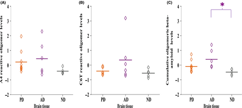
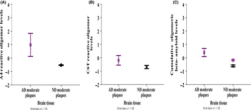
Similarly, we analysed the homogenized human brain tissue samples for the presence of toxic oligomeric β-amyloid aggregates by using A4 and C6T. The mean level of A4-reactive oligomeric β-amyloid variants was highest in the AD samples (mean = 0.439, SD = 1.152), lower in the PD samples (mean = 0.214, SD = 0.768), and lowest in the ND samples (mean = −0.406, SD = 0.203) (Fig. 2A). Similarly with C6T, the AD samples (mean = 0.347, SD = 1.466) were most reactive, followed by the PD samples (mean = −0.407, SD = 0.227), and then the ND samples (mean = −0.550, SD = 0.275) (Fig. 2B). The mean cumulative level of A4-reactive and C6T-reactive oligomeric β-amyloid variants was significantly different between the groups (F2,17 = 5.125, P = 0.018) (Fig. 2C). LSD post hoc analyses revealed that the AD samples (mean = 0.393, SD = 0.635) were significantly more reactive than the ND samples (mean = −0.478, SD = 0.201, P = 0.006), and more reactive compared to the PD samples though just missing the set significance level of P < 0.05 (mean = −0.097, SD = 0.406, P = 0.056). The AD samples contained three cases possessing moderate plaque loads and three possessing high plaque loads; the ND samples also contained three cases possessing moderate plaques and two possessing either no or only light plaques. We compared the three AD brain samples possessing moderate plaque loads with the three ND brain samples possessing moderate plaque loads to determine whether the presence of oligomeric β-amyloid could discriminate between these two groups with similar amyloid plaque loads. A4-reactive oligomeric β-amyloid levels were higher in the AD samples (mean = 0.977, SD = 1.482) than in the ND samples (mean = −0.528, SD = 0.041) (Fig. 3A). As with C6T, the AD samples (mean = −0.192, SD = 0.620) were more reactive than the ND samples (mean = −0.691, SD = 0.211) (Fig. 3B). The cumulative level of A4-reactive and C6T-reactive oligomeric β-amyloid was significantly higher in the AD samples (mean = 0.393, SD = 0.528) than in the ND samples (mean = −0.609, SD = 0.125) (F1,4 = 10.246, P = 0.033) (Fig. 3C).
Human CSF samples
Oligomeric α-synuclein
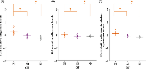
Next, we analysed the available corresponding CSF samples from the PD, AD and ND cases described above for the presence of 10H-reactive and D5-reactive oligomeric α-synuclein. Likewise, the groups were evaluated with one-way anova and LSD post hoc analyses for any resulting statistically significant differences. anova revealed significant differences in the levels of 10H-reactive (F2,13 = 10.407, P = 0.002), D5-reactive (F2,13 = 3.799, P = 0.05) and cumulative 10H-reactive and D5-reactive (F2,13 = 14.637, P = 0.000) oligomeric α-synuclein between the groups (Fig. 4A–C). LSD post hoc analyses revealed that the PD samples (mean = 0.241, SD = 0.221) contained significantly higher levels of 10H-reactive oligomeric α-synuclein than the AD samples (mean = −0.057, SD = 0.120, P = 0.008) and the ND samples (mean = −0.226, SD = 0.113, P = 0.001). Similarly, with D5 the PD samples (mean = 0.014, SD = 0.090) were more reactive than the AD samples (mean = −0.010, SD = 0.088, P = 0.032) and the ND samples (mean = −0.107, SD = 0.054, P = 0.040). The mean cumulative 10H-reactive and D5-reactive oligomeric α-synuclein level was also significantly higher in the PD samples (mean = 0.127, SD = 0.133) than in the AD samples (mean = −0.078, SD = 0.046, P = 0.002) and the ND samples (mean = −0.166, SD = 0.043, P = 0.000).
Oligomeric β-amyloid
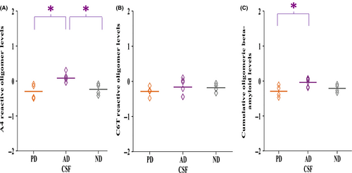
We analysed the CSF samples for the presence of A4-reactive and C6T-reactive oligomeric β-amyloid. Owing to the limited availability of CSF, we were only able to assay four of the PD samples. The level of A4-reactive oligomeric β-amyloid was significantly different between the groups (F2,11 = 7.905, P = 0.007) (Fig. 5A). LSD post hoc analyses showed significantly higher reactivity with the AD samples (mean = 0.083, SD = 0.132) than with both the PD samples (mean = −0.299, SD = 0.214, P = 0.004) and the ND samples (mean = −0.240, SD = 0.160, P = 0.012). With C6T, the average level was higher in the AD samples (mean = −0.163, SD = 0.238) than in the PD samples (mean = −0.286, SD = 0.145) and the ND samples (mean = −0.181, SD = 0.121) (Fig. 5B). The cumulative A4-reactive and C6T-reactive oligomeric β-amyloid level was also significantly different between the groups (F2,11 = 4.839, P = 0.031) (Fig. 5C). The AD samples (mean = −0.040, SD = 0.118) reacted significantly more than the PD samples (mean = −0.293, SD = 0.168, P = 0.013) and almost significantly more than the ND samples (mean = −0.211, SD = 0.110, P = 0.070).
Human serum samples
Oligomeric α-synuclein
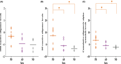
Finally, we analysed the corresponding PD, AD and ND serum samples for the presence of oligomeric α-synuclein by using 10H and D5. Detection in serum involves a less invasive diagnostic process, making serum the preferred test medium in our detection system. The average level of 10H-reactive oligomeric α-synuclein in the serum samples was higher in the PD samples (mean = −0.322, SD = 0.593) than in the AD samples (mean = −0.937, SD = 0.615) and the ND samples (mean = −1.027, SD = 0.513) (Fig. 6A). anova revealed significant differences in the levels of D5-reactive (F2,15 = 4.431, P = 0.031) and cumulative 10H-reactive and D5-reactive (F2,15 = 4.475, P = 0.030) oligomeric α-synuclein between the groups (Fig. 6B and C). LSD post-hoc analyses revealed that the level of D5-reactive oligomeric α-synuclein was significantly higher in the PD samples (mean = 7.217, SD = 8.783) than in the AD samples (mean = −0.984, SD = 4.840, P = 0.038) and the ND samples (mean = −3.540, SD = 1.770, P = 0.019) samples. The same was true for the cumulative level of 10H-reactive and D5-reactive oligomeric α-synuclein in the PD samples (mean = 3.448, SD = 4.638) as compared with the AD samples (mean = −0.960, SD = 2.652, P = 0.036) and the ND samples (mean = −2.283, SD = 0.980, P = 0.019).
Oligomeric β-amyloid
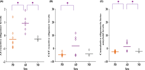
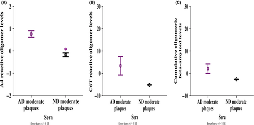
Oligomeric β-amyloid levels in sera were also evaluated and, on average, the levels of A4-reactive oligomeric β-amyloid in the serum samples were significantly different between the AD, PD and ND groups according to anova (F2,15 = 6.992, P = 0.007) (Fig. 7A). LSD post hoc analyses revealed that the AD samples (mean = 0.926, SD = 0.624) contained a higher level of A4-reactive oligomeric β-amyloid than the PD samples (mean = −0.297, SD = 0.787, P = 0.003) and the ND samples (mean = −0.276, SD = 0.138, P = 0.012). The average C6T reactivity was significantly different between the groups (F2,15 = 3.843, P = 0.045): the AD samples (mean = 1.899, SD = 7.854) contained a significantly higher level of oligomeric β-amyloid than the PD samples (mean = −4.833, SD = 1.293, P = 0.018), and an almost significantly higher level than the ND samples (mean = −4.167, SD = 2.084, P = 0.065) (Fig. 7B). Similarly, the mean cumulative level of A4-reactive and C6T-reactive oligomeric β-amyloid in the serum samples was significantly different between the groups (F2,15 = 5.838, P = 0.013) (Fig. 7C). On the basis of LSD post hoc analyses, the AD samples (mean = 1.412, SD = 3.746) had a significantly higher level than the PD samples (mean = −2.565, SD = 0.744, P = 0.005) and the ND samples (mean = −2.222, SD = 1.008, P = 0.025). When we just compared oligomeric β-amyloid levels in the three AD serum samples possessing moderate plaques with those in the three ND samples possessing moderate plaques, the level of A4-reactive oligomeric β-amyloid was significantly higher in the AD samples (mean = 0.758, SD = 0.260) than in the ND samples (mean = −0.173, SD = 0.118) (F1,3 = 20.943, P = 0.020) (Fig. 8A), and the C6T-reactive oligomeric β-amyloid level was also higher in the AD samples (mean = 3.394, SD = 7.192) than in the ND samples (mean = −5.186, SD = 0.057) (Fig. 8B). The mean cumulative A4-reactive and C6T-reactive oligomeric β-amyloid levels were also higher in the AD samples possessing moderate plaques (mean = 2.076, SD = 3.713) than in the ND samples possessing moderate plaques (mean = −2.679, SD = 0.088) (Fig. 8C).
Comparison of cumulative oligomeric α-synuclein and β-amyloid levels
The levels of 10H-reactive and D5-reactive oligomeric α-synuclein and A4-reactive and C6T-reactive oligomeric β-amyloid for each individual human brain tissue, CSF and serum sample are shown in Tables 2 and 3, respectively. The average signal ratio to PBS for each sample was divided by the mean + 2SD of the ND group. Higher oligomeric protein levels are indicated with ‘+’ signs as follows: ‘+’ is an oligomeric protein level greater than the mean + 2SD, ‘++’ is an oligomeric protein level greater than the mean + 4SD, and each additional ‘+’ represents an oligomeric protein level that is an additional 2SD greater than the mean.
Increased levels of 10H-reactive oligomeric α-synuclein were detected in the brain tissue of eight of the nine PD samples and one ND sample (Table 2). The D5 scFv also reacted with eight of the nine PD samples and with the same ND sample that reacted with 10H. Impressively, all nine PD samples were selected between 10H and D5, as the one PD sample not selected by 10H was selected by D5 and vice versa. In CSF, 10H selected all of the tested PD samples, whereas D5 selected four of six PD samples. Therefore, between 10H and D5, all PD samples were identified. Finally, in sera, 10H selected three of the eight PD samples, and D5 selected six of the eight PD samples. Overall, 10H-reactive and D5-reactive oligomeric α-synuclein showed a strong preference for the PD samples as compared with the AD and ND samples.
In brain tissue, A4-reactive oligomeric β-amyloid was detected in three of the AD samples and four of the PD samples, whereas C6T also selected three of the AD samples (Table 3). Together, the A4 and C6T scFvs selected all six AD samples, as the three samples selected by A4 were different from the three selected by C6T. In CSF, A4 selected four of the AD samples, C6T selected two of the AD samples, and, together, A4 and C6T selected five of the six AD samples. In sera, A4-reactive oligomeric β-amyloid was identified in all six AD samples. C6T-reactive oligomeric β-amyloid was present in three AD samples. Therefore, A4 and C6T identified all six AD serum samples.
Mixed protein pathology
With the different anti-oligomeric scFvs, we could distinguish AD, PD and ND cases equally well by using brain tissue, CSF and serum samples, supporting the value of specific protein variants in CSF and/or serum as biomarkers for different neurodegenerative diseases. Further analysis of the CSF and serum samples tested here indicated that mixed pathology was present in several cases, whereby some AD samples also showed reactivity with our anti-oligomeric α-synuclein scFvs, and some PD samples also showed reactivity with our anti-oligomeric β-amyloid scFv. The anti-α-synuclein 10H and D5 scFvs were collectively reactive with three CSF and two serum AD samples (Table 2). The two serum AD samples that showed positive α-synuclein reactivity also showed positive reactivity in the corresponding CSF samples. Similarly, when we performed an analysis for oligomeric β-amyloid, two PD serum samples showed reactivity (Table 3). These results indicate that multiple protein pathologies associated with different neurodegenerative diseases could be present in one individual.
Sensitivity and specificity
To determine the sensitivity and specificity of 10H and D5 for the PD group, we counted cases as positive if the samples showed reactivity with 10H and/or D5, and compared them with the ND samples (Table 4). On analysis of brain tissue samples, all nine PD samples were selected along with one ND sample, yielding 100% sensitivity and 80% specificity. For CSF, all six PD samples and none of the ND samples were selected, yielding 100% sensitivity and 100% specificity. Finally, for sera, six of the eight PD samples and none of the ND samples were selected, yielding 75% sensitivity and 100% specificity. The AD samples were analysed in a similar manner, based on positive reactions with A4 and/or C6T as compared with NDs (Table 4). On analysis of brain tissue samples, all six AD samples and none of the ND samples were selected, yielding 100% sensitivity and 100% specificity. For CSF, five of the six AD samples and none of the ND samples were selected, yielding 83% sensitivity and 100% specificity. Finally, for sera, all six AD samples and none of the ND samples cases were selected, again yielding 100% sensitivity and 100% specificity.
| Sensitivity (%) | Specificity (%) | |
|---|---|---|
| PD brain tissue | 100 | 80 |
| PD CSF | 100 | 100 |
| PD serum | 75 | 100 |
| AD brain tissue | 100 | 100 |
| AD CSF | 83 | 100 |
| AD serum | 100 | 100 |
Discussion
Oligomeric variants of the α-synuclein and β-amyloid proteins have been implicated in the underlying pathology of PD and AD, respectively (Klein et al., 2001; Emadi et al., 2007, 2009; Halliday & McCann, 2008; Lynch et al., 2008; Sinjoanu et al., 2008; Wang et al., 2010; Sierks et al., 2011; Zhou et al., 2012; Chaari et al., 2013; Crimins et al., 2013; Park et al., 2013; Tian et al., 2013; Yang et al., 2013; Cox et al., 2014; Pooler et al., 2014; Salvadores et al., 2014; Wischik et al., 2014). Because of their important roles in these devastating diseases, reagents that can detect specific toxic oligomeric protein variants in human samples have potential value as tools to facilitate the diagnosis of these diseases and to study mechanisms of disease progression. We developed biopanning technology that enables us to isolate scFvs that are reactive with specific protein variants, and a capture ELISA protocol that allows sensitive subfemtomolar detection (Williams et al., 2015a,b). The scFvs 10H and D5 were previously characterized for their specificity for different oligomeric variants of α-synuclein. 10H reacted with in vitro-generated aggregates with molecular masses of 42 kDa and ~ 80 kDa, whereas D5 reacted with in vitro-generated α-synuclein aggregates with molecular masses of 29 kDa and 56 kDa (Emadi et al., 2007, 2009) (Fig. S1A). Detection of multiple oligomeric protein morphological types increases the probability of detecting oligomeric α-synuclein pathology in any given sample. Similarly, A4 and C6T bind different oligomeric variants of β-amyloid, whereby A4 binds an in vitro-generated β-amyloid oligomeric species, and C6T binds an AD brain-derived β-amyloid oligomeric species (Fig. S1B). In this study, we used all four of these morphology-specific scFvs with our phage capture ELISA to analyse postmortem human brain tissue, CSF and serum samples from PD, AD and ND cases to identify samples with oligomeric α-synuclein and/or β-amyloid pathology, in order to distinguish PD cases from controls, AD cases from controls, and PD or AD cases with mixed protein pathology. The use of these morphology-specific scFvs as the capture antibodies and a protein-specific scFv (D10 for α-synuclein; H1v2 for β-amyloid) as a detection antibody provides two different means to ensure that we are selectively detecting α-synuclein or β-amyloid aggregates. First, the capture scFvs were chosen because of their selectivity for the target protein aggregate, and because the scFvs do not show cross-reactivity with other protein aggregates (Emadi et al., 2007, 2009; Zameer et al., 2008; Kasturirangan et al., 2012, 2013). Second, the detection antibody is protein-specific, selectively binding either the α-synuclein (D10) or β-amyloid (H1v2) aggregate already in complex with the capture scFv.
The levels of 10H-reactive and/or D5-reactive oligomeric α-synuclein were statistically greater in the PD samples than in the AD and ND samples (Table 2) of brain homogenates (Fig. 1A, B and C), CSF (Fig. 4A, B and C) and serum (Fig. 6A, B and C). Jointly, 10H and D5 selected all of the PD brain tissue and CSF samples, and six of eight PD serum samples. To determine the sensitivity and specificity of D5 and 10H collectively for PD, the results obtained with the PD samples were compared with those of respective ND samples (Table 4). With brain tissue, our sensitivity was 100% and our specificity was 80% (one ND sample was selected). The sensitivity and specificity were both 100% with CSF, and and they were 75% and 100%, respectively, with serum. Collectively, these results demonstrate that detection of specific oligomeric α-synuclein variants is a powerful tool with which to distinguish PD samples from control samples. Oligomeric α-synuclein has been previously detected in the plasma of PD subjects by the use of undiluted samples (El-Agnaf et al., 2006), providing a precedent for our identification of oligomeric α-synuclein in media such as CSF and serum. Because of the subfemtomolar sensitivity of our capture ELISA (Williams et al., 2015a), we could dilute the CSF and serum samples to 1 : 100 and still obtain significant signal differences. A simple ELISA protocol that can selectively identify PD patients from serum samples has great promise, as it would alleviate painful sample procurement and could facilitate more accurate and widespread diagnosis.
The levels of A4-reactive and C6T-reactive oligomeric β-amyloid clearly distinguish AD brain tissue homogenate (Fig. 2A, B and C), CSF (Fig. 5A, B and C) and serum (Fig. 7A, B and C) samples from the controls and most PD samples. For CSF and sera, the levels of A4-reactive oligomers were statistically higher in the AD samples than in both the PD and ND samples. The cumulative A4-reactive and C6T-reactive oligomeric β-amyloid levels selected all six AD brain tissue samples, five of six CSF samples, and all six serum samples (Table 3). The sensitivity and specificity of A4 and C6T collectively for AD samples as compared with ND samples were also determined. Analysis of both brain tissue and serum samples yielded sensitivity and specificity values of 100% for AD samples as compared with ND samples, and analysis of CSF samples yielded a sensitivity of 83% and specificity of 100% (Table 4). These results provide support for the application of our oligomeric β-amyloid scFvs as potential diagnostic biomarkers for AD pathology. Furthermore, when we compared the levels of oligomeric β-amyloid in AD cases possessing moderate plaque loads with ND cases also possessing moderate plaque loads, we could readily distinguish between these two groups by using either brain tissue or, more impressively, serum samples. These results demonstrate that it may be possible to distinguish between cognitively normal and AD patients with similar plaque loads by using simple blood tests. Additionally, the results provide further evidence for a strong correlation between the presence of specific oligomeric variants of β-amyloid and AD dementia rather than between plaque loads and dementia.
Oligomeric β-amyloid variants have been implicated in the progression of AD (Resende et al., 2008; van Helmond et al., 2009; Klaver et al., 2011; Wisniewski & Goñi, 2014), and we show here that the presence of selected oligomeric β-amyloid variants in serum samples can be used to differentiate AD cases from controls. Similarly oligomeric α-synuclein has been implicated in PD (Emadi et al., 2007, 2009; Halliday & McCann, 2008; Lynch et al., 2008; Wang et al., 2010; Sierks et al., 2011; Chaari et al., 2013; Cox et al., 2014), and we also show that the presence of selected oligomeric α-synuclein variants in CSF or serum can be used to differentiate PD cases from controls. Oligomeric β-amyloid was, however, also detected in two PD serum samples, and oligomeric α-synuclein was detected in three AD CSF and two AD serum samples (Tables 2 and 3). The presence of PD pathology in some AD cases and AD pathology in some PD cases is to be expected, as there is significant overlap between the different neurodegenerative diseases. Interestingly, two of the AD samples contained oligomeric α-synuclein in both CSF and serum samples (Table 2), suggesting that early-stage α-synuclein pathology may be developing in these samples. The presence of α-synuclein-positive LBs was reported in 22% of familial AD cases and in 50–60% of sporadic AD cases, particularly in the amygdala (Lippa et al., 1998; Hamilton, 2000; Wirths & Bayer, 2003; Swirski et al., 2014), so the presence of oligomeric α-synuclein should be expected in a substantial percentage of the AD samples. In a parallel fashion, dementia affects nearly one-third of PD patients (Martí et al., 2007), and there is an increase in cortical Aβ plaque loads in PD with dementia patients (Irwin et al., 2013). Therefore a significant percentage of PD cases should show β-amyloid pathology, as reflected here by the presence of A4-reactive oligomeric β-amyloid in serum. Interestingly, the two PD cases with high oligomeric β-amyloid levels in serum also had a history of dementia, based on postmortem pathological analysis. None of the AD samples analysed here showed the presence of α-synuclein-containing LBs, based on the unified staging system for LB disorders (Beach et al., 2009). However, the presence of oligomeric α-synuclein in two of the AD samples is consistent with the incidence of α-synuclein pathology in AD cases, and suggests that the presence of oligomeric α-synuclein may be a potential early-stage biomarker for α-synuclein pathology, an analysis that will need additional studies using larger sample sizes.
When we analysed oligomeric α-synuclein and β-amyloid contents in our samples, we utilized two scFvs for each protein target to increase the likelihood of binding disease-related variants of the targeted proteins. If a sample does not contain protein variants recognized by one scFv, it may contain variants recognized by the second scFv. Additionally, calculation of cumulative levels of multiple protein variants, e.g. total 10H-reactive and D5-reactive oligomers, may increase the distinction between healthy and affected individuals, since 10H and D5 recognize different protein variants (Emadi et al., 2007, 2009). The cumulative level is based on tallying the results from each scFv tested separately rather than adding both scFvs to a single well, because higher concentrations of each scFv can be immobilized when they are analysed separately, increasing the sensitivity of the assay. Overall, the anti-α-synuclein scFvs reacted strongly with the PD samples, whereas the anti-β-amyloid scFvs reacted strongly with the AD samples, although, occasionally, selected AD samples reacted with the oligomeric α-synuclein scFvs, and selected PD samples reacted with the oligomeric β-amyloid scFvs. These results suggest the possibility that personalized treatment plans may be required to treat different patients. For example, if a PD patient also showed high β-amyloid reactivity, a personalized treatment plan could be created to target both the α-synuclein and β-amyloid aggregates, whereas a PD patient with only α-synuclein would benefit from a different therapy targeting only the identified α-synuclein variants. This customized therapeutic approach may enable more efficient and effective treatment strategies to be administered based primarily on the personalized results of such diagnostic tests.
Some caution should be excercised in interpreting results obtained with brain tissue samples. Here, all brain tissue samples were obtained from the middle temporal gyrus. As both AD and PD progress from one region in the brain to another, but in different patterns, future evaluation of multiple brain regions in both AD and PD subjects may provide further insights into how protein pathologies progress in these diseases. It should also be pointed out that, although we could readily distinguish AD, PD and ND samples in the cases studied here, we expect the results to be even better with more samples taken during disease progression. The different small soluble oligomeric variants of β-amyloid and α-synuclein detected by the scFvs that we used here are probably preferentially generated during the earlier stages of disease progression, and are therefore present at lower concentrations during later stages. As all of the samples tested here were from end-stage postmortem pathologically verified PD and AD cases, which have well-formed fibrillar α-synuclein or β-amyloid aggregates, respectively, we expect that higher concentrations of the relevant oligomeric protein species may be obtained in samples taken during the early stages of disease progression. Also, the average age of our control cases is 87 years, meaning that the chances of any of the cases having an undiagnosed early-stage neurodegenerative disease are relatively high, and therefore the signals that we obtained with the PD and AD cases could be much higher if younger controls were utilized to determine baseline measurements. Table 1 provides some evidence for such undiagnosed pathology, as three of five control cases had microscopic changes of AD in their pathology, which were insufficient for diagnosis, but which may result in the control samples producing higher signals in the ELISA studies. However, even with the postmortem end-stage samples used here and the aged control cases, we could still readily distinguish between the PD, AD and ND cases.
The statistically significant data obtained in this study with our four scFvs and small sample size indicate that future studies with larger sample sizes would have merit. Such significant findings in a larger sample size will help to further validate the potential diagnostic capability of our scFvs with this phage ELISA technology. We therefore performed power analysis to determine the sample size needed in a future cohort, using the results obtained with serum, as diagnosis with serum would be the most widespread application. The power was set to 0.8, the alpha level was set to 0.05, and the effect size was calculated by using the mean and SDs from the current study. Analysis of the data obtained when 10H was used showed that a total of 18 PD and ND samples would be needed. We utilized a total of 12 samples (eight PD and four ND) in the current study, and obtained a P-value of 0.068. Analysis with D5 would require 12 samples in total to obtain sufficient power, which is expected, as our current sample size of 12 samples produced statistically significant results. Similarly, analysis with A4 requires six samples to obtain sufficient power, and we used 10 (six AD and four ND) in the current sample, and obtained statistically significant results. Power analysis for C6T indicated that we would need 24 samples. The small sample size requirements for these studies to obtain sufficient power reflects the proficiency of the oligomeric morphology-selective scFvs in distinguishing between diseased and healthy individuals. Although power analysis of the data indicates we require only relatively small sample sizes, we intend to use substantially larger sample sizes in future studies to validate these results. We also plan to analyse antemortem serum samples to determine the potential use of specific aggregated β-amyloid and α-synuclein variants as early blood-based biomarkers for AD and PD. Analysis of longitudinal antemortem samples may provide valuable information on how the levels of select protein variants change during the course of different neurodegenerative diseases, and provide additional insights into potential toxic mechanisms associated with these diseases. Overall, in this study, we utilized a panel of four scFvs that recognize two different oligomeric variants of α-synuclein and two different oligomeric β-amyloid variants to characterize human postmortem brain tissue, CSF and serum samples of PD cases, AD cases, and cognitively normal control cases. The presence of the oligomeric α-synuclein variants readily identified PD brain tissue, CSF and serum samples, and the presence of oligomeric β-amyloid variants readily identified AD brain tissue, CSF and serum samples. In select cases, the presence of both variants indicated PD cases with dementia or AD cases with α-synuclein pathology. These results indicate the promise of selected protein variants as biomarkers in CSF or serum for individual diagnoses of specific neurodegenerative disorders, and the possibility of developing personalized treatment plans.
Conflict of interest
The authors have no conflict of interest to declare.
Acknowledgements
We would like to thank Ricky Pham and Now Bahar Alam for their contributions to this study. We would also like to thank Dr Geidy E. Serrano at Banner Sun Health Research Institute for her assistance with patient information. We are grateful to the Sun Health Research Institute Brain and Body Donation Program of Sun City, Arizona for the provision of postmortem human samples. The Brain and Body Donation Program is supported by the National Institute of Neurological Disorders and Stroke (U24 NS072026 National Brain and Tissue Resource for Parkinson's Disease and Related Disorders), the National Institute on Aging (P30 AG19610 Arizona Alzheimer's Disease Core Center), the Arizona Department of Health Services (contract 211002, Arizona Alzheimer's Research Center), the Arizona Biomedical Research Commission (contracts 4001, 0011, 05-901 and 1001 to the Arizona Parkinson's Disease Consortium), and the Michael J. Fox Foundation for Parkinson's Research. This research was partially supported by grants from the Michael J. Fox Foundation for Parkinson's Research and from DOD (Grant Number: W81XWH-12-1-0583).
Abbreviations
-
- AD
-
- Alzheimer's disease
-
- CSF
-
- cerebrospinal fluid
-
- ELISA
-
- enzyme-linked immunosorbent assay
-
- LB
-
- Lewy body
-
- ND
-
- cognitively normal control
-
- PBS
-
- phosphate-buffered saline
-
- PD
-
- Parkinson's disease
-
- scFv
-
- single-chain variable fragment
-
- SD
-
- standard deviation


