Peroral digital cholangioscopy-assisted removal of a migrated biliary plastic stent using a novel small dilating balloon
Abstract
Watch a video of this article.
BRIEF EXPLANATION
Biliary plastic stent (PS) migration is occasionally encountered during endoscopic retrograde cholangiopancreatography-related procedures.1 Several removal techniques for migrated stent have been reported.2-4 However, some cases are challenging even with these techniques. Here, we describe a case of successful peroral digital cholangioscopy-assisted removal of a migrated PS using a novel small dilating balloon.
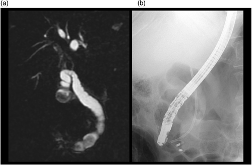
The patient was a 74-year-old man who had undergone biliary drainage using a straight-type 7F PS for cholangitis because of a common bile duct stone at a previous hospital (Fig. 1a).
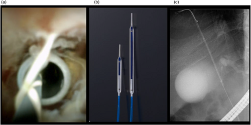
Stone removal was attempted in our hospital, but fluoroscopy showed that the PS had migrated into the bile duct (Fig. 1b). The stone was pushed toward the liver side and papillary balloon dilation was attempted, but this was difficult because of interference from the PS and stone. Therefore, removal of the migrated PS was attempted, first with grasping forceps under fluoroscopic guidance, but was unsuccessful because of the difficulty of grasping the PS. Removal using a basket was predicted to be difficult because of interference from the stone just above the papilla. Therefore, peroral digital cholangioscopy-assisted removal was attempted next. A digital cholangioscope (Spy DS; Boston Scientific, Natick, MA, USA) was inserted into the bile duct and visualized the migrated PS. Then, a 0.025 inch guidewire was passed through the stent's lumen under direct visualization (Fig. 2a). Subsequently, a novel small dilating balloon (3 mm × 6 cm, REN biliary dilation catheter; Kaneka Medix, Osaka, Japan) was inserted into the stent lumen5 (Fig. 2b,c, Video S1). By inflating the balloon, crimping the balloon and the PS, and pulling back slowly, the migrated PS was successfully removed through-the-scope without interference from the balloon catheter or stone. The novel dilating balloon is longer than conventional versions, allowing for stronger crimping. Finally, the stone was removed and the procedure was completed.


CONFLICT OF INTEREST
Author T.I. received lecture fees from Kaneka Medix and Boston Scientific. The other authors declare no conflict of interest for this article.


