Endoscopic ultrasound-assisted removal of an intrahepatic bile duct stone
Abstract
Watch a video of this article.
BRIEF EXPLANATION
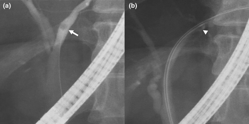
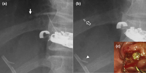
A 41-year-old woman with a left hepatic duct stone underwent endoscopic retrograde cholangiography for stone extraction (Fig. 1a). An over-the-wire type 8 wire basket catheter (Medi-Globe GmbH, Rohrdorf, Germany) failed to catch the stone and rather pushed the stone deeper (Fig. 1b). Because several attempts for stone extraction with a sphincterotome or ultrafine balloon catheter (REN; Kaneka Medix, Osaka, Japan) were unsuccessful, endoscopic retrograde cholangiography combined with endoscopic ultrasound was planned instead of cholangioscopy unfit for nondilated ducts. In the second session, the stone in B2 was depicted from the stomach using a curved linear-array echoendoscope (EG-740UT; FUJIFILM, Tokyo, Japan). Following a puncture of B2 with a 22G needle (SonoTip Pro Control; Medi-Globe) and contrast injection (Fig. 2a), a 0.018 inch guidewire was inserted into the common bile duct. After insertion of a double lumen catheter with a 3.6F tip (Uneven Double Lumen Cannula; Piolax Medical Devices, Kanagawa, Japan) into B2 upstream of the stone, pushing the stone by the guidewire or saline through the second lumen of the catheter was attempted without success. Then an endoscopic introducer (EndoSheather; Piolax Medical Devices) composed of a tapered inner catheter and large-bore outer sheath was inserted into the bile duct upstream of the stone. After removal of the guidewire and inner catheter, the stone was successfully moved to the hilum by flushing with saline through the outer sheath (Fig. 2b). Stone removal was finally accomplished after changing the scope to a duodenoscope without adverse events (Fig. 2c; Video S1). Endoscopic removal of intrahepatic bile duct stones is often challenging because of the difficulty of advancing extraction devices beyond the stone.1 Although the use of a sphincterotome2 or ultrafine balloon catheter3 has been reported, they did not work in the present case. This endoscopic ultrasound-assisted procedure for left intrahepatic bile duct stones may be a useful option when transpapillary attempts have failed.


Authors declare no conflict of interest for this article.


