Endoscopic submucosal dissection with a hand-made traction method for a tumor completely covering the post-appendectomy orifice
Abstract
Watch a video of this article
BRIEF EXPLANATION
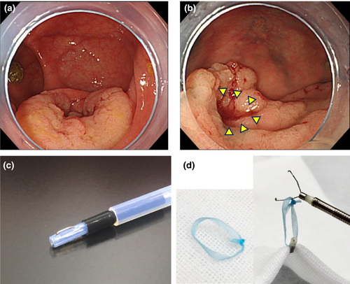
A 78-year-old woman was detected as having a laterally spreading tumor in the cecum, measuring 30 mm in diameter (Fig. 1a). The tumor completely covered the post-appendectomy orifice (Fig. 1b). Although severe fibrosis was expected, endoscopic submucosal dissection (ESD) was scheduled, since it was diagnosed as intramucosal cancer.

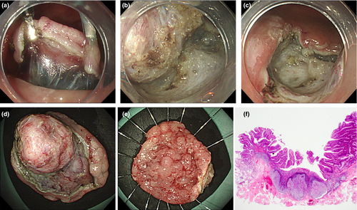
The entire procedure is presented in Video S1. Splash M-Knife and Mucosectom (Fig. 1c) (PENTAX MEDICAL, Tokyo, Japan) were used as the endo-devices, and a local injection of hyaluronic acid was administered. One of the main advantages of Mucosectom is that the insulated wide blade can be rotated against the muscular layer, enabling safe and rapid submucosal dissection. A circumferential incision and trimming sufficiently were performed using Splash M-knife. Subsequently, by applying two tractions with ring-thread made of dental floss on the opposite side, a good field of view was established for dissection (Figs 1d,2a). Although severe fibrosis was observed in the vicinity of the appendiceal orifice (Fig. 2b), the entire appendix could be successfully resected without perforation using Mucosectom (Fig. 2c–e). Histological examination revealed well-differentiated adenocarcinoma, depth Tis, and curative resection was achieved (Fig. 2f).

The efficacy of the traction method for colorectal ESD has been reported,1, 2 and this method is suitable for tumors located at the appendiceal orifice,3, 4 because of the thin luminal wall and higher risk of fibrosis. Usually, tumors completely covering the appendiceal orifice cannot be resected endoscopically, and surgery is required. Although severe fibrosis was expected, we considered that endoscopic treatment was feasible, because of postsurgical adhesion, which helps prevent intraoperative perforation. By using two ring-thread tractions5 against both sides of fibrosis, the endoscopic field of view was maintained despite the severe fibrosis, and the dissection line could be precisely defined.
By using ring-thread traction and Mucosectom®, a tumor covering post-appendectomy orifice, for which endoscopic treatment is technically challenging, was successfully resected by ESD.
Authors declare no conflicts of interest for this article.


