Feasibility of a distal pigtail shaped stent placement above the papilla for biliary obstruction
Abstract
Watch a video of this article
Brief Explanation
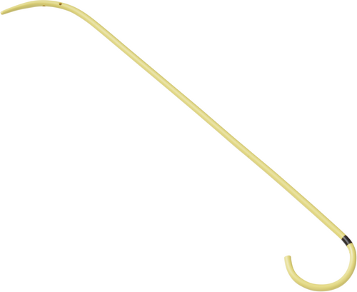
Endoscopic treatment for benign biliary strictures is widely performed because it is safe and repeatable. However, the stent for benign biliary strictures sometimes migrates due to the slippery bile duct. To solve the problem, we placed a unilateral pigtail stent named the “through-the-mesh” (TTM) stent (Fig. 1)1 above the papilla.

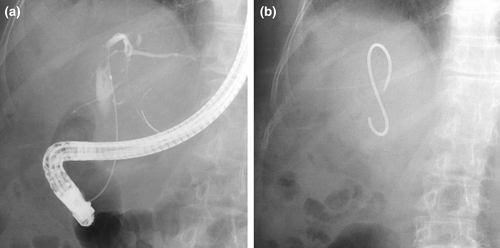
A 66-year-old female was referred to our hospital for biliary stricture and obstructive jaundice as an adverse event after living donor liver transplantation for hepatocellular carcinoma. We performed endoscopic retrograde cholangiography (ERC) which revealed the duct-to-duct anastomotic stricture (Fig. 2a). As the first treatment, we had placed a plastic inside stent with a proximal flap (8.5 Fr, 9-cm length) to bridge the stricture. After 3 months, we attempted stent exchange, however, the stent removal was difficult due to tight anchoring of the flap. To make stent exchange easier, we placed a shortened flap or a proximal pigtail shape stent. However, inside stent removal was subsequently difficult and cholangitis occurred repeatedly by the dislocation. Thus, we planned to deploy the TTM stent above the papilla for prevention of stent dislocation and for making stent exchange easier. After placing a guidewire in the bile duct, we smoothly placed the 7 Fr 14-cm TTM stent above the papilla (Fig. 2b). After that, stent dislocation had not occurred, and four months after the placement, we attempted to exchange the TTM stent. The removal of the TTM stent was easy by using forceps with post-EST status, and we also placed the same type stent without difficulty (Video S1).

This is the first report of a unilateral pigtail stent placement above the papilla for biliary obstruction. A unilateral pigtail stent placement above the papilla can be a rescue in some situations as well as the present case that has both biliary stricture and recurrent stent dislocation.
Authors declare no conflicts of interest for this article.


