Volume 32, Issue 4 pp. e61-e62
DEN Video Article
Free Access

Endoscopic closure of a large-size perforation using a novel through-the-scope twin endoclip in an ex vivo porcine stomach model

Qiang Zhang

Corresponding Author

Qiang Zhang

Guangdong Provincial Key Laboratory of Gastroenterology, Department of Gastroenterology, Nanfang Hospital, Southern Medical University, Guangzhou, China

Corresponding: Qiang Zhang, Guangdong Provincial Key Laboratory of Gastroenterology, Department of Gastroenterology, Nanfang Hospital, Southern Medical University, Guangzhou, Guangdong Province 510515, China. Email: [email protected]

Search for more papers by this author
Zhen Wang

Zhen Wang

Guangdong Provincial Key Laboratory of Gastroenterology, Department of Gastroenterology, Nanfang Hospital, Southern Medical University, Guangzhou, China

Search for more papers by this author
First published: 14 February 2020
Citations: 1

Abstract

Watch a video of this article

Brief Explanation

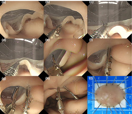
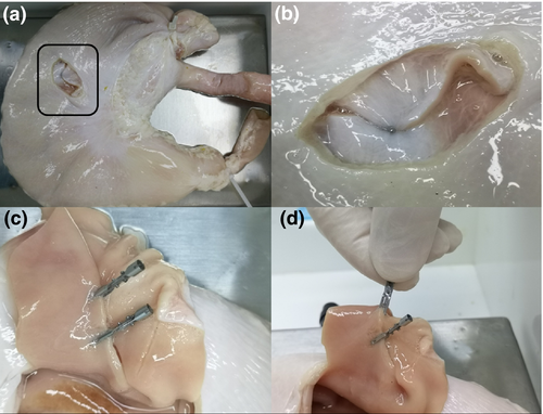
Closure of wound is very important for perforation during endoscopic submucosal dissection (ESD) and endoscopic full-thickness resection.1, 2 Through-the-scope twin endoclip (TTS-TC) was a technique developed by our team.3 In this study, TTS-TC was attempted for suturing a large-size perforation. An ex vivo porcine stomach and a model device for endoscopic submucosal dissection (ESD) in an animal experiment were prepared. Beforehand, a perforation of > 3 cm in size was artificially constructed on the greater curvature close to the anterior wall of porcine stomach with a blade (Fig. 1a), followed by perforation suture with TTS-TCs. The procedures were as follows (Fig. 1 and Video S1): (i) the endoscope (QF-260J; Olympus, Tokyo, Japan) was delivered from the porcine esophagus to the stomach; (ii) TTS-TC was sent to the site of perforation through the endoscopic working channel (Fig. 1b); (iii) With the assistance of operating the handle of TTS-TC, one side of TTS-TC was opened to tightly clamp the mucosal tissue on one side of the perforation (Fig. 1c,d). Then, the clamped mucosa was pulled close to the other side of the perforation, and the other side of TTS-TC was opened subsequently to clamp the mucosal tissue on the other side (Fig. 1e); (iv) After both mucosal tissues were clamped together, TTS-TC was released and the perforation was sutured (Fig. 1f). Two TTS-TCs were used to successfully close the perforation (Fig. 1g,h), and the operation time was 4.5 min. The excised tissue for establishing an artificial perforation was 3.5 × 2.8 cm in size (Fig. 1i). After closing the perforation, the effect of wound closure was observed from the inside and outside of the porcine stomach, respectively, and the closure of perforation was realized using TTS-TC firmly (Fig. 2). TTS-TC has a great potential to effectively suturing a large-sized perforation.
Details are in the caption following the image
Operational case of using through-the-scope twin endoclips (TTS-TCs). (a) A perforation of > 3 cm in an ex vivo porcine stomach model; (b) TTS-TC was delivered to the site of perforation through endoscopic working channel together with the endoscope; (c, d) One side of TTS-TC was opened to tightly clamp the mucosal tissue on one side of perforation; (e) The clamped mucosa was pulled close to the other side of the perforation, and the other side of TTS-TC was opened to clamp the mucosal tissue on the other side; (f) TTS-TC was released to close the perforation; (g) Another TTS-TC was used to close the rest of the perforation; (h) Two TTS-TCs were used to successfully close the perforation. (i) The excised specimen for establishing an artificial perforation.
Details are in the caption following the image
Observation of the closed perforation using through-the-scope twin endoclips (TTS-TCs) from inside and outside of porcine stomach. (a, b) After perforation suturing, it was observed from outside of porcine stomach that the perforation was successfully closed; (c) Following the anatomy of porcine stomach, it was observed from inside of porcine stomach that the perforation was closed smoothly; (d) The tail end of TTS-TC was pinched with fingers and the closed mucosa was lifted, and then TTS-TC detachment was not observed. It indicated the reliability of the effect using TTS-TCs for perforation closure.

Authors declare no conflicts of interest for this article.

    The full text of this article hosted at iucr.org is unavailable due to technical difficulties.