Safe hemostasis method using newly developed hemoclip for post-endoscopic papillary large balloon dilation with sphincterotomy bleeding
Abstract
Watch a video of this article
Brief explanation
This is the first report of successful hemostasis using SureClip (Micro-Tech, Nanjing, China) for hemorrhage in the papillary region.
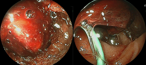
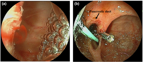
A 77-year-old woman with a history of gastric cancer who had undergone total gastrectomy with Roux-en-Y reconstruction 3 years previously was admitted for acute cholangitis caused by common bile duct (CBD) stones. For CBD stones, endoscopic retrograde cholangiopancreatography using short-type double-balloon enteroscopy (DBE) was carried out. After endoscopic papillary large balloon dilation with endoscopic sphincterotomy (EST), the stones were completely extracted. However, 3 days later, she developed anemia and melena. Emergency endoscope using short-type DBE was carried out, which detected a large amount of coagula and bleeding in the papillary area (Fig. 1). As the oozing bleeding point was clearly confirmed by underwater observation (Fig. 2a), hemostasis using SureClip was done (Fig. 2b), and hemostasis of the bleeding point was completed successfully in one attempt (Video S1). The patient was discharged without any complication such as obstructive cholangitis or acute pancreatitis, including re-bleeding.


Generally, post-EST bleeding occurs in 2–12%, with approximately 0.2% reported as severe cases.1 Hemostasis using hemoclips has been reported to be useful,2, 3 and various types are available for selection according to the patient's condition.4
The biggest advantage of SureClip is an easy re-grasping function which allows a trial gripping to make an accurate release at the target and helps to avoid closing the bile duct and pancreatic duct by mistake. Consequently, accurate hemostasis with a minimum number of clips is possible. In this case study, the bleeding point was gripped for a certain period until it was confirmed that bleeding was stopped and neither the bile duct nor the pancreatic duct was obstructed; it was then released. Hemostasis was completed successfully with one clip only. This method using the feature of SureClip is considered to be a useful method that can safely and accurately carry out hemostasis with the minimum number of clips in the duodenal papillary region especially for post-EST bleeding.
Authors declare no conflicts of interest for this article.


