Successful endoscopic ultrasound-guided gallbladder drainage through the mesh of a duodenal stent for cholecystitis in a patient with pancreatic cancer
Abstract
Watch a video of this article
Brief Explanation
Endoscopic ultrasound-guided gallbladder drainage (EUS-GBD) is an effective drainage technique for cholecystitis.1 EUS-GBD is preferred in patients with advanced malignancy because it obviates the need for an external drainage tube. However, in such cases, patients have often undergone previous placement of a duodenal stent. To the best of our knowledge, there is only one case report of EUS-guided biliary tract intervention after placement of a duodenal stent.2 Herein, we present the first case report of EUS-GBD through the mesh of a duodenal stent for cholecystitis in a patient with pancreatic cancer.
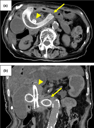
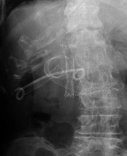
A 91-year-old woman with pancreatic cancer who had previously undergone placement of self-expandable metallic stents (SEMS) in the bile duct and placement of a duodenal stent in the second part of the duodenum experienced cholecystitis. Computed tomography indicated wall thickening of the gallbladder and abscess formation (Fig. 1). EUS-GBD was attempted because percutaneous gallbladder aspiration without tube placement was ineffective (Video S1). After inserting an EUS in the duodenal stent, the gallbladder was successfully imaged and punctured using a 19-gauge needle. Following dilation of the fistula, a fully covered SEMS (Wallflex, 10 × 60 mm; Boston Scientific Japan K.K., Tokyo, Japan) was deployed at the puncture site. In addition, a 7-Fr double-pigtail plastic stent was placed across the SEMS to prevent stent migration. Three days later, the SEMS had expanded between the mesh of the duodenal stent (Fig. 2). The patient recovered with conservative treatments although she suffered from mild peritonitis. Three months later, the patient died from the original disease, but she did not experience recurrence of cholecystitis.


Even in a patient with previous placement of a duodenal stent, EUS-GBD could be conducted without using a special technique. Previous placement of a duodenal stent may not be an obstacle to EUS-guided biliary tract intervention.
Authors declare no conflicts of interest for this article.


