WEO Newsletter
Re-growing the human esophagus
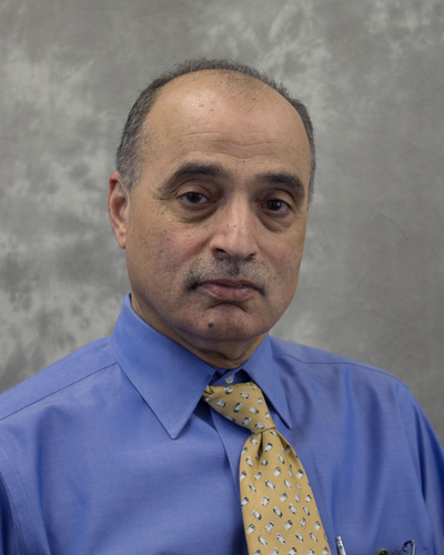
By Dr Kulwinder S. Dua, Professor of Medicine and Pediatrics, Medical College of Wisconsin Milwaukee, US
With an annual incidence of over 400,000 cases worldwide, several patients with esophageal cancers are undergoing esophagectomy. Similar surgery is also required for children born with long-segment esophageal atresia. Gastric pull-up conduits or colon interposition to re-establish luminal continuity after esophagectomy can be associated with significant morbidities rates (up to 70%; stasis of food in the conduit, reflux, fullness, anastomotic stricture and leaks).1 Re-growing the esophagus to restore luminal and functional continuity would be ideal for these patients.
De-novo organogenesis is possible using principles of regenerative medicine, as validated mostly in animal studies.2 These principles include transplanting tissue-engineered extracellular matrix (ECM)3 seeded with autologous pluripotent/stem cells. Researchers have successfully re-grown the human urinary bladder and the trachea.4, 5 Elliott et al. transplanted decellularized cadaveric human trachea populated with recipient's bone marrow stem-cells to re-grow the trachea in a 12 year-old child with congenital tracheal stenosis.5 These techniques can be demanding in terms of expertise, cost, regulations, and time.
Attempts to re-grow the human esophagus have so far been limited to only partial-thickness defects, like after endoscopic sub-mucosal dissection (ESD) or to patchy, non-circumferential transmural defects like perforations and leaks.6-9 Circumferential ESD for superficial cancer can be associated with over 70% risk of developing a stricture. To re-grow the mucosa, Okhi et al. cultured autologous buccal mucosal cells, harvested with a swab into cell-sheets around 2 weeks before planned ESD in nine patients.7 Small disks of these autologous cell-sheet were endoscopically implanted onto the raw esophageal surface immediately after ESD. Only one of the nine patients in this cohort developed a stricture.7 Extracellular matrix (ECM) and autologous stromal cell therapy have also been successfully used to prevent stricture formation after ESD.6, 8, 10, 11 Similar to partial-thickness defects, full-thickness patchy defects like perforation and leaks have also been successfully treated in human beings with devices like expandable stents, clips and suturing.9, 12, 13 Interestingly, the majority of these lesions heal with minimal need for additional measures, such as applying tissue matrix with or without autologous pluripotent cells.
Re-growing the human esophagus after long-segment, full-thickness, circumferential (LFC) defects, such as those present after a esophagectomy, has not been attempted until recently.14 All previous studies concerning re-growth of LFC esophageal defects were done in animal models. The basic principles involved were transplanting ECM molded into a tubular configuration into the recipient animal after populating it with autologous pluripotent cells with or without using a non-biological scaffold (for example, a silicone stent); to maintain the three-dimensional configuration of the esophagus during the slow process of regeneration. Since the pluripotent cells are autologous, immunosuppression to prevent organ rejection is not required. Whether allogenic or xenogeneic, ECM is ideal for tissue re-generation. They provide a biological scaffold on which pluripotent cells can spatially mature into site-specific phenotypic cells resulting in de-novo organogenesis. Suggested mechanisms include release of peptides, ligands and bioactive molecules to attract endogenous stem-cells, alter immune response, induce mitosis, and provide signals to local and migrant cells to mature into a site-specific organ.2, 15, 16
Badylak et al. resected 5-cm long esophagus segments in 22 dogs to create LFC defects. Porcine derived ECM molded into a tubular shape was used to bridge the defects.17 All dogs treated with EMC alone developed strictures while dogs with even as little as 30% of autologous smooth muscle cells which have pluripotent potentials colocalized with the EMC re-grew their esophagus emphasizing the importance of adding pluripotent cells to the matrix. Dogs were sacrificed at various intervals and histology confirmed re-growth of all the layers of the esophageal wall. Moreover, no porcine antigen was identified on immunohistology, thereby suggesting that the porcine ECM used to make the scaffold merely facilitated re-growth of the dog's own tissue.
Until recently, no attempts have been made to re-grow the human esophagus using principles of regenerative medicine as validated in animal models. Availability of less than ideal alternatives like gastric pull-up conduits and the ethical concern on the uncertainty of outcomes using experimental techniques may have been the reason for this. Recently, Dua et al. reported in The Lancet the first human case of re-growing the esophagus in a young adult who had exhausted all other options, including several surgical attempts to repair a long-segment defect.14 The 24-year-old patient presented with a life-threatening abscess, that led to a direct communication between the hypopharynx and the mediastinum. Earlier in his life, the patient had been in a car accident that required stabilization of the cervical spine with metal plates. The anterior metal plate had eroded into the hypopharynx that resulted in the infection and abscess formation. Several attempts at surgical repair failed and gastric pull-up was not possible due to the high location of the esophageal defect. On compassionate grounds, the authors attempted to re-grow a 5 cm LFC defect in the esophagus that extended up to the upper esophageal sphincter level. They used off-the-shelf, readily available, FDA approved for-man-use material. The three-dimensional configuration of the esophagus was maintained by three non-biological fully-covered expandable esophageal stents with the upper end of the proximal stent extending into the hypopharynx. FDA-approved decellularized donated human skin for tissue transplantation (AlloDerm®) was used as the ECM, to cover the defect from outside the stents. The dermal side of the matrix that attracts blood was oriented towards the mediastinum to facilitate neo-vascularization and the epithelial side that repels blood was oriented towards the lumen. To facilitate recruitment of pluripotent cells, the defect region was sprayed with the autologous platelet-rich plasma (PRP) and covered with the patient's sternocleidomastoid muscle. Being autologous PRP, there was no risk of transmitting blood-borne infections. Platelets are known to stimulate growth and regeneration, by releasing platelet-derived growth factors, transforming growth factors, and vascular endothelial growth factors that attract mesenchymal stem-cells, endothelial cells and epidermal cells, which express receptors for these growth factors.18-20.
Initially, the patient refused to give permission to remove the stents for fear of fistula and stricture formation. Eventually all the stents were removed over three-and-half years after placement. Follow-up endoscopy/biopsy, endoscopic-ultrasound, and high-resolution impedance-manometry showed squamous epithelium, normal five-layered esophageal wall, and normal peristaltic motility with bolus transit, respectively. After almost 5 years since the stents were removed, the patient is now eating a normal diet and maintaining his weight. The authors postulate that “by maintaining the three-dimensional morphological configuration of the esophagus in its natural milieu in vivo for an extended period of time and stimulating regeneration with ECM and PRP, it is possible to structurally and functionally re-grow the human esophagus”.14 An added attraction of this case was that rather than using expensive tissue-engineering techniques, using non-FDA approved material followed by surgical transplantation, the authors used readily available, off-the shelf FDA approved materials implanted in-vivo. Research is actively going on in testing various biomaterials seeded with autologous pluripotent cells. Based on the lessons learnt and questions raised from this case, large-scale animal studies, followed by phase I and II clinical trials, will be required. If the results can be replicated, it will have a large impact on patients requiring esophagectomy.14
References
Women's initiative in gastroenterology – WIGNAP – marks presence during World Congress
By Dr Sharmila Sachithanandan
Consultant Gastroenterologist, Ramsay Sime Darby Medical Centre, Kuala Lumpur, Malaysia Chairperson, WIGNAP
Women in Gastroenterology Network Asia-Pacific, known by its acronym WIGNAP, held a historical session during the World Congress of GI Endoscopy – ENDO 2017 in Hyderabad, India, in February 2017, with hundreds of enthusiastic delegates from across 20 countries – literally all corners of the world were in attendance.
WIGNAP was established in June 2014 by a group of pioneer female gastroenterologists from ten Asia-Pacific countries: Australia, China, Hong Kong, India, Japan, Korea, Myanmar, Malaysia, Taiwan and Thailand. The principle aim was to create a professional networking platform for female gastroenterologists across the vast and diverse continent of Asia, specifically with regards to developing training opportunities and mentoring of trainees and to cultivate and hone their leadership skills. In many parts of the world, this is already in place: the American Society of Gastroenterology (ASGE) and the United European Gastroenterology (UEG) have long prioritized the importance of equal opportunities for women. In Asia, prior to WIGNAP, there was no such forum for women specialists to meet, collaborate and share their professional experiences and challenges. The other objective of WIGNAP is to focus on women's GI health – and advocate for improved and adequate access to such care in this part of the world.
Since 2014, WIGNAP has held parallel sessions at the Asia Pacific Digestive Week (APDW) in Bali (2014), Taiwan (2015) and Kobe (2016). WIGNAP has also held training workshops in Endoscopic Ultrasound (EUS) and ERCP across several Asia Pacific countries.
The session held during ENDO 2017 went exceptionally well. Despite his hectic schedule, Dr Nageshwar Reddy, President of the World Congress, kindly graced the occasion with an opening address and his wife Dr Carol Reddy (herself an accomplished dermatologist), said a few inspiring words too. Participants to the session also enjoyed the presence of Dr David Carr-Locke (Past ASGE president), who offered encouraging remarks, as did Dr Dong Wan Seo (WEO Secretary General).
The session included several speakers: Dr Annette Fritscher-Ravens (Germany) and Dr Anand Sahai (Canada), gave candid, life-inspiring lectures. David Devonshire and Payal Saxena (Australia), Than Than Swe and Than Than Aye (Myanmar), Nonthalee Pausawasdi and Thawee Ratanachu-EK (Thailand), Shaesta Mehta (India), Joyce Peetermans (USA), Ryan Ponnudurai and Ida Hilmi (Malaysia) also spoke during the session.
The WIGNAP symposium included an exciting interactive panel discussion, with great participation from those in attendance. Chairpersons included Dr Maryam Al Khatry (UAE), Dr Hafeza Aftab (Bangladesh) and Dr Rupa Banerjee (India). The feedback received was very encouraging, especially from female trainees and residents, who felt more reassured and inspired – highlighting that these types of initiatives are very much needed. The next WIGNAP gathering will be at APDW 2017 in Hong Kong. Hope to see you all there!
WEO President awarded JGES honorary membership
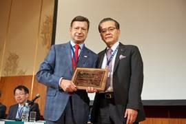
Congratulations to Dr Jean-Francois Rey, WEO President, on being awarded International Honorary Membership of Japan Gastroenterological Endoscopy Society (JGES)!
On May 11, 2017, Dr Rey received the International Honorary Membership of Japan Gastroenterological Endoscopy Society (JGES). This distinguished honor was presented by Professor Hisao Tajiri, JGES President, in recognition of Dr Rey's valuable contribution to JGES over the years. JGES is a preeminent professional organization dedicated to advancing the practice of GI endoscopy. Its mission is to evolve endoscopic procedures and educate endoscopists both in Asia and worldwide.
WEO Upcoming Events
For more information, visit http://www.worldendo.org/events/
- Program for Endoscopic Teachers (PET) RomeSeptember 14–16, 2017 – Rome, Italy
- Colorectal Cancer Screening Committee Meeting 2017 – EuropeOctober 27, 2017 – Barcelona, Spain
WEO Endorsed Events
- Asian EUS Congress 2017September 7–9, 2017 – Mumbai, India
- EUS ENDO 2017 - International Live CourseSeptember 28–29, 2017 – Marseille, France
- International Educational Endoscopy Video Forum - IEEF 2017October 5–6, 2017 – Sochi, Russia
- Jakarta International GI Endoscopy Symposium & Live Demonstration 2017October 6–7, 2017 – Jakarta, Indonesia
- 5th International Symposium on Complications in GI EndoscopyNovember 2, 2017 – Hamburg, Germany
For more news and events covering the world of GI endoscopy, visit WEO's official website: http://www.worldendo.org.
You can sign up to receive the WEO newsletter by email.


